. 1999 Jul 15;19(14):5782-91.
doi: 10.1523/JNEUROSCI.19-14-05782.1999.

alpha-Synuclein shares physical and functional homology with 14-3-3 proteins

Affiliations

alpha-Synuclein shares physical and functional homology with 14-3-3 proteins

N Ostrerova et al. J Neurosci. .

Abstract

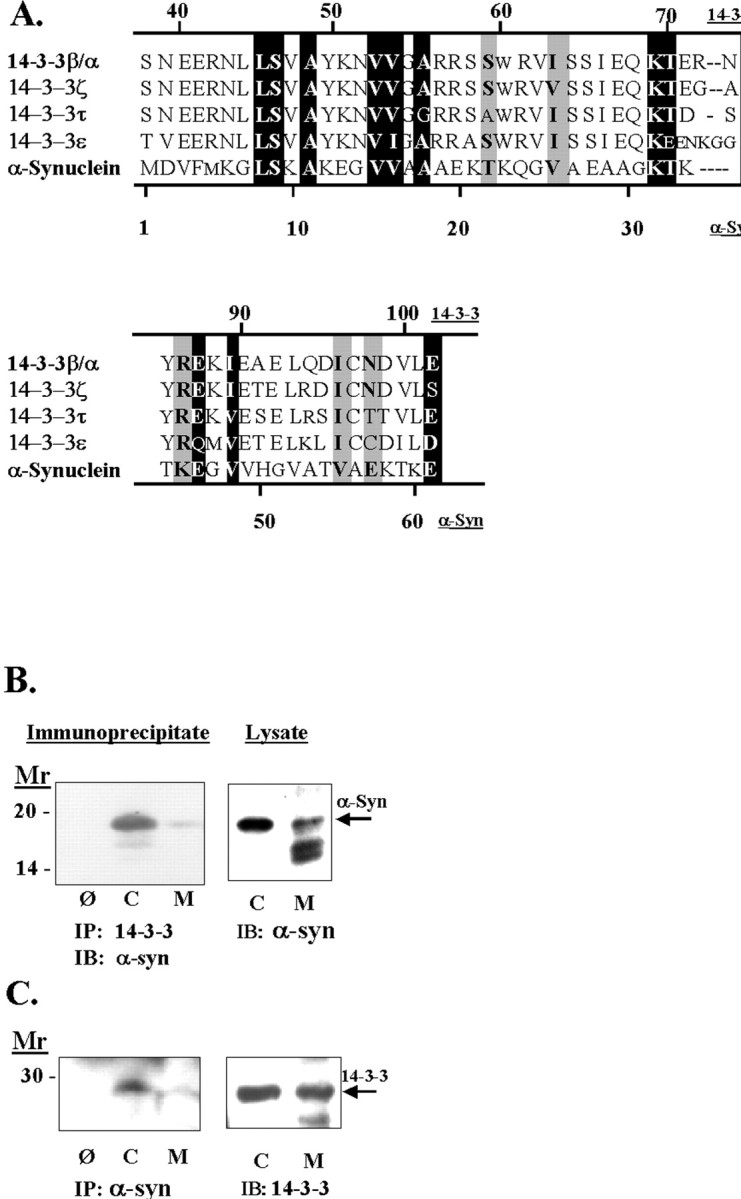
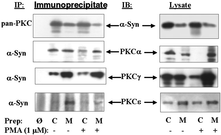
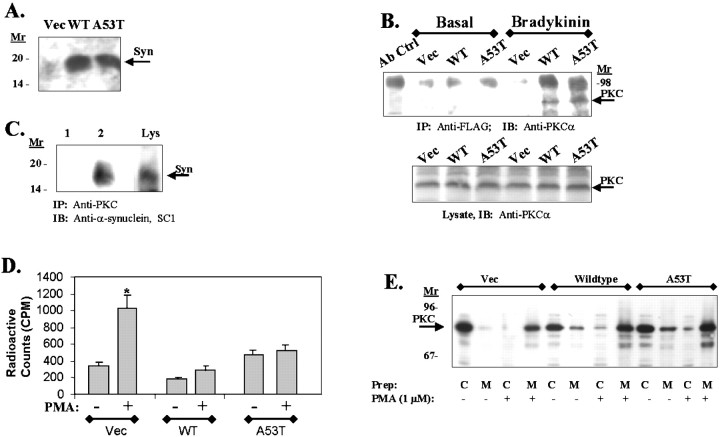
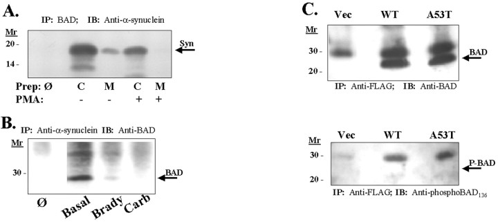
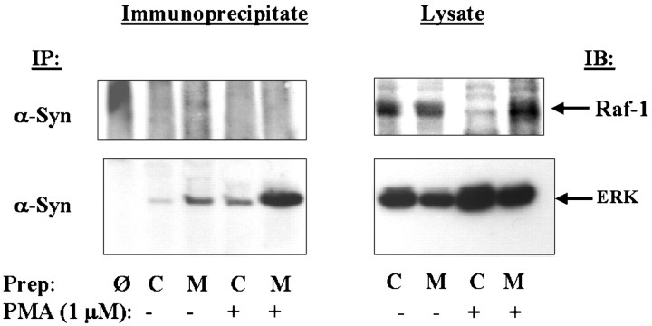
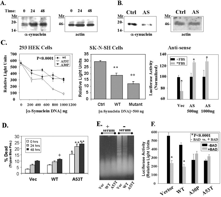
alpha-Synuclein has been implicated in the pathophysiology of many neurodegenerative diseases, including Parkinson's disease (PD) and Alzheimer's disease. Mutations in alpha-synuclein cause some cases of familial PD (Polymeropoulos et al., 1997; Kruger et al., 1998). In addition, many neurodegenerative diseases show accumulation of alpha-synuclein in dystrophic neurites and in Lewy bodies (Spillantini et al., 1998). Here, we show that alpha-synuclein shares physical and functional homology with 14-3-3 proteins, which are a family of ubiquitous cytoplasmic chaperones. Regions of alpha-synuclein and 14-3-3 proteins share over 40% homology. In addition, alpha-synuclein binds to 14-3-3 proteins, as well as some proteins known to associate with 14-3-3, including protein kinase C, BAD, and extracellular regulated kinase, but not Raf-1. We also show that overexpression of alpha-synuclein inhibits protein kinase C activity. The association of alpha-synuclein with BAD and inhibition of protein kinase C suggests that increased expression of alpha-synuclein could be harmful. Consistent with this hypothesis, we observed that overexpression of wild-type alpha-synuclein is toxic, and overexpression of alpha-synuclein containing the A53T or A30P mutations exhibits even greater toxicity. The activity and binding profile of alpha-synuclein suggests that it might act as a protein chaperone and that accumulation of alpha-synuclein could contribute to cell death in neurodegenerative diseases.


Figures

Fig. 1.
Fig. 1.
A, Alignment of α-synuclein and the 14-3-3 family of proteins. The alignment was performed using the Multalin program (http://www.toulouse.inra.fr/multalin.html). To observe homology, we excluded the first 30 amino acids of 14-3-3 proteins while performing the alignment algorithm. Exact matches are shown as white letters on a black background, and matches of proteins with similar properties are shown as black letters on a gray background. In addition, we have noted aligned serines and threonines in black because both amino acids can be phosphorylated by serine/threonine kinases. B, Association of α-synuclein with 14-3-3. Rat brain tissue was fractionated into cytoplasmic (C) and membrane (M) components, taken up in immunoprecipitation buffer, and treated as shown. Theleft shows an immunoblot of a 14-3-3β immunoprecipitate with anti-α-synuclein antibody, and theright shows an immunoblot of the lysates. The omit lane is labeled Ø and refers to immunoprecipitations done using protein A but no 1° antibody. C, Theleft shows an immunoblot of α-synuclein immunoprecipitates with anti-14-3-3ε antibody, and theright shows an immunoblot of the lysates. The omit lane is labeled Ø and refers to an immunoprecipitation done using protein A but no 1° antibody.
Fig. 2.
Fig. 2.
Association of α-synuclein with PKC isoforms in rat brain tissue. Left panels show immunoblots of α-synuclein immunoprecipitates with isoform-specific PKC antibodies. Right panels show immunoblots of each PKC isoform in the membrane and cytoplasmic fractions. Before each immunoprecipitation, half of the brain homogenates were treated with 1 μm PMA plus 1 mm Ca2+ at 30°C for 30 min. The homogenates were then fractionated into membrane and cytoplasmic components taken up in immunoprecipitation buffer, and proteins were isolated as shown. Cytoplasmic, C; membrane, M; omit, Ø, which is an immunoprecipitation done using protein A but no 1° antibody.
Fig. 3.
Fig. 3.
A, An immunoblot of 293 HEK cell lines stably transfected with empty vector (Vec), wild-type (WT), or A53T α-synuclein using α-synuclein antibody SC1. Control 293 HEK cells do endogenously express low levels of α-synuclein (arrow), whereas the transfected cells show increased expression of the 19 kDa α-synuclein proteins. B, Immunoblot of PKCα in α-synuclein immunoprecipitates. 293 HEK cell lines stably transfected with empty vector (Vec), wild-type (WT), or A53T α-synuclein were grown under basal conditions or treated with 20 nm bradykinin for 30 min, and the lysates were immunoprecipitated using agarose-coupled anti-FLAG resin. Immunoblotting of the resulting samples with anti-PKC type III antibody showed coassociation of α-synuclein with PKCα only in the bradykinin-treated samples (top panel). Thebottom panel shows an immunoblot of PKCα in the corresponding total cell lysates. C, Immunoblot of α-synuclein in PKCα immunoprecipitates. Lane 1, Lysates were treated with 1 μm PMA for 30 min, and PKCα was immunoprecipitated using anti-pan-PKC antibody (lane 2) (this antibody recognizes PKCα, β, and γ; Upstate Biotechnology) and immunoblotted with anti-α-synuclein SC1 antibody. Lane 2 shows an immunoprecipitation with the anti-pan-PKC antibody omitted. Lane 3(Lys) shows a parallel anti-synuclein immunoblot of the lysate (30 μg of lysate). No reactivity was seen in absence of PMA stimulation. D, PKC activity does not increase in 293 HEK cell lines overexpressing wild-type or A53T α-synuclein after stimulation with PMA (1 μm, 30 min). *p < 0.001. E, Immunoblots of PKCα in fractionated cell lysates (cytoplasm to membrane) after treatment with PMA (1 μm, 30 min). All cell lines (vector, wild-type α-synuclein, and A53T α-synuclein) showed robust translocation of PKCα after PMA treatment. C, Cytoplasm; M, membrane.
Fig. 4.
Fig. 4.
A, Coimmunoprecipitation of α-synuclein and BAD from brain lysate. Homogenates from rat cortex were fractionated into membrane and cytoplasmic components and taken up in immunoprecipitation buffer. BAD protein was then immunoprecipitated, and precipitates were immunoblotted with monoclonal anti-α-synuclein. Cytoplasmic, C; membrane, M; omit,Ø, which is an immunoprecipitation done using protein A but no 1° antibody. B, Association of α-synuclein with BAD in 293 HEK cells and regulation by agents that stimulate PKC. 293 HEK cells (which express endogenous α-synuclein) were treated with carbachol (1 mm, 30 min) or bradykinin (20 nm, 30 min). α-Synuclein was then immunoprecipitated from total cellular lysates with anti-synuclein SC1 antibody, and the resulting immunoblots were probed with anti-BAD antibody.Ø represents an immunoprecipitation performed without 1° anti-α-synuclein antibody. Immunoblots of the lysates showed that equal amounts of protein were loaded in each lane (data not shown). C, Immunoblot of BAD (arrow,top) or phospho-BAD136(bottom, arrow points to absent band; 1:200; New England Biolabs, Beverly, MA) after immunoprecipitation of FLAG-tagged α-synuclein from HeLa cell lysates. α-Synuclein was immunoprecipitated with agarose-coupled anti-FLAG resin, and the immunoblots were probed with anti-BAD antibody (top) or anti-phospho-BAD136 antibody (bottom). No specific staining for phospho-BAD136 was observed. The bands in the phospho-BAD136 immunoblot were present in the control cell line that was not transfected with FLAG-tagged α-synuclein, and therefore these bands represent nonspecific binding. An immunoblot of the lysates showed equal expression of FLAG-tagged wild-type and A53T α-synuclein (data not shown).
Fig. 5.
Fig. 5.
Association of α-synuclein with members of the ERK cascade. Left panels show immunoblots of α-synuclein immunoprecipitates with antibodies to either Raf-1 or ERK. No association was seen with Raf-1, whereas strong binding was seen with ERK. Right panels show immunoblots with the Raf-1 and ERK antibodies in the membrane and cytoplasmic fractions. Before each immunoprecipitation, half of the rat brain homogenates were treated with 1 μm PMA plus 1 mmCa2+ at 30°C for 30 min. The homogenates were then fractionated into membrane and cytoplasmic components and taken up in immunoprecipitation buffer, and proteins were isolated as shown. Cytoplasmic, C; membrane, M, and omit,Ø, which is an immunoprecipitation done using protein A but no 1° antibody.
Fig. 6.
Fig. 6.
Toxicity of α-synuclein. A, α-Synuclein expression increased during incubation of 293 HEK cells in serum-free medium. The left shows an immunoblot of α-synuclein with SC1 antibody, and the right shows the same immunoblot reprobed with anti-actin antibody (Sigma).B, Transfection of 293 HEK cells with an antisense α-synuclein construct (AS; 2 μg) reduced the amount of endogenous α-synuclein expressed in the cells (left). The control lane(Ctrl) shows lysates from cells transfected with an empty pcDNA3 plasmid under the same conditions at the same time. The immunoblot was then reprobed with anti-actin antibody to show that equal amounts of protein were loaded in each lane(right). C, Transient transfection of 293 HEK (left) or SK-N-SH cells (middle) with a pGL3 luciferase plasmid and vector, wild-type, A53T, or A30P α-synuclein constructs induces a dose-dependent decrease in luciferase activity. In contrast, transfection with antisense α-synuclein increased luciferase expression (right). For the antisense experiments in the right, cells were transfected and then serum-deprived for 24 hr, after which luciferase activity was measured. Cells transfected with antisense α-synuclein showed less toxicity than cells transfected with vector. *p < 0.05; **p < 0.01;n = 4 for each point. D, Similar experiments showed a dose-dependent increase in toxicity as shown by trypan blue staining after serum deprivation for 0, 24, or 48 hr. Parallel experiments with a β-galactosidase vector showed a 40% transfection rate in 293 HEK cells. Based on 20% of the cells being positive for trypan blue after transfection, we estimate that transfection of 1 μg of A53T α-synuclein induced ∼50% cell death in 293 HEK cells. +p < 0.05; *p < 0.01; **p < 0.001.E, Effects of α-synuclein on DNA fragmentation. No fragmentation was seen under basal growth conditions in the control cell line (Vec, lane 1), wild-type α-synuclein overexpresser (WT, lane 2), or A53T α-synuclein expresser (A53T, lane 3). After 24 hr incubation in serum-free medium, oligomeric DNA fragmentation was strong in the control cell line (lane 4), moderate in the wild-type α-synuclein cell line (lane 5), and absent in the A53T α-synuclein cell line (lane 6). A slight increase in highly degraded DNA is apparent in lane 6 above the dye front, suggesting increased necrotic DNA. F, Wild-type α-synuclein increases BAD toxicity, but mutant α-synuclein (A53T and A30P) does not increase BAD toxicity. 293 HEK cells were cotransfected with the constitutively active 1 μg of pGL3 luciferase plasmid with or without 100 ng of BAD, and with or without 500 ng of α-synuclein (wild-type, A53T, or A30P). A β-galactosidase vector was used as a ballast to maintain the DNA amount at 2 μg; this vector does not affect luciferase activity. *p < 0.0001; n = 4; comparing samples with or without BAD. The A53T and A30P transfections alone were also significantly different from vector at p < 0.0001;n = 4.

References

    1. Aitken A, Howell S, Jones D, Madrazo J, Martin H, Patel Y, Robinson K. Post-translationally modified 14-3-3 isoforms and inhibition of protein kinase C. Mol Cell Biochem. 1995;149:41–49. - PubMed
    1. Atwood C, Moir R, Huang X, Scarpa R, Bacarra N, Romano D, Hartshorn M, Tanzi R, Bush A. Dramatic aggregation of Alzheimer abeta by Cu(II) is induced by conditions representing physiological acidosis. J Biol Chem. 1998;273:12817–12826. - PubMed
    1. Behl C, Davis J, Klier F, Schubert D. Amyloid β peptide induces necrosis rather than apoptosis. Brain Res. 1994;645:253–264. - PubMed
    1. Broadie K, Rushton E, Skoulakis E, Davis R. Leonardo, a Drosophila 14-3-3 protein involved in learning, regulates presynaptic function. Neuron. 1997;19:391–402. - PubMed
    1. Clayton D, George J. The synucleins: a family of proteins involved in synaptic function, plasticity, neurodegeneration and disease. Trends Neurosci. 1998;21:249–254. - PubMed

Publication types

MeSH terms