. 1999 Jul 15;19(14):5792-801.
doi: 10.1523/JNEUROSCI.19-14-05792.1999.

Estrogen stimulates a transient increase in the number of new neurons in the dentate gyrus of the adult female rat

Affiliations

Estrogen stimulates a transient increase in the number of new neurons in the dentate gyrus of the adult female rat

P Tanapat et al. J Neurosci. .

Abstract

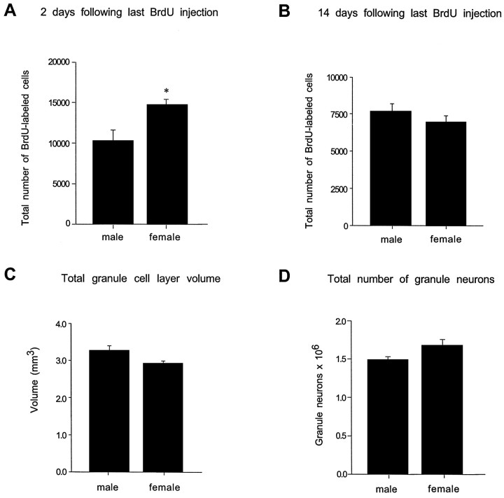
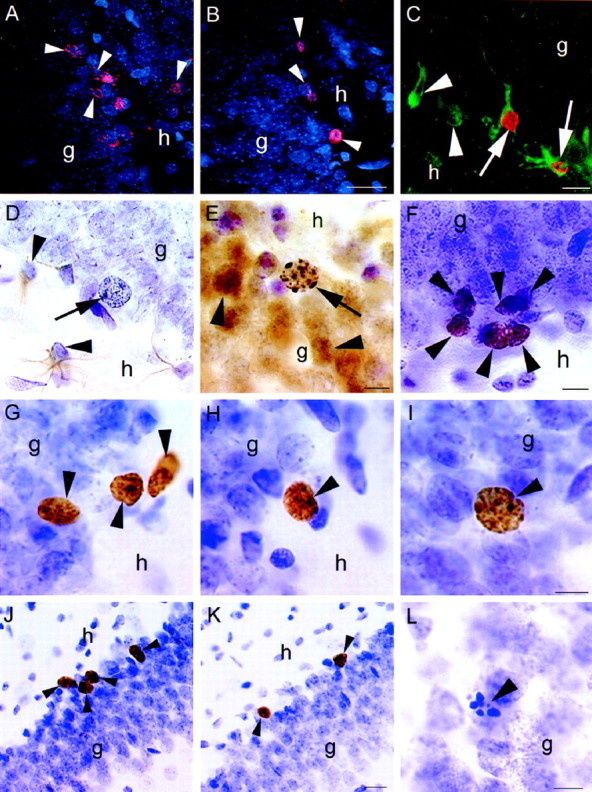
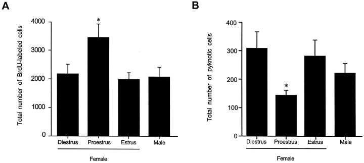
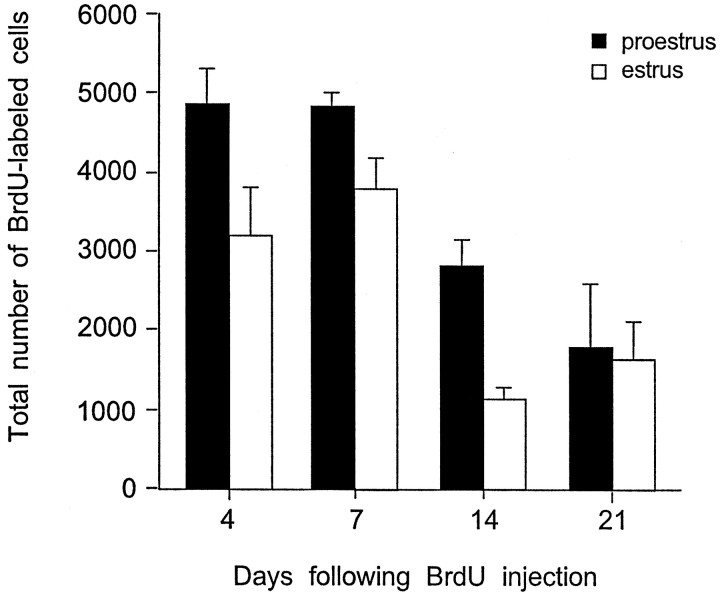
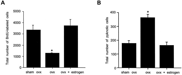
To determine whether a sex difference exists in the production of hippocampal cells during adulthood, we examined proliferating cells and their progeny in adult rats using the thymidine analog bromodeoxyuridine (BrdU) combined with immunohistochemistry for markers of neurons and glia. Additionally, to determine whether ovarian hormones affect cell proliferation, we examined the numbers of BrdU-labeled cells at different estrous cycle stages and after ovarian steroid manipulation. Stereological analyses of the numbers of BrdU-labeled cells revealed that females produced more cells than males in the dentate gyrus but not in the subventricular zone. The production of new hippocampal cells in females appears to be affected by ovarian hormone levels; ovariectomy diminished the number of BrdU-labeled cells, an effect reversed by estrogen replacement. A natural fluctuation in cell proliferation was also noted; females produced more cells during proestrus (when estrogen levels are highest) compared with estrus and diestrus. Many of these cells acquired neuronal characteristics, including the formation of dendrites and expression of Turned-On-After-Division 64 kDa, a marker of immature granule neurons, and the calcium-binding protein calbindin, a marker of mature granule neurons. However, examination of the numbers of pyknotic cells and the numbers of BrdU-labeled cells at longer survival times revealed that many new cells in the dentate gyrus eventually degenerate. Consistently the number of labeled cells in females is no longer higher than that observed in males by 2 weeks after the last BrdU injection. These findings suggest that estrogen-enhanced cell proliferation during proestrus results in more immature neurons in the hippocampal formation of females compared with males and present the possibility that these new cells exert an important influence on hippocampal function.


Figures

Fig. 1.
Fig. 1.
Stereological estimates of the total numbers of BrdU-labeled cells in the dentate gyrus of adult male and female rats.A, Two days after a period of five daily BrdU injections, female rats had significantly more BrdU-labeled cells in the dentate gyrus compared with age-matched males. B, By 14 d after the last BrdU injection, this sex difference was no longer evident. No significant differences in the granule cell layer volume (C) or total number of granule neurons (D) were observed between males and females. Bars represent mean + SEM, each obtained from four to six animals.Asterisk indicates a significant difference (p < 0.05).
Fig. 2.
Fig. 2.
Confocal laser-scanning images of BrdU-labeled cells (arrowheads) in the dentate gyrus of female (A) and male (B) rats injected with BrdU five times and perfused 2 d after the last injection. More BrdU-labeled cells were observed in females compared with males. C, Confocal image of BrdU-labeled cells colabeled with TOAD-64, a marker of immature neurons (arrow), in the dentate gyrus of an adult female.Arrowheads indicate TOAD-64-labeled cells not labeled with BrdU. D, Light microscopic image of a BrdU-labeled cell (arrow) that is not colabeled with GFAP, a marker of astroglia, in the dentate gyrus of an adult female.Arrowheads indicate GFAP-labeled cells not labeled with BrdU. E, BrdU-labeled cell (arrow) that is colabeled with calbindin, a marker of mature neurons, in the dentate gyrus of an adult female. Arrowheads indicate calbindin-labeled cells not labeled with BrdU. F, Ki67-labeled cells (arrowheads) in the subgranular zone of the dentate gyrus. These cells were similar in morphology and location to BrdU-labeled cells at early time points. BrdU-labeled cells (arrowheads) at 4 d (G), 7 d (H), and 21 d (I) after BrdU injection in the dentate gyrus of the adult female. With increasing time after BrdU injection, many BrdU-labeled cells become incorporated into the granule cell layer and express the morphological characteristics of mature granule neurons. More BrdU-labeled cells (arrowheads) were observed in animals injected during proestrus (J) compared with those injected during estrus (K). These animals were perfused 2 hr after injection with BrdU. L, Pyknotic cell (arrowhead) in the dentate gyrus of an adult female. These cells were more numerous in females than males during the majority of the estrous cycle, g, Granule cell layer;h, hilus. Scale bars: B, 25 μm (applies to A, B); C, 10 μm;E, 10 μm (applies to D,E); F, 10 μm; I, 10 μm (applies to G–I); K, 25 μm (applies toJ, K); L, 10 μm.
Fig. 3.
Fig. 3.
Stereological estimates of the total number of BrdU-labeled cells in the dentate gyrus (A) and pyknotic cells in the sgz (B) of adult female rats injected with BrdU during diestrus, proestrus, or estrus and in age-matched male rats. All animals were perfused 2 hr after BrdU injection. Female rats injected during proestrus had significantly more BrdU-labeled cells compared with females in estrus or diestrus, as well as compared with age-matched males. Conversely, female rats in proestrus on the day of perfusion had significantly fewer pyknotic cells compared with females in estrus or diestrus, as well as compared with age-matched males. Bars represent mean + SEM each obtained from five or six animals. Asterisk indicates a significant difference from all other means (p < 0.05).
Fig. 4.
Fig. 4.
Stereological estimates of the total number of BrdU-labeled cells in the dentate gyrus of adult female rats 4, 7, 14, and 21 d after a single BrdU injection administered during proestrus (black bars) or estrus (white bars). The numbers of BrdU-labeled cells decreased over time in both groups but were greater in females injected during proestrus up to 14 d after BrdU labeling. By 21 d, this difference was no longer detectable. Bars represent mean + SEM each obtained from three animals. Asterisk indicates significant difference from proestrus (p < 0.05).
Fig. 5.
Fig. 5.
Stereological estimates of the total number of BrdU-labeled cells (A) and pyknotic cells (B) in the dentate gyrus of sham ovariectomized, ovariectomized, and estrogen-replaced ovariectomized adult female rats. Ovariectomy produced a significant decrease in the numbers of BrdU-labeled cells in the dentate gyrus of adult female rats compared with both sham-operated controls (the majority of which were in proestrus at the time of BrdU injection) and estrogen-replaced, ovariectomized animals. Conversely, ovariectomy doubled the numbers of pyknotic cells in the dentate gyrus of adult female rats compared with both sham-operated controls and estrogen-replaced, ovariectomized animals. Bars represent mean + SEM each obtained from four or five animals. Asterisk indicates significant difference from other means (p < 0.05).

References

    1. Altman J, Das GD. Autoradiographic and histological evidence of postnatal hippocampal neurogenesis in rats. J Comp Neurol. 1965;124:319–335. - PubMed
    1. Arnold AP, Breedlove SM. Organizational and activational effects of sex steroids on brain and behavior: a reanalysis. Horm Behav. 1985;19:469–498. - PubMed
    1. Barnea A, Nottebohm F. Seasonal recruitment of hippocampal neurons in adult free-ranging black-capped chickadees. Proc Natl Acad Sci USA. 1994;91:11217–11221. - PMC - PubMed
    1. Bayer SA. Changes in the total number of dentate granule cells in juvenile and adult rats: a correlated volumetric and 3H-thymidine autoradiographic study. Exp Brain Res. 1982;46:315–323. - PubMed
    1. Bucci DJ, Chiba AA, Gallagher M. Spatial learning in male and female Long–Evans rats. Behav Neurosci. 1995;109:180–183. - PubMed

Publication types

LinkOut - more resources