. 1999 Jul 15;19(14):5823-33.
doi: 10.1523/JNEUROSCI.19-14-05823.1999.

Subcellular localization of full-length and truncated Trk receptor isoforms in polarized neurons and epithelial cells

Affiliations

Subcellular localization of full-length and truncated Trk receptor isoforms in polarized neurons and epithelial cells

D Kryl et al. J Neurosci. .

Abstract

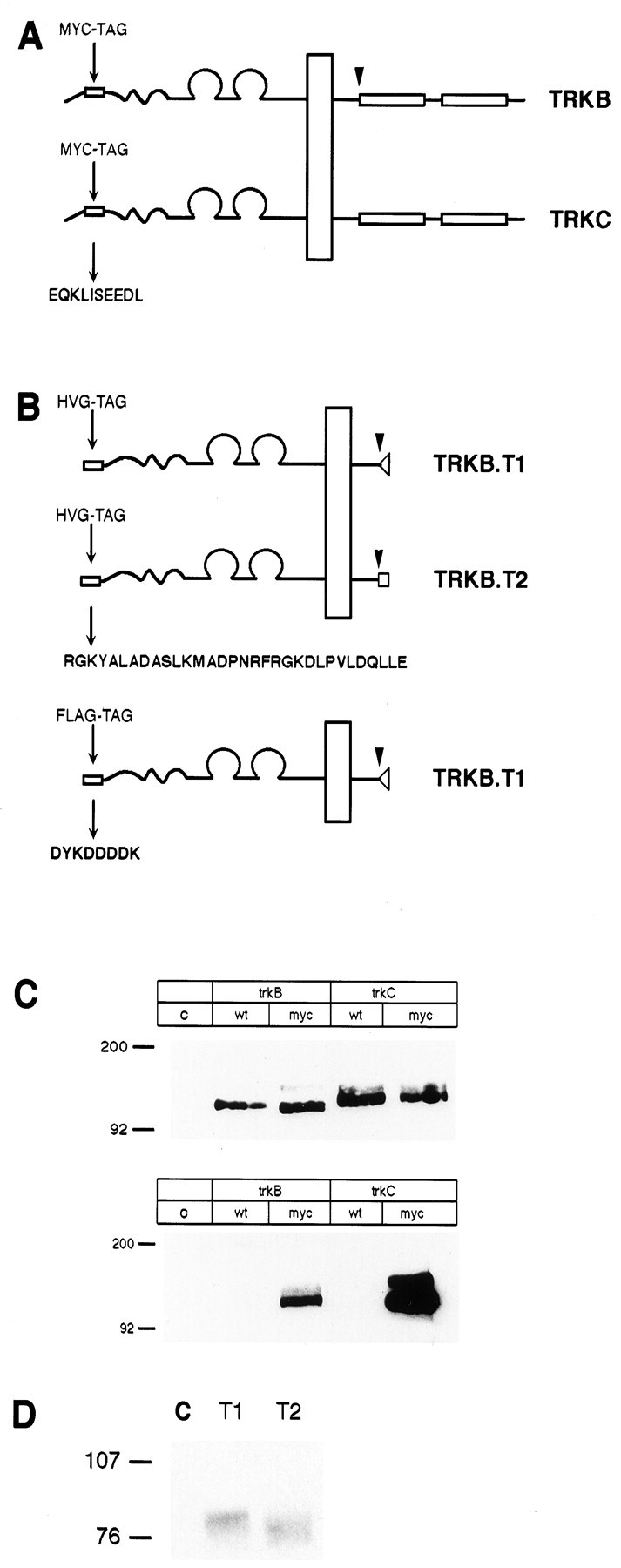
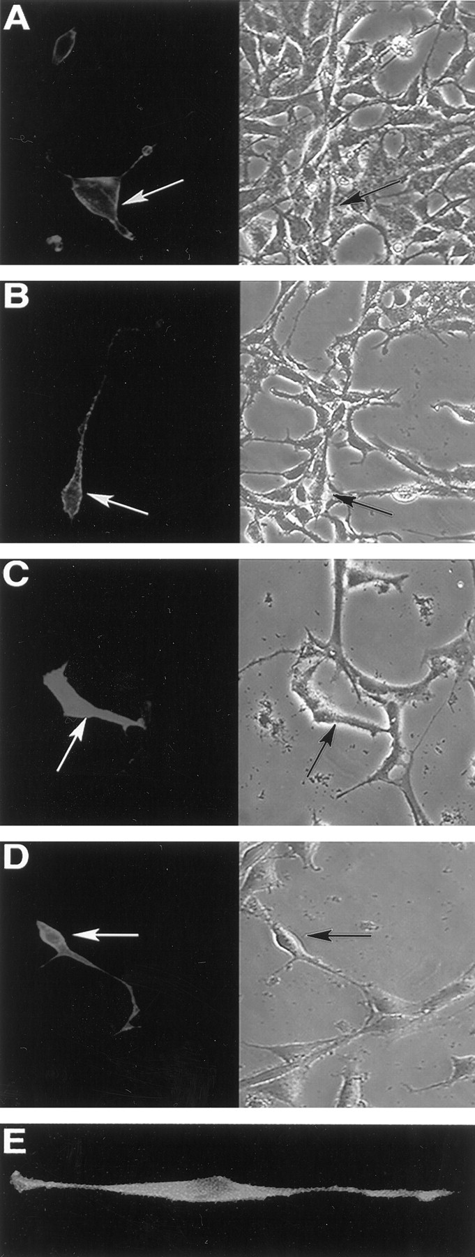
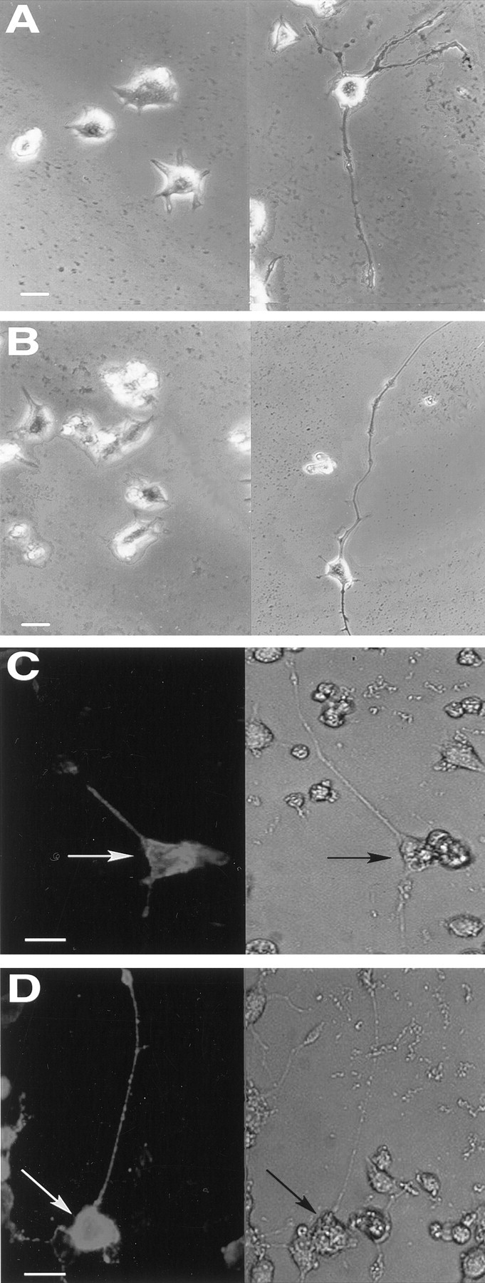
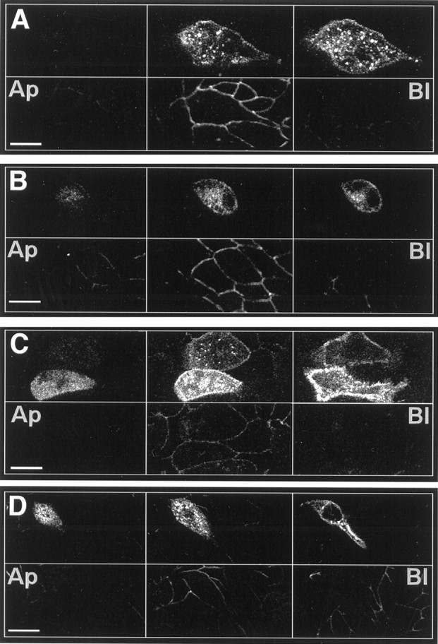
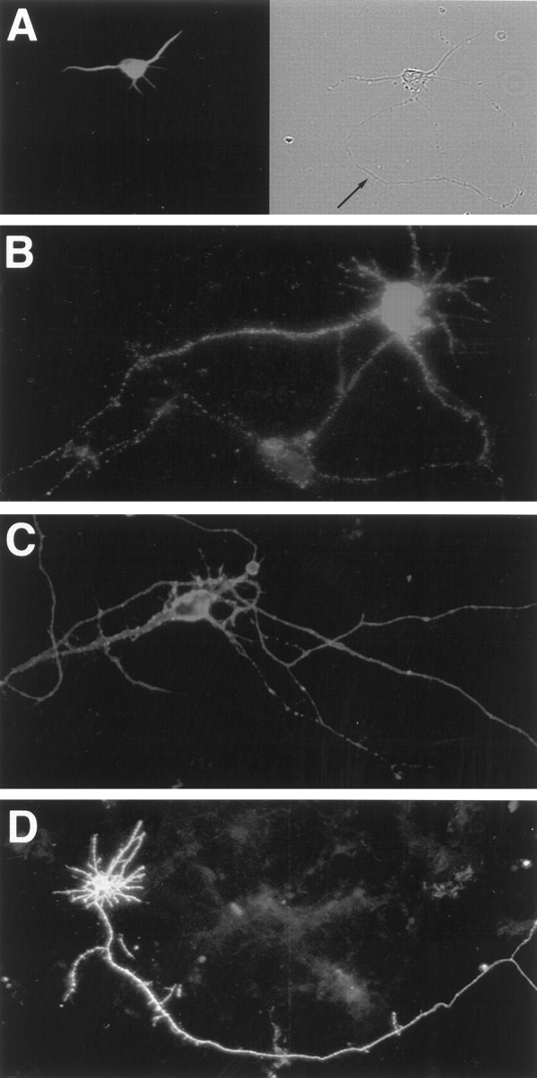
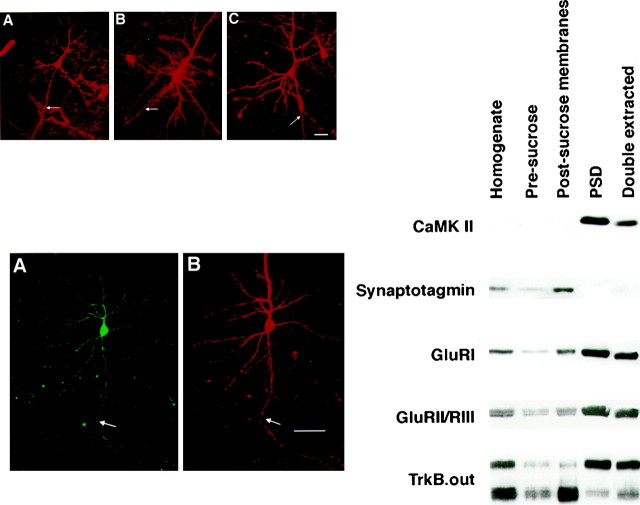
Neurotrophins affect neuronal development and plasticity via spatially localized effects, yet little is known about the subcellular distribution of the Trk neurotrophin receptors and the impact of this distribution on neurotrophin action. To address this, we examined the subcellular location of full-length TrkB and TrkC tyrosine kinase receptors and truncated TrkB isoforms after transfection of Madin-Darby canine kidney (MDCK) cells, dissociated primary hippocampal neurons, and cortical neurons within intact brain slices. Myc-, herpes virus glycoprotein (HVG)-, or FLAG-derived epitope-tagged receptor isoforms were created to allow their unambiguous identification and localization after transfection. All tagged receptors were appropriately synthesized, and full-length myc-TrkB and myc-TrkC mediated appropriate neurotrophin-signaling events. We found that full-length TrkB receptors were excluded from the apical domain of MDCK cells but that TrkC receptors were present in both apical and basolateral domains. Full-length TrkB and TrkC were found throughout transfected primary cultured hippocampal neurons and transfected neurons in neocortical brain slices and showed no evidence of vectorial sorting. Truncated forms of TrkB were also homogeneously distributed in MDCK cells, dissociated hippocampal neurons, and cortical neurons within slice preparations. Levels of full-length and truncated TrkB were examined in postsynaptic densities; both receptor isoforms were present but only moderately enriched in these structures. Together, these findings suggest that Trk receptors are uniformly distributed in both axonal and dendritic compartments and that local neurotrophin responses are controlled by other mechanisms.


Figures

Fig. 1.
Fig. 1.
Epitope-tagged TrkB and TrkC receptors are fully processed and recognized by epitope tag–specific antibodies.A, B, Schematic representations of the placement of c-myc, HVG, and FLAG epitope tag insertion into full-length TrkB and TrkC (A) and truncated TrkB iso forms (B) are shown.Arrowheads show the point of sequence divergence between full-length and truncated TrkB isoforms. C, HEK293 cells were transiently transfected with myc-TrkB and myc-TrkC, and the receptors were immunoprecipitated with pan-Trk antibody 203 and immunoblotted with antibody 203 (top) or with 9E10 (bottom). D, HEK293 cells transiently transfected with tagged TrkB-T1 or -T2 were immunoprecipitated with 5B6 and immunoblotted with 5B6. Lane C (lane c in C) represents immunoprecipitate from control, nontransfected HEK293 cells.wt, Wild type.
Fig. 2.
Fig. 2.
Epitope-tagged receptors are expressed at the cell surface. Nonpermeabilized HEK293 cells were fixed in 4% paraformaldehyde and immunostained with 9E10 (A, B), 5B6 (C, D), or M2 (E) to detect transiently transfected epitope-tagged TrkB (A), epitope-tagged TrkC (B), epitope-tagged TrkB-T1 (C), and epitope-tagged TrkB-T2 (D). NIH 3T3 fibroblasts expressing FLAG–TrkB-T1 (E) are shown. Bright-field images of the fields are shown on the rightin A–D. Arrows indicate the same transfected cell in adjacent panels. Control experiments (data not shown) confirmed that 4% paraformaldehyde alone does not permeabilize HEK293 cells; intracellular epitopes are detected after fixation with 4% paraformaldehyde only if cells were treated with 0.3% Triton X-100.
Fig. 3.
Fig. 3.
Epitope-tagged TrkB and TrkC are functional and distributed throughout cell bodies and neurites of differentiated PC12 cells. A, B, PC12 cells were transiently transfected with either tagged TrkB (A) or TrkC (B), treated with 100 ng/ml BDNF (A; right) or NT-3 (B;right) for 3 d, and then examined for neurite outgrowth. Left, Cells without neu rotrophin are shown. C, D, PC12 cells were transfected with either myc-TrkB (C) or myc-TrkC (D), treated with BDNF (C) or NT-3 (D) for 3 d, and then fixed and immunostained using 9E10. Scale bars, 20 μm.
Fig. 4.
Fig. 4.
Myc-TrkC, HVG–TrkB-T1, and HVG–TrkB-T2 are not preferentially distributed in MDCK cells, but full-length myc-TrkB is excluded from the apical domain. MDCK cells transiently transfected with full-length TrkB (A), TrkC (B), TrkB-T1 (C), or TrkB-T2 (D) were immunostained with 9E10 (A, B; top panels) or with 5B6 (C, D; top panels) and with anti-ZO-1 (A–D; bottom panels) to define tight junctions. Each set of panels shows confocal optical sections that progress from apical (Ap;right) to basolateral (Bl;left). Scale bars, 20 μm.
Fig. 5.
Fig. 5.
Neither full-length nor truncated receptors are preferentially sorted in dissociated hippocampal neurons.A, MAP2 immunostaining of a typical pyramidal cell demonstrates polarization in our culture system. Thearrow (right) indicates the axonal process in the phase panel. B–D, Hippocampal neurons transiently transfected with myc-TrkB (B), myc-TrkC (C), or FLAG–TrkB-T1 (D) were fixed in paraformaldehyde and immunostained with 9E10 or M2. Scale bars, 20 μm.
Fig. 6.
Fig. 6.
Left, top. Full-length TrkB and TrkC and truncated TrkB-T1 show no preferential sorting in pyramidal neurons in visual cortical brain slices. Ferret cortical slices were transfected with myc-TrkB (A), myc-TrkC (B), or FLAG-tagged TrkB-T1 (C) and immunostained with either 9E10 (A, B) or M2 (C). Arrowsindicate axonal processes. Scale bar, 20 μm.

References

    1. Acheson A, Lindsay RM. Non target-derived roles of the neurotrophins. Philos Trans R Soc Lond [Biol] 1996;351:417–422. - PubMed
    1. Acheson A, Conover JC, Fandl JP, DeChiara TM, Russell M, Thadani A, Squinto SP, Yancopoulos GD, Lindsay RM. A BDNF autocrine loop in adult sensory neurons prevents cell death. Nature. 1995;374:450–453. - PubMed
    1. Ahn J, Mundigl O, Muth TR, Rudnick G, Caplan MJ. Polarized expression of GABA transporters in Madin-Darby canine kidney cells and cultured hippocampal neurons. J Biol Chem. 1996;271:6917–6924. - PubMed
    1. Akaneya Y, Tsumoto T, Hatanaka H. Brain-derived neurotrophic factor blocks long-term depression in rat visual cortex. J Neurophysiol. 1996;76:4198–4201. - PubMed
    1. Allendoerfer KL, Cabelli RJ, Escandón E, Kaplan DR, Nikolics K, Shatz CJ. Regulation of neurotrophin receptors during the maturation of the mammalian visual system. J Neurosci. 1994;14:1795–1811. - PMC - PubMed

Publication types

MeSH terms

Substances

LinkOut - more resources