. 1999 Jul 15;19(14):5847-60.
doi: 10.1523/JNEUROSCI.19-14-05847.1999.

The stoned proteins regulate synaptic vesicle recycling in the presynaptic terminal

Affiliations

The stoned proteins regulate synaptic vesicle recycling in the presynaptic terminal

T Fergestad et al. J Neurosci. .

Abstract

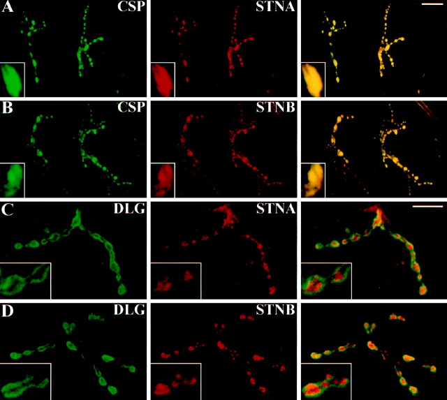
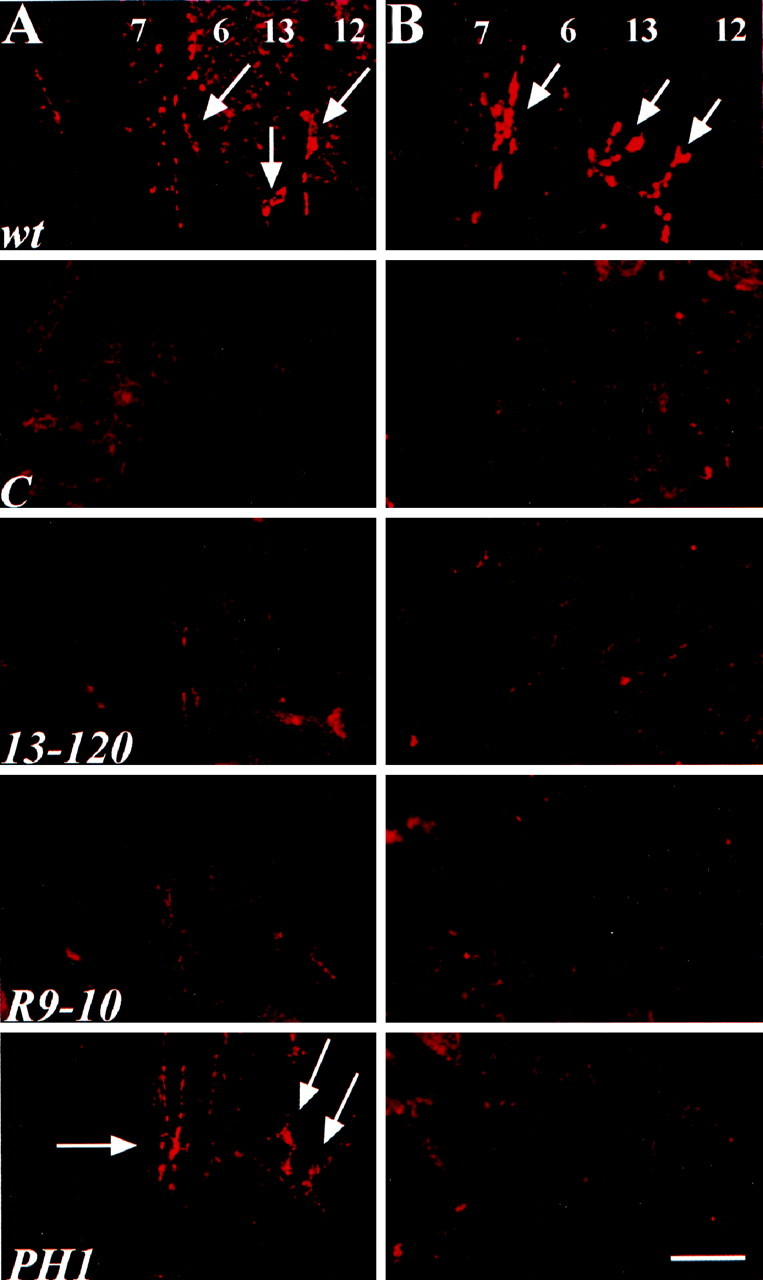
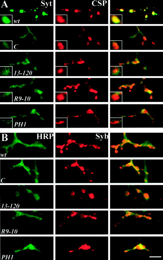
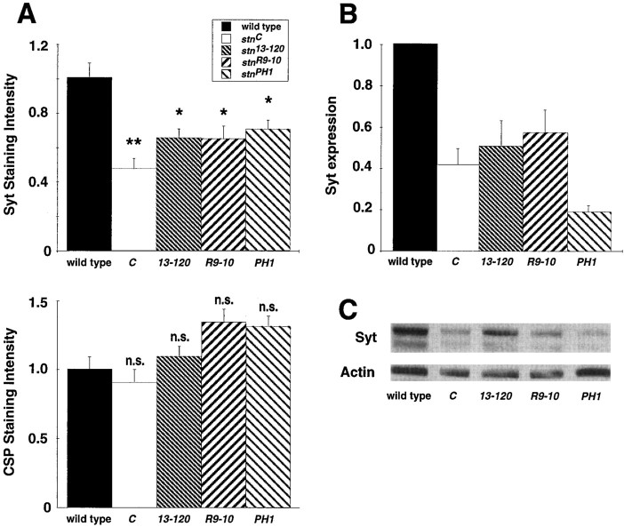
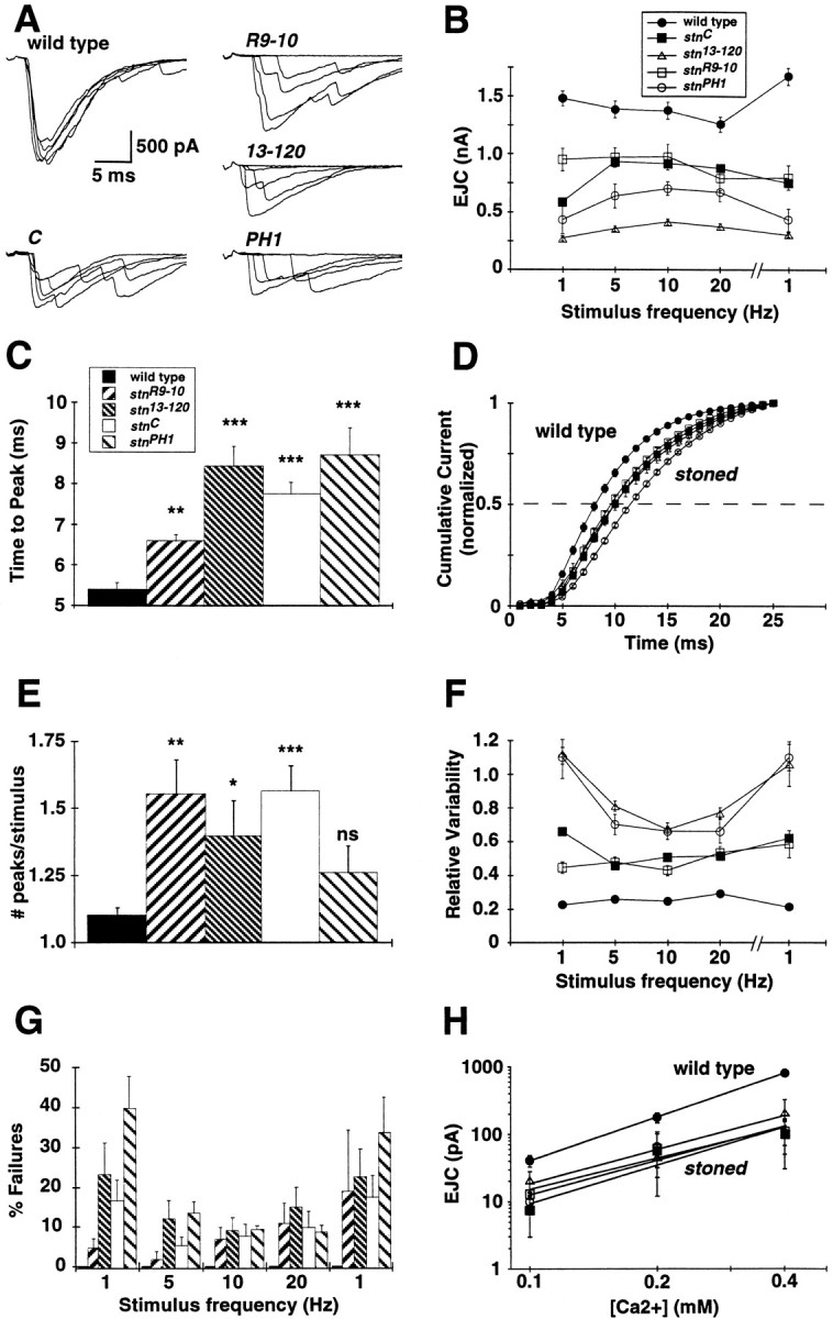
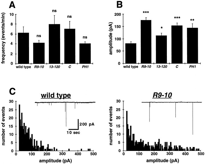
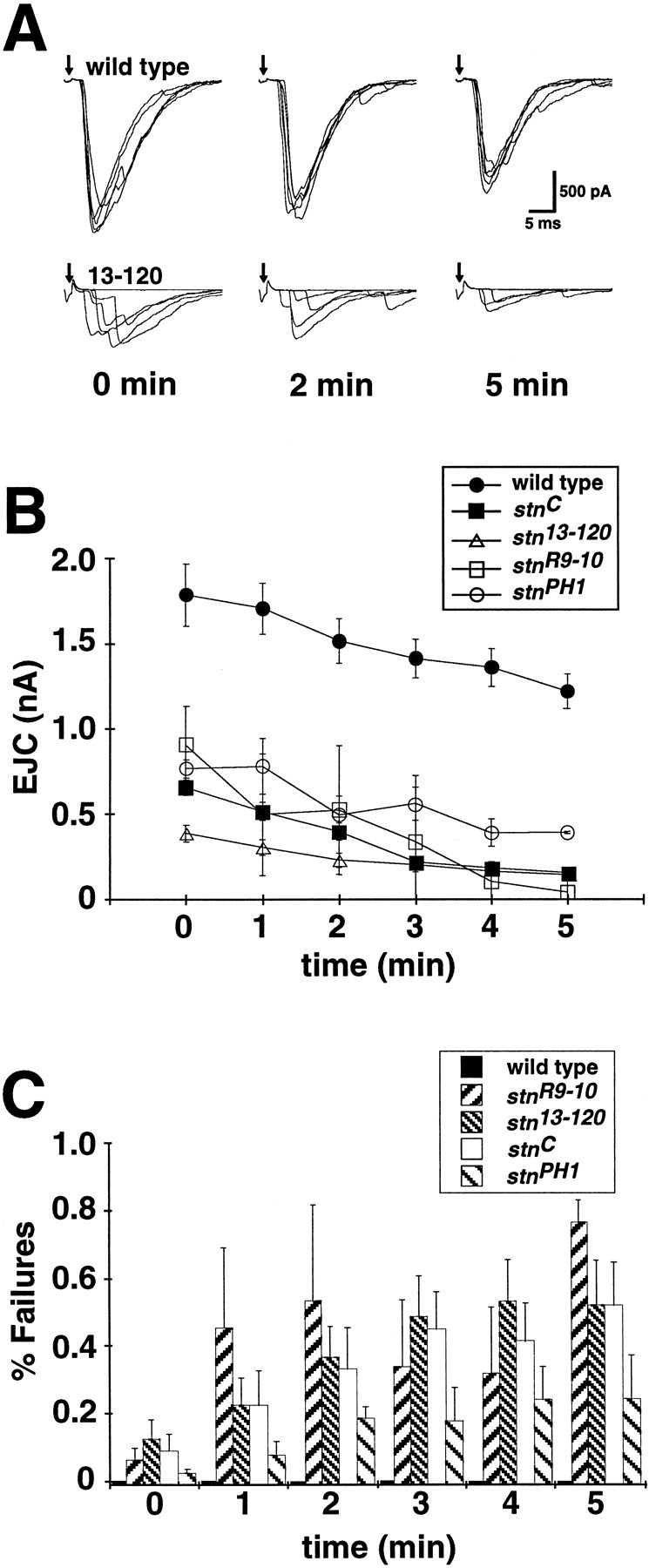
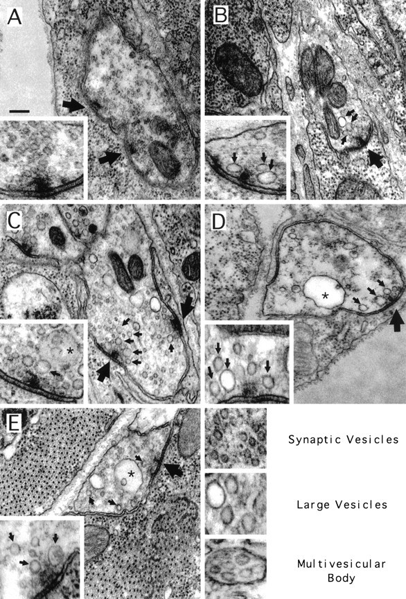
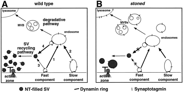
The Drosophila stoned locus was identified 25 years ago on the basis of stress-sensitive behavioral mutants (Grigliatti et al., 1973). The locus is dicistronic and encodes two distinct proteins, stoned A and stoned B, which are expressed specifically in presynaptic terminals at central and peripheral synapses. Several stoned mutant alleles cause embryonic lethality, suggesting that these proteins are essential for synaptic function. Physiological analyses at the stoned synapse reveal severe neurotransmission defects, including reduced and asynchronous neurotransmitter release and rapid fatigue after repetitive stimulation. At the EM level, stoned synapses show a depletion of synaptic vesicles and a concomitant increase in membrane-recycling intermediates. Mutant terminals also display a specific mislocalization of the synaptic vesicle protein synaptotagmin. These results suggest that the stoned proteins are essential for the recycling of synaptic vesicle membrane and are required for the proper sorting of synaptotagmin during endocytosis.


Figures

Fig. 1.
Fig. 1.
The stoned proteins are expressed presynaptically at synaptic terminals. A, B, Third instar larval NMJs double-labeled for the synaptic vesicle-associated CSP (green) and STNA (A,red) or STNB (B, red).Right, Merged images showing the colocalization of each of the stoned proteins with CSP. Insets, Boutons at higher magnification with CSP and the stoned proteins precisely colocalized. C, D, Larval NMJs double-labeled for the postsynaptic DLG (green) and STNA (C, red) or STNB (D, red). Right, Merged images displaying the presynaptic expression of the stoned proteins (red) surrounded by a halo of the postsynaptic DLG (green). Insets, Magnified boutons with the red stoned proteins surrounded by thegreen postsynaptic DLG. Scale bars: A,B, 50 μm; C, D, 30 μm.
Fig. 2.
Fig. 2.
All stoned mutant alleles eliminate STNB expression, and most reduce or eliminate STNA expression. A, STNA antibody labeling of embryonic NMJ shows virtually undetectable STNA expression instnC (C),stn13-120 (13-120), and stnR9-10 (R9-10) alleles. The stnPH1(PH1) allele displays near wild-type levels of STNA expression. B, STNB antibody labeling shows that allstoned mutant alleles lack detectable protein levels at the embryonic NMJ. wt, Wild type. Scale bar, 20 μm.
Fig. 3.
Fig. 3.
Synaptotagmin is reduced and mislocalized atstoned presynaptic boutons. A, Embryonic NMJs double-labeled with antibodies to the SV protein Syt (green) and the SV-associated protein CSP (red). The Syt-staining pattern in wild-type embryos shows distinct punctate expression in the boutons that colocalizes with CSP expression. Mutant synapses have reduced and mislocalized Syt expression but maintain punctate CSP expression. Syt expression in mutants appears dispersed throughout the presynaptic terminal including innervating axons. B, Embryonic NMJs double-labeled with antibodies against the SV protein Syb (red) and a neuronal membrane marker recognized by antibodies against HRP (green). The entire nerve terminal is labeled by HRP, and wild-type synapses display strong Syb expression at the boutons. Mutant animals display properly localized Syb that has punctate expression at the boutons, substantially different from the sparse expression pattern of Syt. Scale bar, 5 μm.
Fig. 4.
Fig. 4.
Synaptotagmin levels are reduced instoned mutant embryos. A, Confocal images of embryonic NMJs (Fig. 3) double-labeled with antibodies to Syt and CSP were analyzed for pixel intensity. All animals were dissected and processed with wild-type animals, and images were acquired with identical settings. The amount of Syt and CSP signal intensity in single boutons was determined for all strains and normalized to that of wild type for that same experiment. All stoned mutant boutons show a significant ∼50% reduction in the amount of Syt (Mann–Whitney U test, p < 0.001 for all strains). In contrast, CSP levels were not significantly different from wild-type levels in anystoned allele. n.s., Not significant.B, C, Western blot analysis ofstoned mutant whole embryos shows a strong reduction in Syt levels. B, The plot displays the average synaptotagmin intensity for the stoned mutants after actin normalization for deviations in protein loading from independent trials. All values represent the mean ± SEM. Syt levels were significantly decreased in all stoned alleles (Mann–Whitney U test, p < 0.001 for all strains). C, A sample gel is shown. Thelower band identified by the DSYT2 antibody represents a previously characterized Syt breakdown product (Littleton et al., 1993).
Fig. 5.
Fig. 5.
All four stoned mutant alleles exhibit strongly impaired presynaptic function. A, Neurotransmission was assayed by stimulating the embryonic nerve with a suction electrode at 1 Hz while recording EJCs in the voltage-clamped muscle (−60 mV). Five superimposed representativetraces are shown for each allele. B, Average EJC amplitudes are shown for basal stimulation (1 Hz) up to high-frequency stimulation (20 Hz), followed by repeat basal stimulation. All mutant EJC amplitudes are significantly decreased from that of wild type at all stimulation frequencies. The average area of the current responses was also analyzed, and similar significant decreases in mutant transmission were found (data not shown). Mean EJC amplitudes in each animal were determined from 25 consecutive EJCs evoked at each frequency. Average amplitudes were determined for at least six animals per strain. C, Allstoned mutants exhibit a significant delay in evoked transmission. The duration from stimulation to the EJC peak is increased in all mutants, representative of delayed transmitter release (Mann–Whitney U test, **p < 0.005; ***p < 0.001). D, Normalized cumulative current amplitude distributions for allstoned mutants are significantly delayed. The time toI50 for all mutants is significantly delayed (C, p < 0.0001;13-120, p = 0.038;R9-10, p = 0.042;PH1, p = 0.0004). E, Control transmission responses to stimulation usually result in a single synchronous release of transmitter, whereas asynchronous release resulting from delayed vesicle fusion is frequently observed in allstoned mutants. The asynchronous release of transmitter was quantified by analysis of the number of 100+ pA peaks per stimulus (*p < 0.05; **p < 0.005; ***p < 0.0001). F, EJC amplitudes vary greatly in stoned mutants. The poor fidelity of mutant transmission is displayed as high variability at different stimulation frequencies. G, Mutant NMJs show a significant transmission failure rate, whereas control synapses never fail. Failure rate is quantified in a stimulation series of 1–20 Hz, followed by repeat stimulation at 1 Hz. H, The calcium dependence of transmission was determined for all thestoned mutants. The calcium cooperativity of release was similar for all stoned alleles and is not significantly different from the control as measured by the power relationship of the slope for wild type (2.15) and for the mutants (C = 1.87; 13-120 = 1.66; R9-10 = 1.54; PH1 = 1.70). Each value represents the mean ± SEM.
Fig. 6.
Fig. 6.
Miniature EJC amplitudes are increased in allstoned mutants. A, Spontaneous EJC events were collected at 0.5 mm Ca2+ in the presence of 0.1 μm TTX. The frequency of events in allstoned alleles is not significantly (ns) different from that of wild-type embryos. B, The average type I MEJC amplitude is significantly increased for allstoned mutants (*p < 0.05; **p < 0.005; ***p < 0.0005).C, Current amplitude distributions of type I MEJCs in wild-type and stnR9-10 embryos in 0.5 mm Ca2+ are shown. The smallest quantal size event distribution is similar for both wild-type and mutant embryos; however, all stoned mutants display a significant increase in the number of abnormally large events (>200 pA). Current amplitudes are divided into 5 pA bins.
Fig. 7.
Fig. 7.
The stoned mutant synapses fatigue during prolonged stimulation. A, The NMJ was stimulated at 10 Hz in normal calcium (1.8 mm) over a 5 min period, and EJC amplitude was quantified throughout the protocol. Sampletraces are shown for t = 0, 2, and 5 min. Transmission becomes severely impaired in stonedmutants by the end of the stimulus protocol, and wild-type transmission remains relatively robust. B, Wild-type EJC amplitude decreases slightly (∼25%) over the 5 min stimulus protocol, whereas stoned mutants display a relatively severe decrease (50+%) in transmission amplitude. EJC amplitudes at each time point are averaged from 25 consecutive stimuli. C, The decreased EJC amplitude in stoned alleles is accompanied by a significant increase in failure rate. In each plot, points represent the mean ± SEM for at least four embryos per genotype.
Fig. 8.
Fig. 8.
All stoned mutant alleles display a reduction of synaptic vesicles and accumulation of other membrane structures. Images of typical boutons (background) and active zones (insets) for wild-type (A), stn13-120(B), stnR9-10(C), stnPH1(D), and stnC(E) mutants. Regular synaptic vesicles comprise the most prominent membrane structure in the wild-type bouton, and clusters of these vesicles are observed clustered around active zone t-bars (large arrows). In the mutant embryos, fewer synaptic vesicles are clustered around t-bars, and an increase in large, translucent vesicles (small arrows) was observed both in boutons and in regions proximal to t-bars. In some strains, an increase in multivesicular bodies (asterisks) was observed in boutons. Scale bar, 200 nm.
Fig. 9.
Fig. 9.
Schematic model of putative synaptic vesicle-recycling mechanisms at the presynaptic terminal. Synaptic vesicles release their contents by fusing with the plasma membrane at the active zone and are retrieved by a dynamin-mediated process that may recycle single vesicles under light stimulation (fast component) and may mediate bulk membrane retrieval under high-stimulus conditions (slow component). A, Numerous studies have suggested that synaptic vesicle-recycling mechanisms may require an endosomal-sorting step (arrows 1, 2), although there is also evidence of a more direct mechanism (arrow 3). The hypothesized role of a sorting endosome is to retain functional synaptic vesicle proteins for the generation of new mature vesicles and to send old proteins to the somatic lysosomes for degradation via MVBs. B, Impaired vesicle recycling in stoned mutants results in a decrease of synaptic vesicles and an increase in recycling intermediates such as cisternae/endosomes and MVBs. Such data strongly suggest a role for stoned in the recycling of synaptic vesicles. The stonedmutants also display mislocalization and reduced levels of Syt, possibly because of loss of Syt to somatic lysosomes.

References

    1. Andrews J, Smith M, Merakovsky J, Coulson M, Hannan F, Kelly LE. The stoned locus of Drosophila melanogaster produces a dicistronic transcript and encodes two distinct polypeptides. Genetics. 1996;143:1699–1711. - PMC - PubMed
    1. Artalejo CR, Henley JR, McNiven MA, Palfrey HC. Rapid endocytosis coupled to exocytosis in adrenal chromaffin cells involves Ca2+, GTP, and dynamin but not clathrin. Proc Natl Acad Sci USA. 1995;92:8328–8332. - PMC - PubMed
    1. Atwood HL, Govind CK, Wu CF. Differential ultrastructure of synaptic terminals on ventral longitudinal abdominal muscles in Drosophila larvae. J Neurobiol. 1993;24:1008–1024. - PubMed
    1. Bennett MK, Scheller RH. A molecular description of synaptic vesicle membrane trafficking. Annu Rev Biochem. 1994;63:63–100. - PubMed
    1. Betz WJ, Angleson JK. The synaptic vesicle cycle. Annu Rev Physiol. 1998;60:347–363. - PubMed

Publication types

MeSH terms