. 1999 Jul 15;19(14):5861-74.
doi: 10.1523/JNEUROSCI.19-14-05861.1999.

Ca(2+)-permeable AMPA receptors induce phosphorylation of cAMP response element-binding protein through a phosphatidylinositol 3-kinase-dependent stimulation of the mitogen-activated protein kinase signaling cascade in neurons

Affiliations

Ca(2+)-permeable AMPA receptors induce phosphorylation of cAMP response element-binding protein through a phosphatidylinositol 3-kinase-dependent stimulation of the mitogen-activated protein kinase signaling cascade in neurons

M S Perkinton et al. J Neurosci. .

Abstract

Ca(2+)-permeable AMPA receptors may play a key role during developmental neuroplasticity, learning and memory, and neuronal loss in a number of neuropathologies. However, the intracellular signaling pathways used by AMPA receptors during such processes are not fully understood. The mitogen-activated protein kinase (MAPK) cascade is an attractive target because it has been shown to be involved in gene expression, synaptic plasticity, and neuronal stress. Using primary cultures of mouse striatal neurons and a phosphospecific MAPK antibody we addressed whether AMPA receptors can activate the MAPK cascade. We found that in the presence of cyclothiazide, AMPA caused a robust and direct (no involvement of NMDA receptors or L-type voltage-sensitive Ca(2+) channels) Ca(2+)-dependent activation of MAPK through MAPK kinase (MEK). This activation was blocked by GYKI 53655, a noncompetitive selective antagonist of AMPA receptors. Probing the mechanism of this activation revealed an essential role for phosphatidylinositol 3-kinase (PI 3-kinase) and the involvement of a pertussis toxin (PTX)-sensitive G-protein, a Src family protein tyrosine kinase, and Ca(2+)/calmodulin-dependent kinase II. Similarly, kainate activated MAPK in a PI 3-kinase-dependent manner. AMPA receptor-evoked neuronal death and arachidonic acid mobilization did not appear to involve signaling through the MAPK pathway. However, AMPA receptor stimulation led to a Ca(2+)-dependent phosphorylation of the nuclear transcription factor CREB, which could be prevented by inhibitors of MEK or PI 3-kinase. Our results indicate that Ca(2+)-permeable AMPA receptors transduce signals from the cell surface to the nucleus of neurons through a PI 3-kinase-dependent activation of MAPK. This novel pathway may play a pivotal role in regulating synaptic plasticity in the striatum.


Figures
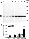
Fig. 1.
Fig. 1.
Cyclothiazide unmasks an AMPA-mediated activation of MAPK in striatal neurons. A, Crude homogenates (15 μg of each), prepared from striatal neurons preincubated for 5 min in the absence (lanes1,3, 5, 7) or presence (lanes 2, 4, 6,8) of 50 μm cyclothiazide (Cyz), and then exposed for 5 min to vehicle (lanes 1, 5), 50 μm Cyz (lanes 2, 6), 50 μmAMPA (lanes 3, 7), or 50 μm AMPA and 50 μm Cyz (lanes 4, 8), were immunoblotted with an antibody that specifically recognizes the dually phosphorylated Thr/Glu/Tyr region within the catalytic core of the active form of ERK1 and ERK2 (anti-ACTIVE MAPK pAb) (lanes 1-4), or with an antibody that recognizes total levels of ERK1 and ERK2 (anti-ERK1/ERK2 pAb) (lanes 5-8). B, Data obtained from immunoblot experiments represented in lanes 1-4 was analyzed using BioImage Intelligent Quantifier software. Each column is the mean ± SEM value of 6–12 independent cultures (n = 6–12). **AMPA/Cyz was significantly different from control (Con) (p < 0.001, unpaired two-tailed Student’s t test).
Fig. 2.
Fig. 2.
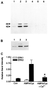
Ca2+ dependence of AMPA receptor-evoked MAPK activation in striatal neurons. A, Crude homogenates (15 μg of each), prepared from striatal neurons preincubated for 5 min in the absence (lanes 1,2, 4, 5) or presence (lanes 3, 6) of 50 μm cyclothiazide (Cyz), and then exposed for 5 min to vehicle (lanes 1, 4), 100 μm glutamate (lanes 2, 5), or 50 μm AMPA and 50 μm Cyz (lanes 3, 6) in the presence (lanes 1-3) or absence (lanes 4-6) of CaCl2 (1 mm), were immunoblotted with an antibody that specifically recognizes the dually phosphorylated Thr/Glu/Tyr region within the catalytic core of the active form of ERK1 and ERK2 (anti-ACTIVE MAPK pAb). In experiments in which CaCl2 was omitted from the incubation medium, EGTA (200 μm) was added 1 min before stimulation. B, Crude homogenates (15 μg of each), prepared from striatal neurons exposed to vehicle (lane 1), 100 μm glutamate (lane 2), or 5 μm ionomycin (lane 3) for 5 min, were immunoblotted with anti-ACTIVE MAPK pAb. C, Data obtained from immunoblot experiments with AMPA/Cyz was analyzed using BioImage Intelligent Quantifier software. Each column is the mean ± SEM value of six independent cultures (n = 6). *AMPA/Cyz in the absence of CaCl2(AMPA/CyzCa2+) was significantly different from AMPA/Cyz in the presence of CaCl2 (AMPA/Cyz) (p < 0.05, unpaired two-tailed Student’st test).
Fig. 3.
Fig. 3.
AMPA receptor-evoked increase in intracellular Ca2+ is enhanced by cyclothiazide and blocked by GYKI 53655. Fura-2-loaded striatal neurons were exposed to 50 μm AMPA, 50 μm AMPA in the presence of 50 μm cyclothiazide (AMPA/Cyz), or AMPA/Cyz in the presence of 100 μm GYKI 53655 added 5 min before stimulation. Fura-2 fluorescence (340 nm/380 nm ratio) was monitored as described in Materials and Methods. Results are means ± SEM of responses from the number of neurons (n) shown in parentheses.
Fig. 4.
Fig. 4.
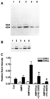
AMPA/cyclothiazide activation of MAPK through MEK is direct and independent of NMDA receptor and/or L-type voltage-sensitive Ca2+ channel activation.A, Crude homogenates (15 μg of each), prepared from striatal neurons preincubated for 5 min in the absence (lanes 1, 2) or presence (lanes 3-5) of 50 μm cyclothiazide (Cyz), and then exposed for 5 min to vehicle (lane 1), 50 μm AMPA (lane 2), 50 μm AMPA and 50 μm Cyz (lane 3), 50 μm AMPA and 50 μm Cyz in the presence of 100 μm GYKI 53655 added 5 min before AMPA/Cyz (lane 4), or 50 μm AMPA and 50 μm Cyz in the presence of 50 μm PD 98059 added 5 min before AMPA/Cyz (lane 5), were immunoblotted with an antibody that specifically recognizes the dually phosphorylated Thr/Glu/Tyr region within the catalytic core of the active form of ERK1 and ERK2 (anti-ACTIVE MAPK pAb). B, Crude homogenates (15 μg of each), prepared from striatal neurons preincubated for 5 min in the absence (1) or presence (lanes 2-5) of 50 μm Cyz and then exposed for 5 min to vehicle (lane 1), 50 μm AMPA and 50 μm Cyz (lane 2), 50 μm AMPA and 50 μm Cyz in the presence of 100 μm GYKI 53655 added 5 min before AMPA/Cyz (lane 3), 50 μm AMPA and 50 μm Cyz in the presence of 2 μm (+)-MK 801 added 5 min before AMPA/Cyz (lane 4), or 50 μm AMPA and 50 μm Cyz in the presence of 5 μm nimodipine added 5 min before AMPA/Cyz (lane 5), were immunoblotted with anti-ACTIVE MAPK pAb.C, Data obtained from immunoblot experiments represented in A (lanes 1-5) was analyzed using BioImage Intelligent Quantifier software. Each column is the mean ± SEM value of five to eight independent cultures (n = 5–8). *AMPA/Cyz in the presence of GYKI 53655 or PD 98059 was significantly different from AMPA/Cyz (p < 0.05, unpaired two-tailed Student’st test).
Fig. 5.
Fig. 5.
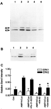
Involvement of a Src family protein tyrosine kinase and CaM-KII, but not protein kinase C, in AMPA receptor-evoked activation of MAPK. A, Crude homogenates (15 μg of each), prepared from striatal neurons preincubated for 5 min in the absence (lane 1) or presence (lanes 2-5) of 50 μm cyclothiazide (Cyz) and then exposed for 5 min to vehicle (lane 1), 50 μm AMPA and 50 μm Cyz (lane 2), 50 μm AMPA and 50 μm Cyz in the presence of 5 μm Ro-31–8220 added 5 min before AMPA/Cyz (lane 3), 50 μm AMPA and 50 μm Cyz in the presence of 5 μm KN-62 added 5 min before AMPA/Cyz (lane 4), or 50 μm AMPA and 50 μmCyz in the presence of 50 μm genistein added 5 min before AMPA/Cyz (lane 5), were immunoblotted with an antibody that specifically recognizes the dually phosphorylated Thr/Glu/Tyr region within the catalytic core of the active form of ERK1 and ERK2 (anti-ACTIVE MAPK pAb). B, Crude homogenates (15 μg of each), prepared from striatal neurons preincubated for 5 min in the absence (lane 1) or presence (lanes 2-4) of 50 μm Cyz, and then exposed for 5 min to vehicle (lane 1), 50 μm AMPA and 50 μm Cyz (lane 2), or 50 μm AMPA and 50 μm Cyz in the presence of either 10 μm PP2 (lane 3) or 10 μm PP3 (lane 4) both added 5 min before AMPA/Cyz, were immunoblotted with anti-ACTIVE MAPK pAb.C, Data obtained from immunoblot experiments represented in A (lanes 1-5) was analyzed using BioImage Intelligent Quantifier software. Each column is the mean ± SEM value of four to six independent cultures (n = 4–6). *AMPA/Cyz in the presence of KN-62 or genistein was significantly different from AMPA/Cyz (p < 0.05, unpaired two-tailed Student’st test).
Fig. 6.
Fig. 6.
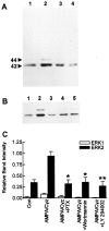
Essential role for PI 3-kinase and a Gi/Go-type G-protein in the activation of MAPK by Ca2+-permeable AMPA receptors.A, Crude homogenates (15 μg of each), prepared from striatal neurons preincubated for 5 min in the absence (lane 1) or presence (lanes 2-4) of 50 μm cyclothiazide (Cyz) and then exposed for 5 min to vehicle (lane 1), 50 μm AMPA and 50 μm Cyz (lane 2), 50 μm AMPA and 50 μm Cyz in the presence of 1 μg/ml pertussis toxin (PTX) added 24 hr before AMPA/Cyz (lane 3), or 50 μm AMPA and 50 μm Cyz in the presence of either 100 nm wortmannin (lane 4) or 50 μm LY 294002 (C) both added 5 min before AMPA/Cyz, were immunoblotted with an antibody that specifically recognizes the dually phosphorylated Thr/Glu/Tyr region within the catalytic core of the active form of ERK1 and ERK2 (anti-ACTIVE MAPK pAb). B, Crude homogenates (15 μg of each), prepared from striatal neurons exposed for 5 min to vehicle (lane 1), 100 μm KA (lane 2), 100 μm KA in the presence of 100 μm GYKI 53655 added 5 min before KA (lane 3), 100 μm KA in the presence of 1 μg/ml PTX added 24 hr before KA (lane 4), or 100 μm KA in the presence of 100 nm wortmannin added 5 min before KA (lane 5), were immunoblotted with anti-ACTIVE MAPK pAb. C, Data obtained from immunoblot experiments represented in A (lanes 1-4) was analyzed using BioImage Intelligent Quantifier software. Each column is the mean ± SEM value of five to six independent cultures (n = 5–6). *AMPA/Cyz in the presence of PTX or wortmannin was significantly different from AMPA/Cyz (p < 0.05, unpaired two-tailed Student’s t test) and **AMPA/Cyz in the presence of LY 294002 was significantly different from AMPA/Cyz (p < 0.001, unpaired two-tailed Student’st test).
Fig. 7.
Fig. 7.
AMPA receptor-evoked mobilization of arachidonic acid in striatal neurons is independent of the MAPK cascade. Striatal neurons were preincubated for 10 min in HEPES-buffered medium containing fatty acid-free BSA (2 mg/ml; HBM/BSA) in the presence or absence of enzyme inhibitors (100 nm wortmannin or 50 μm PD 98059), with or without 50 μmcyclothiazide (Cyz) and then incubated for 15 min in HBM/BSA containing either vehicle (Basal), or 50 μm AMPA and 50 μm Cyz (AMPA/Cyz) in the continued presence or absence of enzyme inhibitors as shown. Both AMPA and Cyz alone failed to release [3H]AA (data not shown). Results are expressed as the percent release of [3H]AA relative to basal values, which was defined as 100%. Data are the mean ± SEM values of three to five independent cultures (n = 3–5), each performed in triplicate.
Fig. 8.
Fig. 8.
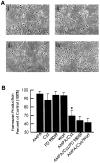
Onset of Ca2+-permeable AMPA receptor-induced neurotoxicity is neither reversed nor accelerated by inhibition of MAPK/PI 3-kinase activity. A, Striatal neurons were exposed to vehicle (i), 50 μm AMPA (ii), 50 μm AMPA in the presence of 50 μm cyclothiazide (Cyz) (iii), or 50 μm AMPA and 50 μm Cyz in the presence of 100 μm GYKI 53655 added 5 min before AMPA/Cyz (iv) for 4 hr and then photographed under phase contrast microscopy (320× magnification).B, Striatal neurons were exposed to vehicle or AMPA, with or without Cyz (as described above), in the presence or absence of 50 μm PD 98059 or 100 nm wortmannin (Wort) which were added 5 min before treatments. After 4 hr exposure, the culture medium was removed and MTT added as described in Materials and Methods. Results are expressed as the percent of formazan production relative to control, which was defined as 100%. Each column is the mean ± SEM value of four independent cultures (n = 4), each performed in triplicate. *AMPA/Cyz was significantly different from AMPA (p < 0.05, unpaired two-tailed Student’s t test).
Fig. 9.
Fig. 9.
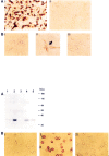
Localization of MAPK and activation by AMPA receptor stimulation in cultured striatal neurons. A, Striatal neurons were immunostained with an antibody to the MAPK enzymes ERK1 and ERK2 (anti-ERK1/ERK2 pAb) (i), or without primary antibody (ii), as described in Materials and Methods. White arrows indicate intense cytosolic staining of ERK1 and ERK2. B, Striatal neurons were either unstimulated (i) or, after incubation with 50 μm cyclothiazide for 5 min, stimulated with 50 μm AMPA (in the presence of 50 μmcyclothiazide) for 5 min in the absence (ii) or presence (iii) of 100 μm GYKI 53655 added 5 min before stimulation. After stimulation, neurons were immunostained with an antibody that specifically recognizes the dually phosphorylated Thr/Glu/Tyr region within the catalytic core of the active form of ERK1 and ERK2 (anti-ACTIVE MAPK pAb) as described in Materials and Methods.Black arrow indicates neurons that are immunopositive for anti-ACTIVE MAPK pAb.

References

    1. Alessi DR, Cuenda A, Cohen P, Dudley DT, Saltiel AR. PD 098059 is a specific inhibitor of the activation of mitogen-activated protein kinase kinase in vitro and in vivo. J Biol Chem. 1995;270:27489–27494. - PubMed
    1. Bading H, Ginty DD, Greenberg ME. Regulation of gene expression in hippocampal neurons by distinct calcium signaling pathways. Science. 1993;260:181–186. - PubMed
    1. Bading H, Segal MM, Sucher NJ, Dudek H, Lipton SA, Greenberg ME. N-methyl-d-aspartate receptors are critical for mediating the effects of glutamate on intracellular calcium concentration and immediate early gene expression in cultured hippocampal neurons. Neuroscience. 1995;64:653–664. - PubMed
    1. Barria A, Muller D, Derkach V, Griffith LC, Soderling TR. Regulatory phosphorylation of AMPA-type glutamate receptors by CaM-KII during long-term potentiation. Science. 1997;276:2042–2045. - PubMed
    1. Bernard V, Somogyi P, Bolam JP. Cellular, subcellular, and subsynaptic distribution of AMPA-type glutamate receptor subunits in the neostriatum of the rat. J Neurosci. 1997;17:819–833. - PMC - PubMed

Publication types

MeSH terms

Substances