. 1999 Jul 15;19(14):5980-9.
doi: 10.1523/JNEUROSCI.19-14-05980.1999.

Targeted mutagenesis of the POU-domain gene Brn4/Pou3f4 causes developmental defects in the inner ear

Affiliations

Targeted mutagenesis of the POU-domain gene Brn4/Pou3f4 causes developmental defects in the inner ear

D Phippard et al. J Neurosci. .

Abstract

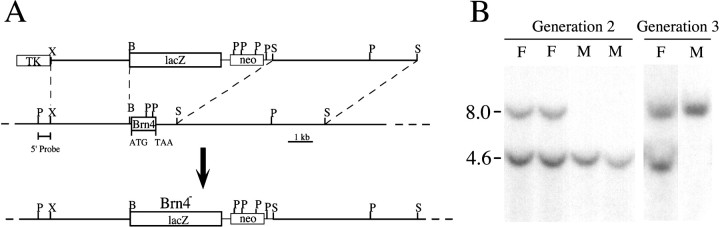
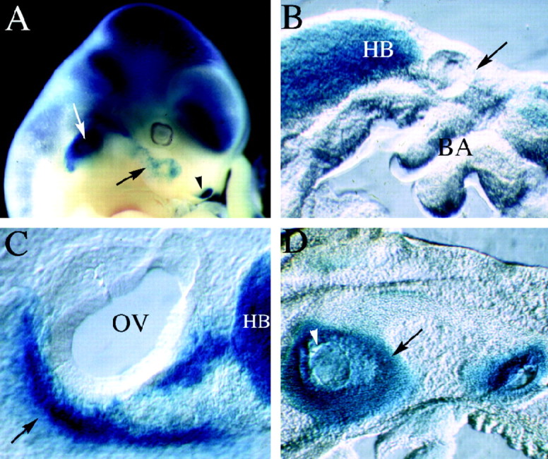
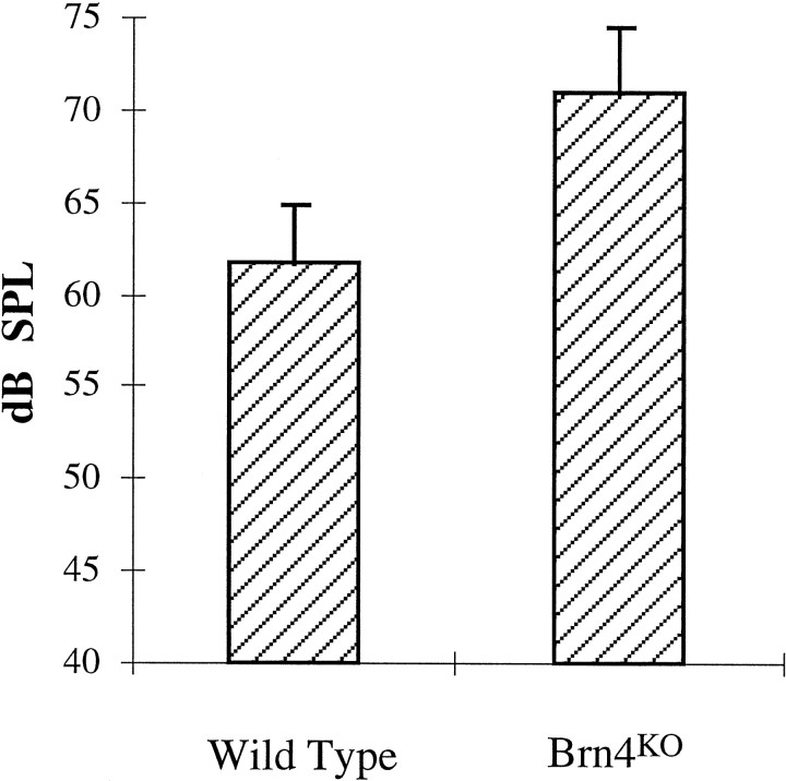
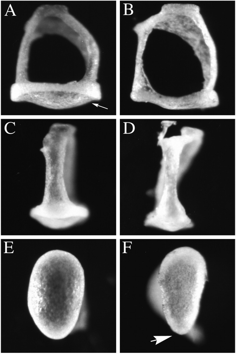
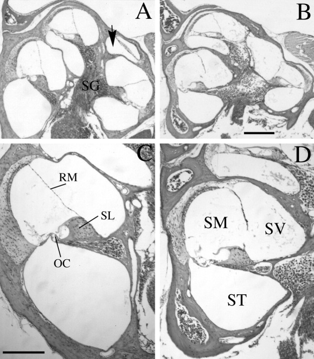
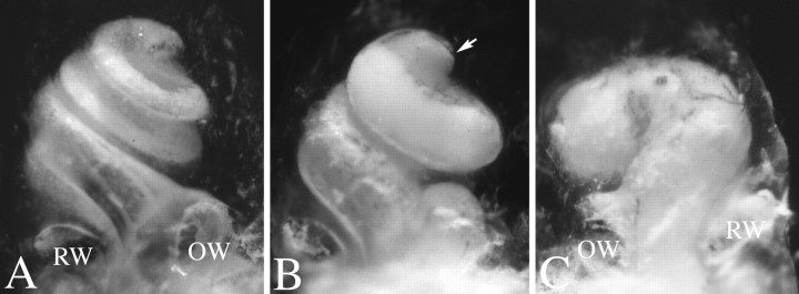
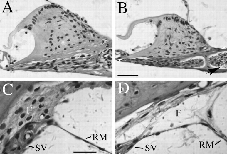
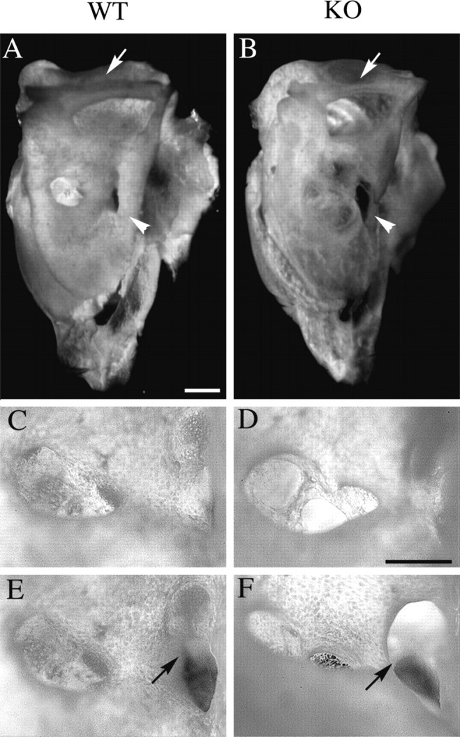
Targeted mutagenesis in mice demonstrates that the POU-domain gene Brn4/Pou3f4 plays a crucial role in the patterning of the mesenchymal compartment of the inner ear. Brn4 is expressed extensively throughout the condensing mesenchyme of the developing inner ear. Mutant animals displayed behavioral anomalies that resulted from functional deficits in both the auditory and vestibular systems, including vertical head bobbing, changes in gait, and hearing loss. Anatomical analyses of the temporal bone, which is derived in part from the otic mesenchyme, demonstrated several dysplastic features in the mutant animals, including enlargement of the internal auditory meatus. Many phenotypic features of the mutant animals resulted from the reduction or thinning of the bony compartment of the inner ear. Histological analyses demonstrated a hypoplasia of those regions of the cochlea derived from otic mesenchyme, including the spiral limbus, the scala tympani, and strial fibrocytes. Interestingly, we observed a reduction in the coiling of the cochlea, which suggests that Brn-4 plays a role in the epithelial-mesenchymal communication necessary for the cochlear anlage to develop correctly. Finally, the stapes demonstrated several malformations, including changes in the size and morphology of its footplate. Because the stapes anlage does not express the Brn4 gene, stapes malformations suggest that the Brn4 gene also plays a role in mesenchymal-mesenchymal signaling. On the basis of these data, we suggest that Brn-4 enhances the survival of mesodermal cells during the mesenchymal remodeling that forms the mature bony labyrinth and regulates inductive signaling mechanisms in the otic mesenchyme.


Figures

Fig. 1.
Fig. 1.
Targeted mutagenesis of the Brn4 locus.A, Diagram of the targeting vector (top line), the wild-type allele (middle line), and the mutated allele (bottom line). The entire intronless coding region (box labeled Brn4) and a small region of 3′-flanking sequences have been replaced by the lacZ reporter gene and the PGK-neo gene. The lacZ-coding sequences have been introduced into the Brn4 locus such that theBrn4 promoter drives the transcription of the reporter gene from the mutated allele. Homologous recombination was detected with the 5′ P/X probe that detects the conversion of a 4.6 kbPstI fragment to 8.0 kb. P,PstI; S, SalI;X, XbaI. B, Southern blot analysis of the genetic transmission of the X-linked locus. Analysis of progeny from the founder chimeric male animals indicated that the mutated allele is transmitted to only the female progeny in generation 2. Genomic DNA was digested with PstI and probed with the 5′ P/X probe. F, Females; M, males.
Fig. 2.
Fig. 2.
LacZ expression in mutant animals. Mutant embryos were stained with X-gal to visualize the expression of the lacZ gene, which was inserted into the Brn4 locus during the generation of the knock-out allele. Unless specified otherwise the embryos are oriented with rostral to the right and dorsal toward the top. A, The whole-mount preparation of a chimeric founder animal that corresponds to an 11.5 dpc embryo (chimeric embryos are developmentally delayed for ∼1 day because of experimental manipulation of the blastocysts). The expression of lacZ recapitulates precisely the pattern of endogenousBrn4 gene expression detected by hybridization histochemical analyses (Le Moine and Young, 1992; Mathis et al., 1992;Alvarez-Bolado et al., 1995; Phippard et al., 1998). Expression is found throughout most of the neuraxis and in a handful of mesodermally derived tissues in the head, including the otic capsule (white arrow), a small population of first branchial arch mesenchyme (black arrow), and the lateral nasal recess (black arrowhead). B, A parasagittal vibratome section (150 μm) through the otic vesicle of a 9.5 dpc embryo. Expression of lacZ is not detected in the mesenchyme surrounding the otic vesicle (black arrow), which lies dorsal to the branchial arches. However, expression is detected in the hindbrain of these embryos. C, A parasagittal vibratome section through the otic vesicle of a 10.5 dpc embryo. Expression of lacZ is detected in the condensing mesenchyme of the otic vesicle (black arrow), which lies ventral to the otic vesicle at this stage of embryogenesis. In this panel, the black arrow indicates the dorsal–ventral axis, with dorsal corresponding to the upper right-hand corner of the panel.D, Expression patterns of lacZ in a parasagittal section of a 14.5 dpc embryo. At this stage of development, lacZ expression is detected throughout the otic capsule but not in the otic epithelium (some regions of the otic epithelium appear blue, because they are covered with lacZ staining the otic capsule in these thick vibratome sections). BA, Branchial arches;HB, hindbrain; OV, otic vesicle.
Fig. 3.
Fig. 3.
Preyer’s reflex in Brn4 knock-out (KO) mutants. The Brn4 null mutants demonstrate approximately a 10 dB SPL hearing loss in comparison with that in wild-type littermates, as assessed by Preyer’s reflex. Wild-type animals, 62 ± 3.1 dB SPL (n= 12). Brn4 null mutants, 71 ± 3.6 dB SPL (n = 12). Error bars represent SD. The Preyer’s reflex threshold is measured in decibels SPL relative to a standard of 20 μPa. One-tailed Student’s t test,p < 0.00001. If female animals are excluded from the comparison, then the wild-type male animals demonstrate a threshold of 62 ± 3.3 dB SPL (n = 9). Furthermore, there is no statistically significant difference in the threshold between male and female animals, whether they are mutant or wild type (data not shown).
Fig. 4.
Fig. 4.
Malformations of the stapes observed inBrn4 null mice. The stapes of the mutant animals demonstrate several malformations, particularly in the stapedial footplate. A, C, E, Wild-type stapes are depicted. B, D,F, Mutant stapes are depicted. A,B, The footplate of the stapes (arrow inA) in mutant embryos (B) is flatter in comparison with that in wild-type stapes (A). C, D, A lateral view of the stapes demonstrates that the crus from which the stapedial ligament is attached is thinner in the mutant animals (D) than in the wild-type animals (C). E, F, Examination of the sole of the stapes footplate illustrates the slightly eccentric ovoid shape of the wild-type footplate (E). The mutant footplate adopts a more polygonal shape with an acutely angled tip on one end of the footplate (arrow in F). Stapes (n = 10) isolated from six mutant male animals were examined. Wild-type stapes (n = 15) were examined from eight male animals, including six wild-type littermates, two males from the inbred strain 129/SvJ, and two CBA/2J male animals.
Fig. 5.
Fig. 5.
Brn4 knock-out mutants demonstrate cochlear dysplasias in adult mice. B, D, Midmodiolar sections from an adult (6-week-old) Brn4hemizygous null mutant are shown. A, C, Similar sections from a wild-type littermate are shown.A, B, The cochlea of the mutant mouse (B) demonstrates an overall hypoplastic structure compared with that of the wild-type animal (A). The arrow in A indicates the most apical turn of the normal cochlea, which is rarely detected in similar sections of the mutant animal. C, D, The scala tympani of the mutant mouse (D) is flattened and elliptical in comparison with that of the wild-type control mouse (C). Additionally, Reissner’s membrane displays the distended morphology seen in D in the mutant embryos, consistent with a hydrops condition in the mutant animals. In all cases examined, similar phenotypes were found in homozygous knock-out female animals (data not shown).OC, Organ of Corti; RM, Reissner’s membrane; SG, spiral ganglion; SL, spiral limbus; SM, scala media; ST, scala tympani; SV, scala vestibuli. Scale bars:A, B, 300 μm; C,D, 200 μm.
Fig. 6.
Fig. 6.
Cleared temporal bone preparations demonstrate cochlear hypoplasia. Temporal bone preparations were perfused through the oval window with white paint to visualize cochlear morphology. Cochleae are oriented such that the apical turn is at thetop of the photograph, as indicated by thearrow in B. A, A cochlea from a wild-type animal with one and three-fourth turns is shown.B, C, Mutant cochlea displayed a reduced number of turns, which could vary between less than one complete turn to one and one-half turns. Additionally, the amount of coiling can differ between left and right cochlea of the same mutant animal. For example, C demonstrates the right cochlea of a homozygous knock-out female animal, which has less than one coil. By contrast, B demonstrates the left cochlea of the same animal, which has one and one-half turns.
Fig. 7.
Fig. 7.
Histological analysis of spiral limbus and strial fibrocyte dysplasias. B,D, Midmodiolar sections from an adult (6-week-old)Brn4 hemizygous null mutant are shown. A,C, Similar sections from a wild-type littermate are shown. A, B, This view demonstrates that the spiral limbus of mutant animals (B) is smaller than that detected in the wild-type animals (A). The height of the spiral limbus was calculated by drawing a baseline (height = 0) at the widest part of the spiral limbus from the tympanic lip of the internal spiral sulcus to the point where the spiral limbus meets the bony wall of the cochlea (see arrowheads in B). Measurements were made from photomicrographs, and the highest point attained by the interdental cells at the crest of the spiral limbus was measured in a plane perpendicular to the baseline. An additional aspect of the mutant phenotype seen in this figure includes acellular gaps that are often detected between the spiral ganglion cells of the mutants and the modiolus (arrow in B). In wild-type animals, such an acellular gap is rarely detected (B). C, D, This view demonstrates that the strial fibrocytes are not tightly adher-ent in the mutant (D) compared with the wild-type (C) mouse. This phenotype was fully penetrant in all 12 mutant animals examined. F, Fibrocytes; RM, Reissner’s membrane; SV, stria vascularis. Scale bars: A, B, 50 μm; C, D, 20 μm.
Fig. 8.
Fig. 8.
Malformations of the temporal bone inBrn4 null mice. Analysis of whole-mount preparations of adult temporal bone demonstrates dysplasia of the three foramina of the internal auditory meatus. A, C,E, A wild-type temporal bone is depicted.B, D, F, A mutant temporal bone is depicted. A, B, A medial view of the temporal bone demonstrates that the bony tissue appears thinner in the mutant (B) than in the wild type (A). The bone encompassing the superior semicircular canal is thinner in the mutant than in the wild-type animal (arrow), and the rostromedial ridge of the temporal bone is thinner in the mutant (arrowhead).C, D, The cochlear foramen of the internal auditory meatus (IAM) is enlarged in the mutant (D) animal when compared with that in the wild-type animal. E, F, The second and third foramen of the mutant IAM (F) appear to be fused (arrow) when compared with that in the wild type (E). Scale bars: A,B, 1 mm; C–F, 0.5 mm.
Fig. 9.
Fig. 9.
Constriction of the superior semicircular canal inBrn4 null mice. Temporal bone preparations were filled with latex paint and cleared in methyl salicylate to examine the structure of the bony labyrinth. A, Medial view of the left superior semicircular canal of a heterozygous female animal. The specimen is oriented with anterior to the right and dorsal toward the top of the photograph.B, The left superior semicircular canal of a homozygous female knock-out animal. The arrow indicates the typical constriction in the most dorsal extent of the superior semicircular canal. The narrowest constriction points of the mutant canals are ∼2.5 times smaller than are those of the wild-type mice. Scale bar, 300 μm.
Fig. 10.
Fig. 10.
Schematic illustration of development of the cochlea. AC, The formation of the coiled form of the cochlea during embryogenesis is depicted. Theseschematics illustrate the morphogenetic changes in the epithelial sac that is derived from the otic vesicle. The illustrated morphogenetic movements all occur within the surrounding condensed mesenchyme of the otic capsule, which expresses the Brn4gene. A, Formation of the cochlea is initiated by a tubular outgrowth of the pars inferior (ventral region) of the otic vesicle. B, Further extension of this tubular outgrowth gives rise to the cochlear duct. C, The cochlear duct coils progressively during development to give rise ultimately to one and three-fourth turns in the mouse, whereas the adjacent region of the otic just superior to the cochlea becomes the saccule (Sher, 1971;Morsli et al., 1998). The cochlear duct is the anlage of the middle fluid-filled sound-conducting chamber in the cochlea, referred to as the scala media. DF, The mesenchymal remodeling of the otic capsule that gives rise to the two additional sound conduction compartments in the cochlea, the scala vestibuli and the scala tympani, is depicted. These panels depict a cross section through the cochlear duct. D, When the cochlear duct is initially formed, it is surrounded by the condensed mesenchyme of the otic capsule. E, The scala tympani and the scala vestibuli are formed by cavitation of the otic capsule. A time point during which these compartments are cavitating but are still filled with trabeculae of mesenchymal material is depicted inE. F, Illustration of the adult cochlea depicts the fully formed scala vestibuli and scala tympani. Both of these structures exhibit defects in the Brn4 mutant animals. This figure was schematized on the basis of figures found inLangman (1982) and modified to reflect mouse development more accurately.

References

    1. Alvarez-Bolado G, Rosenfeld MG, Swanson LW. Model of forebrain regionalization based on spatiotemporal patterns of POU-III homeobox gene expression, birthdates, and morphological features. J Comp Neurol. 1995;355:237–295. - PubMed
    1. Bermingham JR, Scherer SS, O’Connell S, Arroyo E, Kalla KA, Powell FL, Rosenfeld MG. Tst-1/Oct-6/SCIP regulates a unique step in peripheral myelination and is required for normal respiration. Genes Dev. 1996;10:1751–1762. - PubMed
    1. Curthoys IS. Scarpa’s ganglion in the rat and guinea pig. Acta Otolaryngol (Stockh) 1981;92:107–113. - PubMed
    1. de Kok YJ, Merkx GF, van der Maarel SM, Huber I, Malcolm S, Ropers HH, Cremers FP. A duplication/paracentric inversion associated with familial X-linked deafness (DFN3) suggests the presence of a regulatory element more than 400 kb upstream of the POU3F4 gene. Hum Mol Genet. 1995a;4:2145–2150. - PubMed
    1. de Kok YJ, van der Maarel SM, Bitner-Glindzicz M, Huber I, Monaco AP, Malcolm S, Pembrey ME, Ropers HH, Cremers FP. Association between X-linked mixed deafness and mutations in the POU domain gene POU3F4. Science. 1995b;267:685–688. - PubMed

Publication types

MeSH terms

LinkOut - more resources