. 1999 Jul 15;19(14):6027-36.
doi: 10.1523/JNEUROSCI.19-14-06027.1999.

Impairment of AMPA receptor function in cerebellar granule cells of ataxic mutant mouse stargazer

Affiliations

Impairment of AMPA receptor function in cerebellar granule cells of ataxic mutant mouse stargazer

K Hashimoto et al. J Neurosci. .

Abstract

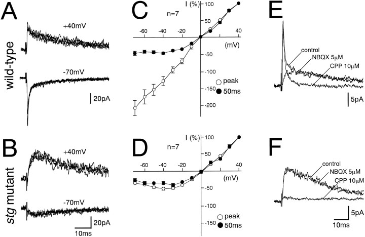
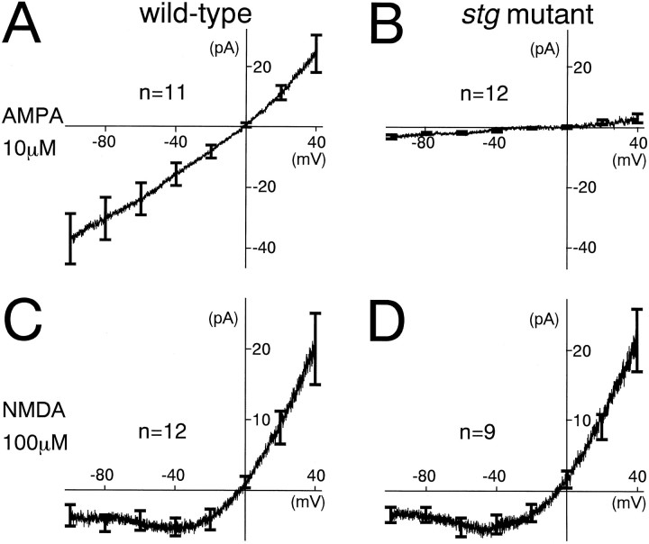
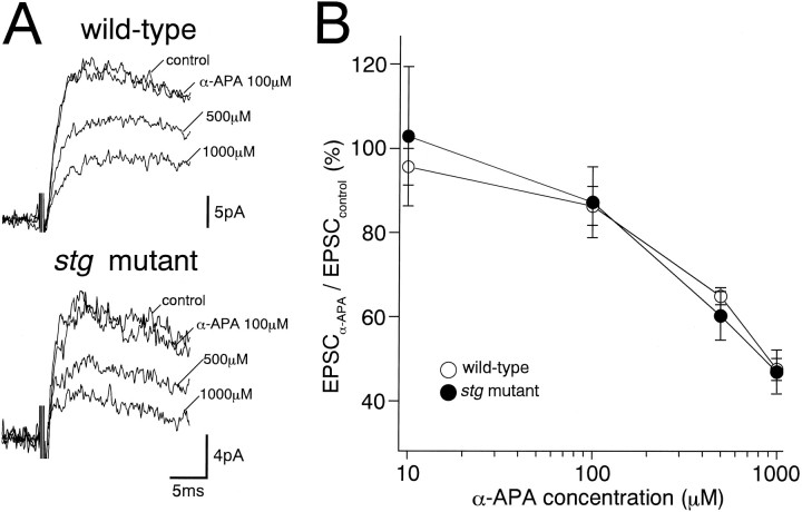
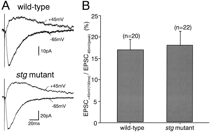
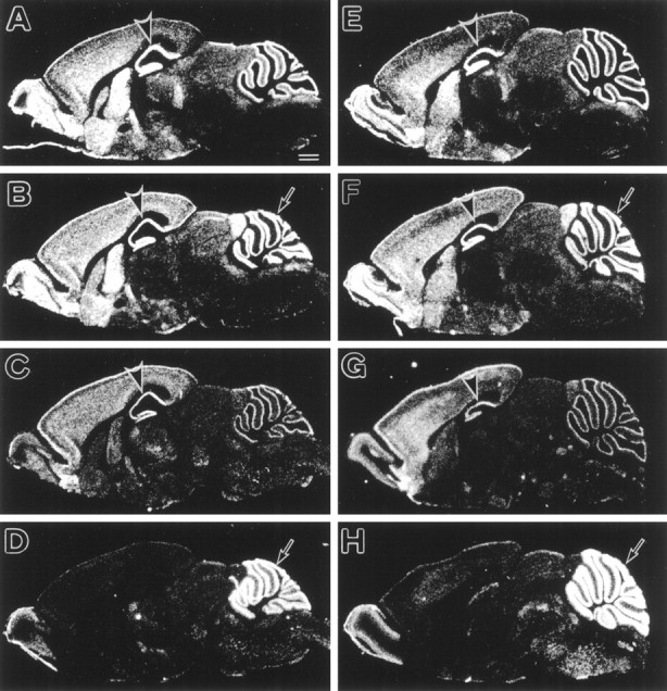
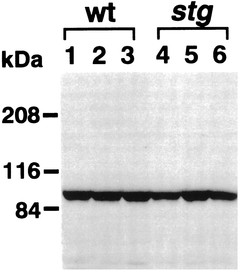
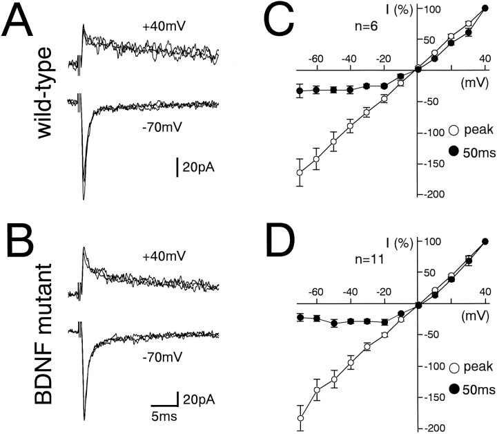
The spontaneous recessive mutant mouse stargazer (stg) begins to show ataxia around postnatal day 14 and display a severe impairment in the acquisition of classical eyeblink conditioning in adulthood. These abnormalities have been attributed to the specific reduction in brain-derived neurotrophic factor (BDNF) and the subsequent defect in TrkB receptor signaling in cerebellar granule cells (GCs). In the stg mutant cerebellum, we found that EPSCs at mossy fiber (MF) to GC synapses are devoid of the fast component mediated by AMPA-type glutamate receptors despite the normal slow component mediated by NMDA receptors. The sensitivity of stg mutant GCs to exogenously applied AMPA was greatly reduced, whereas that to NMDA was unchanged. Glutamate release from MF terminals during synaptic transmission to GCs appeared normal. By contrast, AMPA receptor-mediated EPSCs were normal in CA1 pyramidal cells of the stg mutant hippocampus. Thus, postsynaptic AMPA receptor function was selectively impaired in stg mutant GCs, although the transcription of four AMPA receptor subunit genes in the stg GC was comparable to the wild-type GC. We also examined the cerebellum of BDNF knockout mice and found that their MF-GC synapses had a normal AMPA receptor-mediated EPSC component. Thus, the impaired AMPA receptor function in the stg mutant GC is not likely to result from the reduced BDNF-TrkB signaling. These results suggest that the defect in MF to GC synaptic transmission is a major factor that causes the cerebellar dysfunction in the stg mutant mouse.


Figures

Fig. 1.
Fig. 1.
Lack of non-NMDA receptor-mediated fast component in MF–GC excitatory synaptic transmission in the stgmutant mice. A, B, EPSCs elicited by MF stimulation in GCs from the wild-type (A) and stgmutant (B) mice at holding potentials of +40 mV (top panels) and −70 mV (bottom panels). Each trace is a single-sweep record, and several traces are superimposed for each record. C, D,IV relationships of MF–EPSCs from wild-type (C) and stg mutant (D) mice measured at the peak (○) and 50 msec after (●) the stimulus. The EPSC amplitudes were normalized to the mean value at +40 mV in each experimental condition. Each data point and attached error bar represent mean and SEM. E, F, Effects of NBQX (5 μm) and CPP (10 μm) on MF–EPSCs in GCs from wild-type (E) andstg mutant (F) mice at a holding potential of +40 mV. Each trace is an average of 10 consecutive sweeps. Records in the control external solution, in the solution containing NBQX (5 μm), and in that containing CPP (10 μm) are superimposed. MF stimulation was repeated at 0.2 Hz.
Fig. 2.
Fig. 2.
Significantly reduced sensitivity to exogenous AMPA in GCs of the stg mutant mice. A, B, Instantaneous IVrelationships of the AMPA (10 μm)-induced current evoked in GCs from wild-type (A) and stgmutant (B) mice. Currents were measured during voltage ramp from +40 to −110 mV (duration, 2000 msec). C, D, Instantaneous IVrelationships of the NMDA (100 μm)-induced current evoked in GCs from wild-type (C) and stgmutant (D) mice. Currents were measured and illustrated as in A and B. Records were taken in the presence of tetrodotoxin (0.5 μm) and strychnine (1 μm). For leak subtraction, evoked currents measured in the control solution were subtracted from those in the presence of AMPA or NMDA. Each IV curve is an average of data from 9–12 different GCs. Error bars at −100, −80, −60, −40, −20, 0, +20, and +40 mV represent SEM.
Fig. 3.
Fig. 3.
Effects of a low-affinity NMDA receptor blocker, α-amino pimelic acid (α-APA), on the NMDA receptor-mediated components of MF–EPSCs. Specimen records (A) and summary graph show concentration–inhibition curves (B) for the wild-type (top record in A; open circles in B) and stg mutant (bottom record in A; closed circles in B) GCs. Each trace inA is an average of 20 consecutive sweeps. Theordinate in B indicates percentage of the control EPSC amplitude before application of α-amino pimelic acid (mean ± SEM). Records were taken at a holding potential of +40 mV and in the presence of CNQX (10 μm) and bicuculline (10 μm).
Fig. 4.
Fig. 4.
Normal excitatory synaptic responses in hippocampal CA1 pyramidal cells of the stg mutant mice.A, EPSCs elicited by stimulation of Schaffer collateral/commissural afferents in CA1 pyramidal cells from the wild-type (top traces) and stg mutant mice (bottom traces) measured at holding potentials of +45 mV (upward traces) and −65 mV (downward traces). B, Ratios of EPSC at +45 mV measured at 100 msec after the stimulus [EPSC(+45)] to peak EPSC at −65 mV [EPSC(−65)]. Error bars represent SEM.
Fig. 5.
Fig. 5.
In situ hybridization showing the expressions of four AMPA receptor subunit mRNAs in the adult stg mutant (A–D) and wild-type (E–H) mice. A, E, GluRα1; B, F, GluRα2; C, G, GluRα3;D, H, GluRα4. No differences in the distribution and levels of each AMPA subunit mRNA are found between thestg mutant and wild-type mice. Rostral is to the left, and dorsal is to the top. Scale bar, 1 mm.
Fig. 6.
Fig. 6.
Immunoblot analysis of the GluRα4 subunit proteins from the cerebella of three wild-type (lanes 1–3) and three stg mutant (lanes 4–6) mice. Fifty micrograms each of the postnuclear proteins of the cerebellum were loaded on each lane of SDS-PAGE (7% gel). The anti-GluRα4 antibody was used at 1 μg/ml. Molecular mass markers are indicated on the left.
Fig. 7.
Fig. 7.
MF–GC excitatory synaptic transmission in BDNF knockout mice is normal. A, B, EPSCs elicited by MF stimulation in GCs from the wild-type (A) and BDNF knockout (B) mice at holding potentials of +40 mV (top panels) and −70 mV (bottom panels). Each trace is single-sweep record, and several traces are superimposed for each record. C, D,IV relationships of MF–EPSCs from wild-type (C) and BDNF knockout (D) mice measured at the peak (○) and at 50 msec after the stimulus (●). The EPSC amplitudes were normalized to the mean value at +40 mV in each experimental condition. Each data point and attached error bar represent mean and SEM.

References

    1. Aiba A, Kano M, Chen C, Stanto ME, Fox GD, Herrup K, Zwingman TA, Tonegawa S. Deficient cerebellar long-term depression and impaired motor learning in mGluR1 mutant mice. Cell. 1994;79:377–388. - PubMed
    1. Boulter J, Hollmann M, O’Shea-Greenfield A, Hartley M, Deneris E, Maron C, Heinemann S. Molecular cloning and functional expression of glutamate receptor subunit genes. Science. 1990;249:1033–1037. - PubMed
    1. D’Angelo E, Rossi P, Taglietti V. Voltage-dependent kinetics of N-methyl-d-aspartate synaptic currents in rat cerebellar granule cells. Eur J Neurosci. 1994;6:640–645. - PubMed
    1. D’Angelo E, De Filippi G, Rossi P, Taglietti V. Synaptic excitation of individual rat cerebellar granule cells in situ: evidence for the role of NMDA receptors. J Physiol (Lond) 1995;484:397–413. - PMC - PubMed
    1. Di Pasquale E, Keegan KD, Noebels JL. Increased excitability and inward rectification in layer V cortical pyramidal neurons in epileptic mutant mouse stargazer. J Neurophysiol. 1997;77:621–631. - PubMed

Publication types

MeSH terms

Substances

LinkOut - more resources