. 1999 Jul 15;19(14):6200-12.
doi: 10.1523/JNEUROSCI.19-14-06200.1999.

Interdependence of multiple theta generators in the hippocampus: a partial coherence analysis

Affiliations

Interdependence of multiple theta generators in the hippocampus: a partial coherence analysis

B Kocsis et al. J Neurosci. .

Abstract

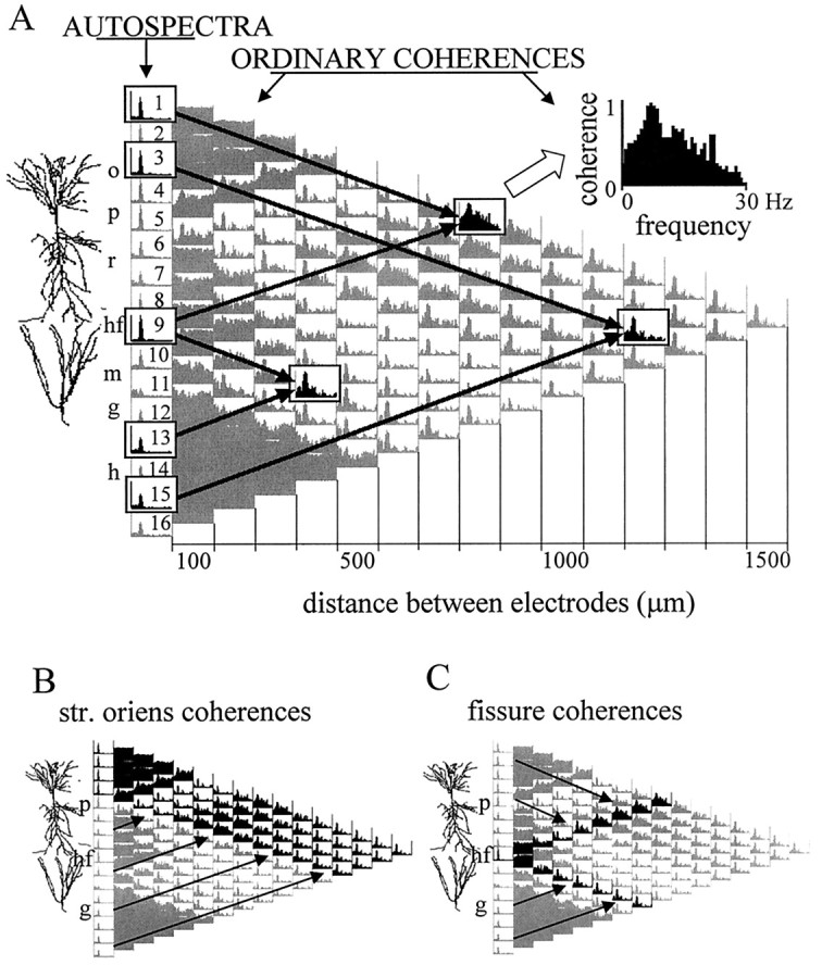
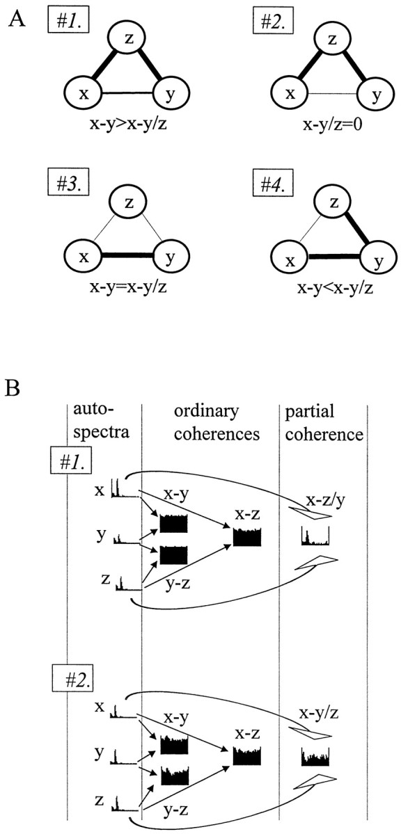
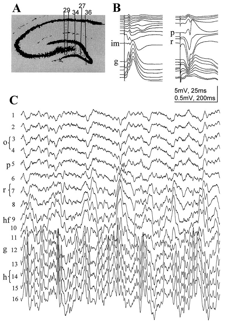
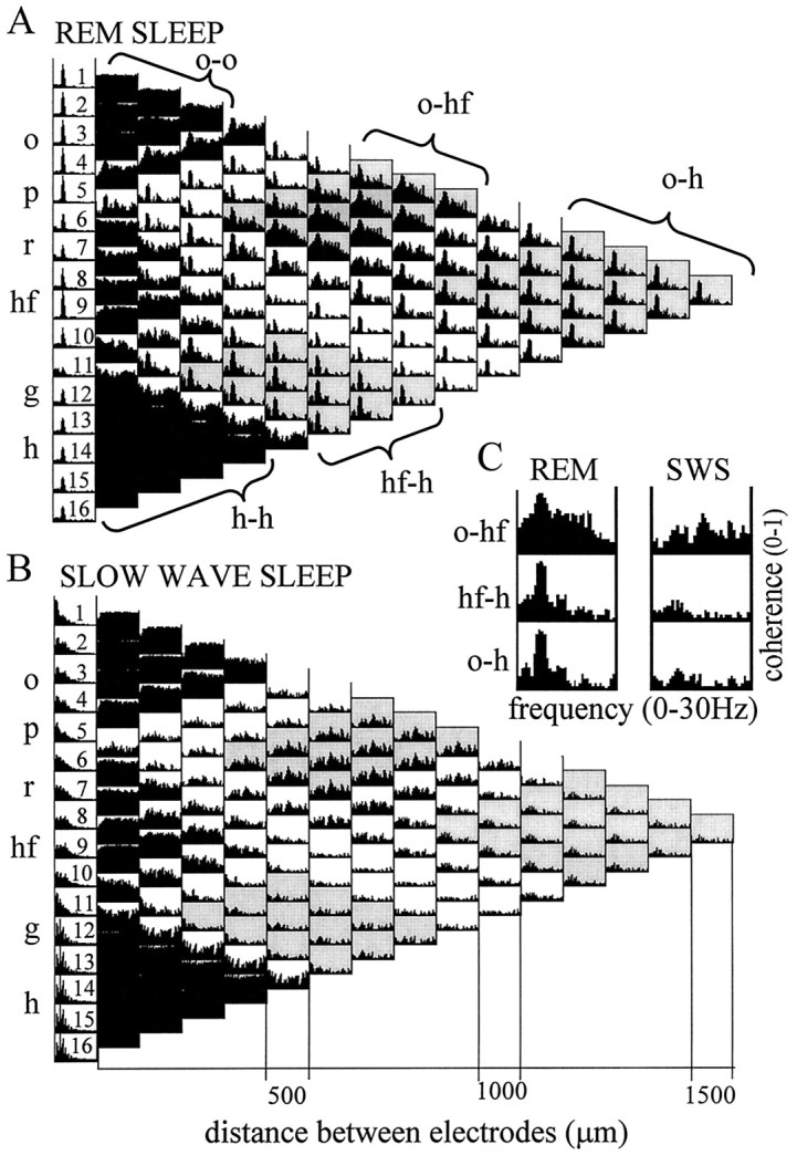
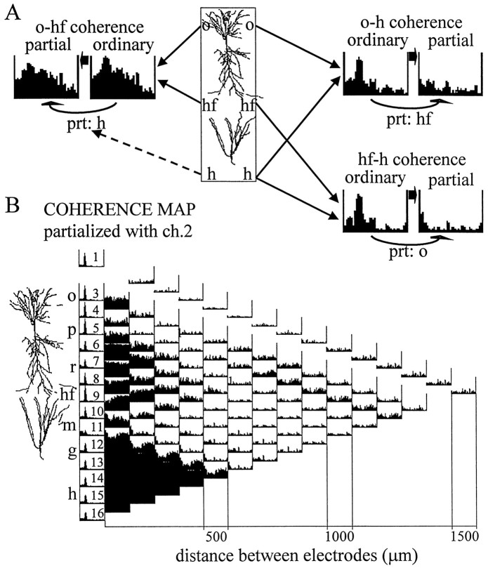
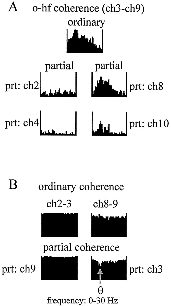
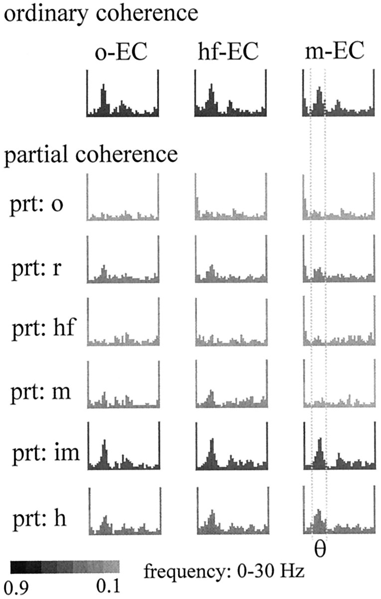
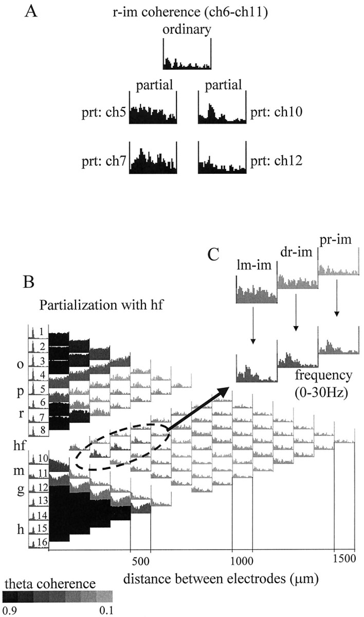
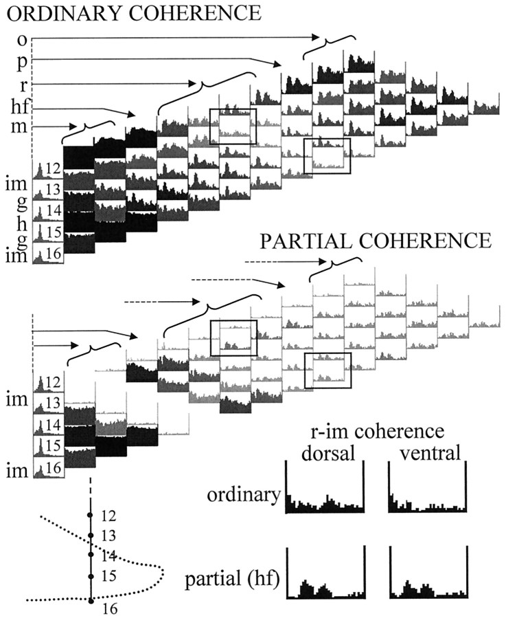
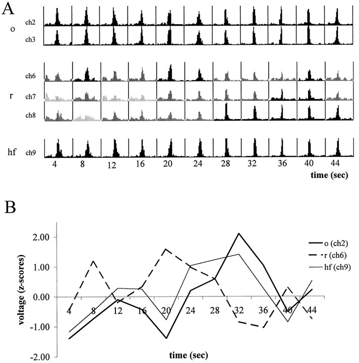
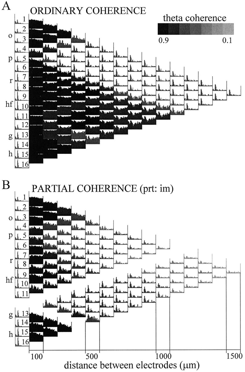
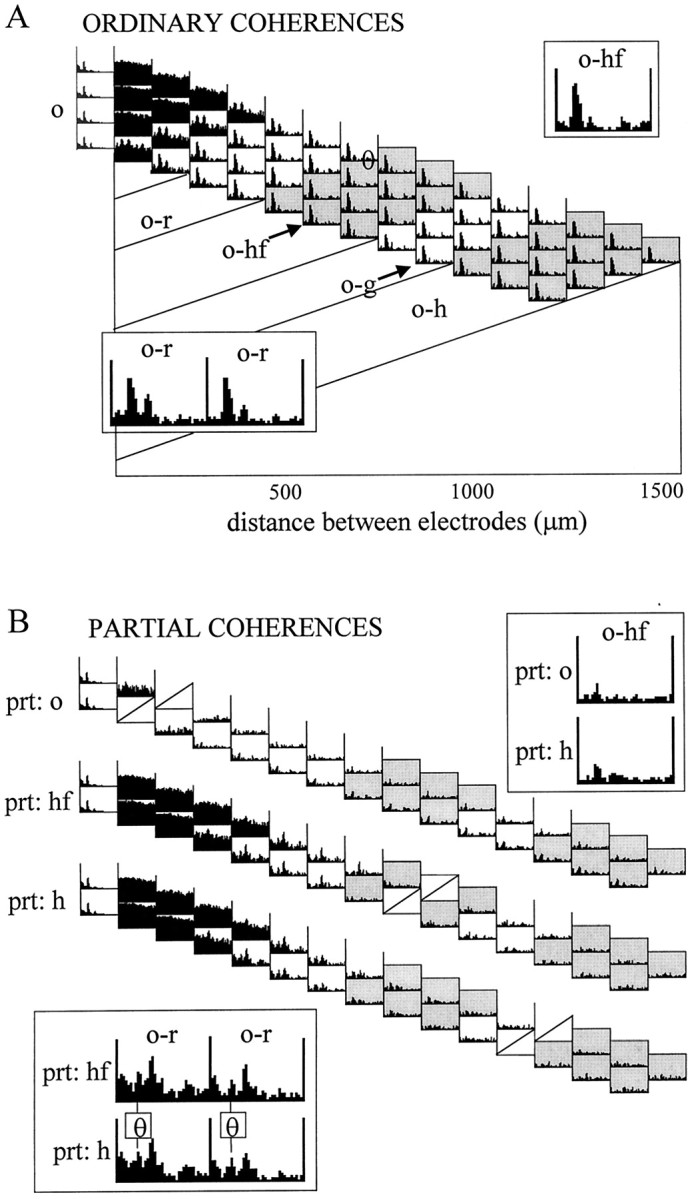
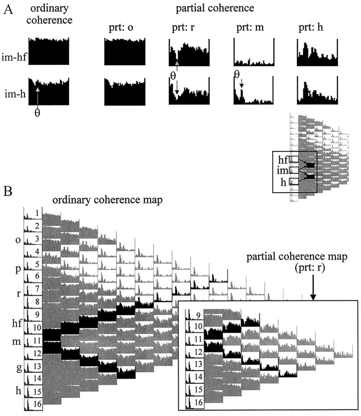
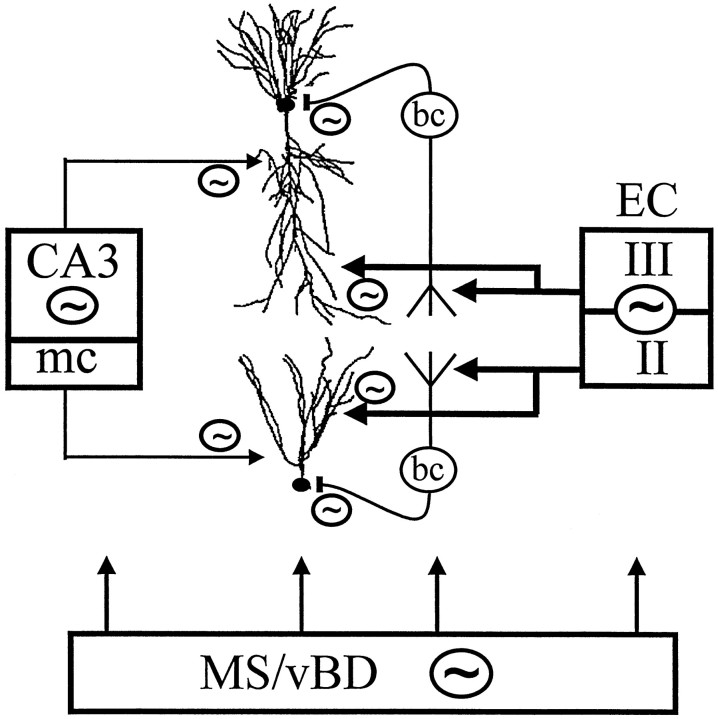
The extracellularly recorded theta oscillation reflects a dynamic interaction of various synaptic and cellular mechanisms. Because the spatially overlapping dipoles responsible for the generation of theta field oscillation may represent different mechanisms, their separation might provide clues with regard to their origin and significance. We used a novel approach, partial coherence analysis, to reveal the various components of the theta rhythm and the relationship among its generators. Hippocampal field activity was recorded by a 16-site silicon probe in the CA1-dentate gyrus axis of the awake rat. Field patterns, recorded from various intrahippocampal or entorhinal cortex sites, were used to remove activity caused by a common source by the partialization procedure. The findings revealed highly coherent coupling between theta signals recorded (1) from the hippocampal fissure and stratum (str.) oriens of the CA1 region and (2) between CA1 stratum radiatum and the dentate molecular layer. The results of partial coherence analysis indicated that rhythmic input from the entorhinal cortex explained theta coherence between signals recorded from the hippocampal fissure and str. oriens but not the coherence between signals derived from str. radiatum and the dentate molecular layer. After bilateral lesions of the entorhinal cortex, all signals recorded from both below and above the CA1 hippocampal pyramidal cell layer became highly coherent. These observations indicate the presence of two, relatively independent, theta generators in the hippocampus, which are mediated by the entorhinal cortex and the CA3-mossy cell recurrent circuitry, respectively. The CA3-mossy cell theta generator is partially suppressed by the dentate gyrus interneuronal output in the intact brain. We suggest that timing of the action potentials of pyramidal cells during the theta cycle is determined by the cooperation between the active CA3 neurons and the entorhinal input.


Figures

Fig. 1.
Fig. 1.
Ordinary coherence map of EEG activity and overview of data presentation. A, The 16 recording sites were equally spaced along a line between the CA1 and dentate gyrus regions. The different layers (o, str. oriens;p, pyramidal layer; r, str. radiatum;hf, hippocampal fissure; m, dentate gyrus molecular layer; g, granule cell layer;h, hilus) and the relative position of the principal cells is indicated left of the coherence map. Theleft column of the matrix shows the distribution of power (autospectrum) between 0 and 30 Hz for each signal, scaled to the channel with the highest power peak in the theta band (usually at the hippocampal fissure). The 120 pairwise coherence functions were arranged in a triangular matrix so that their position on the map indicates the location of the sites compared. Individual coherence functions for any signal pair are found at the point of crossing of arrows departing from theboxes containing the autospectra of these signals (inA, three examples are highlighted on the map). Thefirst column of coherence functions (right of the autospectra) contains 15 traces representing the relationship between neighboring signals (100 μm spacing); the second column (14 spectra) compares second neighbors (i.e., electrode tips separated by 200 μm), etc. The coherence spectrum at the peak of the triangle map connects electrodes 1 and 16 (CA1 str. oriens–alveus and dentate hilar region, respectively; 1.5 mm distance). Each coherence function spans from 0 to 30 Hz and is scaled from 0 to 1 (insert attop). B, C, Two different zones of the coherence map representing all coherence functions related to field potentials recorded at any location above the CA1 pyramidal layer (highlighted in black in B) and those related to the signal recorded from the hippocampal fissure (highlighted in black in C).
Fig. 2.
Fig. 2.
Features of the partial coherence analysis.A, Various combinations of anatomical connections can be revealed by the partialization technique. Different patterns of coupling (#1–#4) between three signals (x, y, and z). Partialization with z (x−y/z) may either decrease (#1, #2) or increase (#4) x−y coherence or leave unchanged (#3) the ordinary coherence betweenx and y, depending on the relationships between the three signals. B, Separation of an oscillatory component (theta) between two closely placed electrodes (x and z) from the wide-band noise using two different auxiliary signals. Signal y inB, #1 shares only the wide-band component with x and z. In B,#2, only the rhythmic component is common. Accordingly, partialization selectively eliminates the noise (B,#1) or theta pattern coherence (B,#2).
Fig. 3.
Fig. 3.
Simultaneous recording of evoked and spontaneous field activity in the CA1–dentate gyrus axis. A, Positions of the silicon probes in the rats included in this study. Thetraces shown in B and Cwere recorded with probe 29. Spacing of recording sites, 100 μm.B, Evoked field potentials in response to commissural (right) and perforant path (left) stimulation. These evoked potential profiles were used to determine the vertical location of the recording sites. C, Theta activity recorded during REM sleep. o, Str. oriens;p, pyramidal layer; r, str. radiatum;hf, hippocampal fissure; im, inner third of the molecular layer; g, granule cell layer;hi, hilus.
Fig. 4.
Fig. 4.
Autospectra (1–16) and maps of pairwise ordinary coherence functions computed from two 50 sec segments recorded during REM sleep (A) and SWS (B). Coherence spectra related to three major anatomical regions are highlighted: alveus–CA1 str. oriens (signals 1–4), distal dendritic regions of CA1 pyramidal cells and dentate granule cells, straddling the hippocampal fissure (8–10), and the dentate hilar region (12–16). Note similarity of the coherence functions within the respective zones. Note also that coherence functions of signals recorded within the same anatomical region showed high values over the entire 0–30 Hz frequency band (compare the threeblack triangles on the left of both maps). The three zones were separated by coherence spectra with relatively low values at all frequencies, corresponding to CA1 str. radiatum (6, 7) and dentate inner molecular layer (11) recordings. C, Pairwise coherence functions between three sample signals, recorded in different layers (o-hf, hf-h,o-h), for frequencies between 0 and 30 Hz during REM and SWS. During REM sleep, all coherence spectra were dominated by a large peak at theta frequency. Abbreviations as in Figure 1.
Fig. 5.
Fig. 5.
Coupling between theta field oscillations in str. oriens (o) and hippocampal fissure (hf). A, The pattern of relationships between recordings from str. oriens, hippocampal fissure, and hilus (h) during REM sleep was tested by comparing theta coherences of each possible signal pairs before (ordinary) and after (partial) partialization with the third signal. High ordinary coherence was found at theta frequency for all three signal pairs, which could be eliminated by partialization with either hippocampal fissure (prt: hf) or str. oriens (prt: o) signals. High theta coherence remained between str. oriens and hippocampal fissure recordings, however, after partialization with hilus signal (prt: h). B, Partial coherence map showing pairwise residual coherence after elimination of the components, which were coherent with field potentials recorded in str. oriens (signal 2). Compare the partial coherence map with the ordinary coherence map computed from the same data (Fig.3A).
Fig. 6.
Fig. 6.
Spatial resolution of the partialization technique. A, Effect of partialization on the str. oriens–hippocampal fissure coherence using neighboring signals recorded at 100 μm distance on either side of the str. oriens and hippocampal fissure electrodes. High theta peak present in the ordinary coherence function between hippocampal fissure (ch9) and any one of the str. oriens sites (ch3,top) was eliminated by partialization with signals recorded from within str. oriens (ch2 andch4, left) but not with signals recorded in distal str. radiatum (ch8) or dentate molecular layer (ch10, right). B, The high coherence, homogeneous in the entire 0–30 Hz range, between neighboring signals in the str. oriens were not affected by partialization with signals recorded from outside str. oriens, including that in hippocampal fissure (ch9,left). On the other hand, theta oscillations in the str. oriens explained some of the coherence between hippocampal fissure and its closest neighbors because partialization with ch3 (o) specifically decreased the coherence between ch9 (hippocampal fissure) and ch8 (str. lacunosum moleculare) at theta frequency.
Fig. 7.
Fig. 7.
Coherence in the theta band between a recording site in the entorhinal cortex (EC) and different layers of the hippocampus. The ordinary coherence peak at theta frequency could be eliminated by partialization with signals in str. oriens (prt: o) or hippocampal fissure (prt: hf). Significant residual theta coherence remained, however, after partialization with signals in the str. radiatum (prt: r), the midmolecular layer (prt: m), the inner molecular layer (prt: im), or the hilus (prt: h). Note that partialization with the inner molecular signal did not affect theta coherence between entorhinal cortex and at site 100 μm from the inner molecular layer (m-EC).
Fig. 8.
Fig. 8.
Coherence between signals recorded from CA1 str. radiatum (r) and dentate inner molecular (im) layers. A, Effect of partialization on the str. radium–inner molecular ordinary coherence using neighboring signals recorded 100 μm on either side of radiatum (left) or inner molecular (right) electrodes. Ordinary coherence between ch6 and ch11 was relatively low in the entire 0–30 Hz frequency range (top), including the theta band. Elimination of the locally shared components (with ch5 and ch7) from the r (ch6) recording increased radiatum–inner molecular pairwise theta coherence (see Results for more details). Similar enhancement of theta coherence was obtained after elimination of the local components with its neighbors (ch10 and ch12) from the inner molecular (ch11) recording. The largest increase in theta coherence occurred after partialization with ch7 or ch10. B, Coherence map after partialization with hippocampal fissure signal (ch 9). Grayscale indicates the magnitude of the coherence value at the theta frequency. The lowest ordinary coherence values (inset) were found between the inner molecular and the recordings from different locations in radiatum (pr, proximal radiatum; dr, distal radiatum; lm, str. lacunosum moleculare). These ordinary coherence functions did not have peaks at theta, whereas all the others exhibited strong theta coherence (Fig. 3A), which could be eliminated by partialization with the hippocampal fissure signal. On the other hand, partialization with signals close to inner molecular (including ch9 as shown here) eliminated a substantial part of the inner molecular signal (see relatively high ch9–ch11 ordinary coherence in the entire 0–30 Hz range in Fig. 3A), thereby uncovering a weak theta component unrelated to the hippocampal fissure signal. This “uncovered” theta was specifically localized to inner molecular and was coherent only with the radiatum recordings.
Fig. 9.
Fig. 9.
Coherent theta oscillation between CA1 str. radiatum (r) and dentate inner molecular (im) revealed by partialization with hippocampal fissure signal (i.e., after eliminating the entorhinal cortex-mediated theta) in a rat with the silicon probe placed medially (BA36 in Fig. 1).Grayscale indicates the magnitude of the coherence value at the theta frequency. Coherence between signals recorded from str. radiatum (r) and inner molecular layer (im) of both the dorsal and ventral blades of the dentate gyrus showed increased coherence at theta frequency after partialization with the hippocampal fissure signal.
Fig. 10.
Fig. 10.
Relative independence of theta activity in different layers. A, Autospectra of theta activity during successive 4 sec epochs. In each row, the values are expressed as a percentage of maximum theta power (integrated 6–8 Hz). Note similar variability of theta power in str. oriens (o) and hippocampal fissure (hf) recordings and different variability in the str. radiatum (r). B,z-Scores of theta voltage (root mean square) in the different layers. Note reciprocal relationship between hippocampal fissure and str. radiatum.
Fig. 11.
Fig. 11.
Effect of entorhinal cortex lesion on the coherence map of intrahippocampal signals. A, Ordinary coherence map of pairwise coherence functions in an animal after entorhinal cortex lesion (same rat as shown in Figs. 3 and 4). The largest power theta peaks were recorded in the str. radiatum and the hilus. High coherence was found between all signals recorded below the CA1 pyramidal layer, in the entire 0–30 Hz frequency band. As a consequence, clear theta peaks could be identified only in coherence functions comparing signals within this apparently homogeneous zone (5-16) with signals outside of this zone (1-4), i.e., above and below the CA1 pyramidal layer. B, Theta coherence buried in the homogeneous ordinary coherence functions could be revealed, however, by selectively eliminating the wide-band noise using the partialization technique. Partialization with the signal recorded from the inner molecular layer of dentate gyrus (prt: im), for example, uncovered strong theta coherence between the hilar region and the different layers of the CA1–dentate fields. Partialization with inner molecular also decreased the theta peak between str. oriens versus other recordings.
Fig. 12.
Fig. 12.
Coherence between theta oscillations in str. oriens (o) and other hippocampal layers after entorhinal cortex lesion. A, The shape of all ordinary coherence functions between str. oriens (o) and other recordings were similar, consisting of a relatively narrow peak at theta frequency. In contrast to the intact rat (Fig.3A), coherence between str. oriens and hippocampal fissure was not stronger than str. oriens and radiatum coherence.B, As in the intact rat, theta coherence between str. oriens and other layers could be eliminated by partialization with another str. oriens signal. After entorhinal cortex lesion, however, the effect of partialization with signals outside the str. oriens was different. Partialization with hippocampal fissure and hilus signals now had a similar effect (insets).
Fig. 13.
Fig. 13.
Coherence between theta oscillations in str. radiatum (r) and dentate inner molecular layer (im) after entorhinal cortex lesion. A, Changes in coherence function between inner molecular and locations 200 μm above (hippocampal fissure, hf) or below (hilar region, h). Ordinary coherence values were high in the entire 0–30 Hz band and were not affected by partialization with signals in str. oriens (prt: o). Partialization with radiatum (prt: r) selectively decreased inner molecular–hippocampal fissure and inner molecular–hilus coherence at theta frequency but did not change the wide-band coherence. Inner molecular–hippocampal fissure coherence values, on the other hand, were eliminated at all frequencies by partialization with a signal recorded from between the hippocampal fissure and the inner molecular layer in the dentate molecular layer (prt: m). Partialization with hilus signal (prt: h) decreased the wide-band component of the coherence function but had no effect on the theta coherence.B, Partialization with radiatum (prt: r, inset) selectively eliminated theta coherence between the inner molecular and all other signals.
Fig. 14.
Fig. 14.
Cooperation of extrahippocampal (septum, entorhinal cortex, EC) and intrahippocampal (CA3, mossy cells, mc) theta generators. The septal pacemaker [medial septum/vertical limb of diagonal band (MS/vBD)] may affect each member of the circuitry and is a requisite of theta oscillation. The main current generator of extracellular theta is the entorhinal input from layers II and III. The rhythmic entorhinal input discharges basket (bc) and chandelier cells (data not shown) and depolarizes pyramidal and granule cells. In the intact hippocampus, the intrahippocampal theta generator (CA3, mc) is suppressed by the inhibitory output of the dentate gyrus (data not shown). Only a few CA3 and mossy cells are selectively active. Coherent and converging activity of the entorhinal and intrahippocampal association inputs are critical for the timing of discharges of CA1 pyramidal and granule cells during the theta cycle.

References

    1. Acsády L, Kamondi A, Sik A, Buzsáki G. GABAergic cells are the major postsynaptic targets of mossy fibers in the rat hippocampus. J Neurosci. 1998;18:3386–3403. - PMC - PubMed
    1. Alonso A, Garcia-Austt E. Neuronal sources of the theta rhythm in the entorhinal cortex of the rat. II. Phase relations between unit discharges and theta field potentials. Exp Brain Res. 1987;67:502–509. - PubMed
    1. Amaral DG, Witter MP. The three-dimensional organization of the hippocampal formation: a review of anatomical data. Neuroscience. 1989;31:571–591. - PubMed
    1. Andersen P. Interhippocampal impulses. II. Apical dendritic activation of CA1 neurons. Acta Physiol Scand. 1960;48:178–208. - PubMed
    1. Apostol G, Creutzfeldt OD. Crosscorrelation between the activity of septal units and hippocampal EEG during arousal. Brain Res. 1974;67:65–75. - PubMed

Publication types

LinkOut - more resources