

Review
doi: 10.1128/MCB.19.8.5237.
Regulation of DNA-dependent activities by the functional motifs of the high-mobility-group chromosomal proteins
Affiliations
- PMID: 10409715
- PMCID: PMC84367
- DOI: 10.1128/MCB.19.8.5237
Item in Clipboard
Review
Regulation of DNA-dependent activities by the functional motifs of the high-mobility-group chromosomal proteins
Mol Cell Biol.
1999 Aug.
No abstract available
Figures
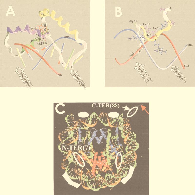
Interaction of HMG functional motifs with their binding site. (A) HMG-1 domain. Shown is a representation of the interaction of the HMG-1 domain in LEF-1 with a 16-bp oligonucleotide containing its cognate sequence. The red and blue tubes represent the DNA, the colored sticks represent the base pairs, and the ribbon represents the polypeptide backbone. Helices I, II, and III of the HMG-1 domain are shown in violet, green, and yellow, respectively. The view illustrates the distortion caused by Ile13, the hydrophobic pocket generated by the region containing Trp15 and Trp43, and the distortion of the base pair stacking in the minor groove. (B) AT hook. Shown is a surface representation of the interaction of the second DNA binding domain of HMG-I with a region of the PRDII element of the IFN-β enhancer. The red and blue tubes represent the DNA backbone, the ribbon represents the polypeptide backbone, and the AT hook domain is shown in yellow. Note the central Arg 10-Gly 11-Arg 12 sequence positioned in the minor DNA groove. (C) Nucleosomal binding domain. Shown is a model of the HMG-14 binding sites in nucleosome cores. The DNA and histones in the core particles are shown as ribbon traces (71). The solid white symbols in the two major grooves flanking the dyad axis and approximately 25 bp from the end of the DNA indicate the regions where HMG-14/-17 proteins protect the DNA from hydroxyl radical cleavage. The open white symbols represent the approximate location of the cross-links between the N terminus and C terminus of the HMG. The red arrow points to the amino-terminal region of histone H3. Histones H2B and H3 are represented by red and blue ribbons, respectively. Panels A and B were generated by David Landsman using GRASP software (76); panel C is reproduced from reference . The coordinates of the structures in panels A to C are available in the protein data bank at Brookhaven National Laboratory (PDB) under 2EZE, 1LEF, and 1AOI, respectively.
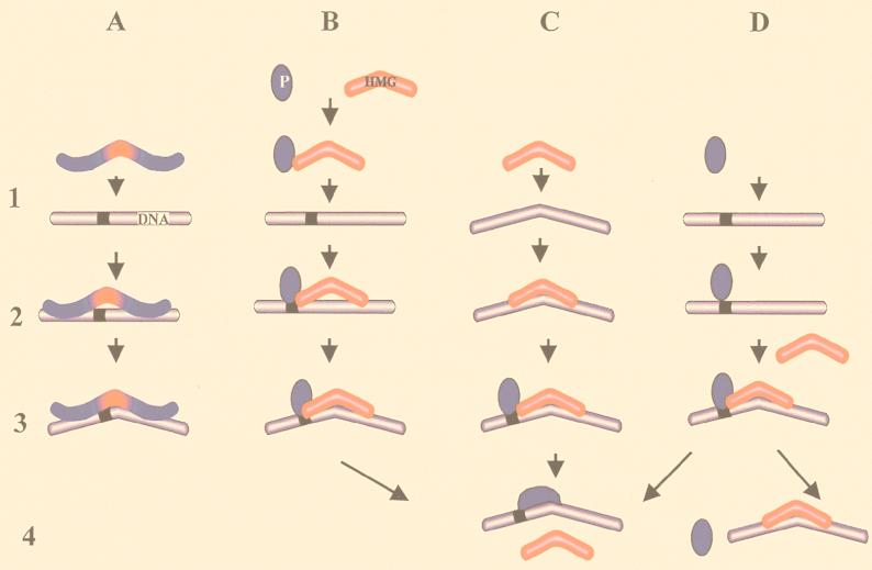
Multiple modes of sequence recognition and DNA bending by the HMG-1 domain. The HMG-1 domains are shown in red, the DNA recognition elements are shown in blue, and the black ribbon on the DNA represents a specific oligonucleotide sequence. The numbers on the left indicate four arbitrarily chosen steps involved in the interaction of an HMG-1 domain with its target. Step 1 represents targeting of a molecule to its binding site, step 2 represents binding, step 3 represents the induction of a conformational change in the target DNA, and step 4 represents possible changes in the protein contacts in the DNA-protein complex. The letters represent four different binding modes. (A) Binding by a sequence-specific HMG-1 domain protein. The HMG-1 domain targets the protein to a specific sequence and induces a conformational change in that locus. (B to D) Binding by the archetypal HMG-1/-2 proteins. (B) A preformed binary complex of a sequence-specific protein or protein complex (P) and an HMG molecule bind to a specific sequence. (C) The HMG-1/-2 protein targets distorted DNA structures. In step 3 a specific protein may target the DNA-HMG complex. (D) A sequence-specific protein binds to its cognate site and triggers the subsequent binding of an HMG-1/-2 protein. The arrows point out possible dissociation pathways of the resulting DNA-protein complexes. For further details, see the text.
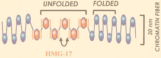
Model of the organization of HMG-17 in chromatin. Nucleosomes containing two molecules of HMG-17 (pink) are clustered into a domain whose average size is six contiguous nucleosomes. In this domain the entry-exit angles of the linker DNA are altered (double-headed arrow) and the higher-order structure of the chromatin is unfolded. HMG-17 may unfold the higher-order structure by interacting with the amino termini of the core histone, with histone H1, or with both. The structure of the 30-nm chromatin fiber is not known. The model presented was inspired by cryoelectron microscope studies, which suggested a zigzag organization of nucleosomes in the 30-nm fiber (9). However, it is equally possible that the 30-nm chromatin fiber has a solenoidal structure (34). The effect of HMG-14/-17 on the higher-order chromatin structure is the same for both the zigzag and solenoid organizations of nucleosomes.
References
-
- Aizawa S, Nishino H, Saito K, Kimura K, Shirakawa H, Yoshida Y. Stimulation of transcription in cultured cells by high mobility group protein 1: essential role of the acidic carboxyl-terminal region. Biochemistry. 1994;33:14690–14695. - PubMed
-
- Alfonso P J, Crippa M P, Hayes J J, Bustin M. The footprint of chromosomal proteins HMG-14 and HMG-17 on chromatin subunits. J Mol Biol. 1994;236:189–198. - PubMed
-
- Arlotta P, Rustighi A, Mantovani F, Manfioletti G, Giancotti V, Tell G, Damante G. High mobility group I proteins interfere with the homeodomains binding to DNA. J Biol Chem. 1997;272:29904–29910. - PubMed
-
- Ashar H R, Cherath L, Przybysz K M, Chada K. Genomic characterization of human HMGIC, a member of the accessory transcription factor family found at translocation breakpoints in lipomas. Genomics. 1996;31:207–214. - PubMed
Publication types
MeSH terms
Substances
LinkOut - more resources
Full Text Sources
Other Literature Sources