. 1999 Aug;19(8):5289-97.
doi: 10.1128/MCB.19.8.5289.

Bradykinin B(2) receptor-mediated mitogen-activated protein kinase activation in COS-7 cells requires dual signaling via both protein kinase C pathway and epidermal growth factor receptor transactivation

Affiliations

Bradykinin B(2) receptor-mediated mitogen-activated protein kinase activation in COS-7 cells requires dual signaling via both protein kinase C pathway and epidermal growth factor receptor transactivation

A Adomeit et al. Mol Cell Biol. 1999 Aug.

Abstract

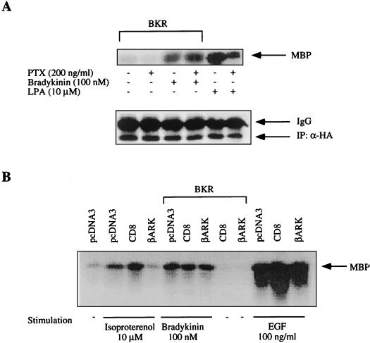
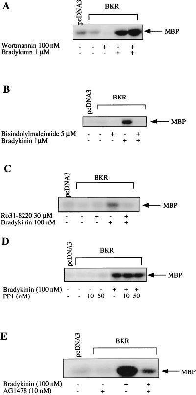
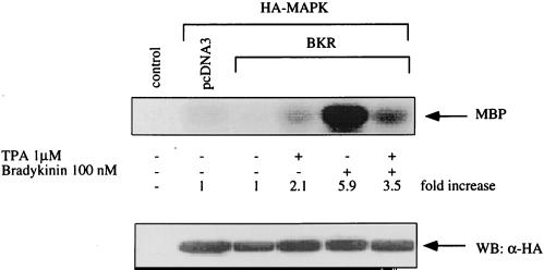
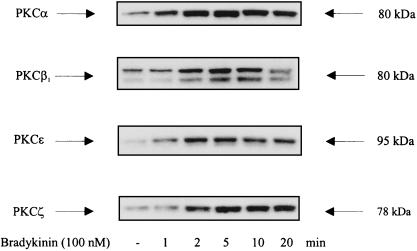
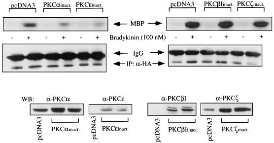
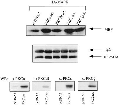
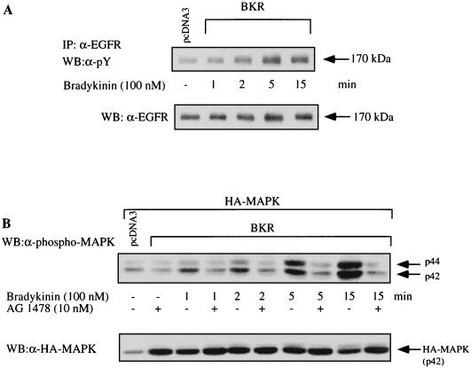
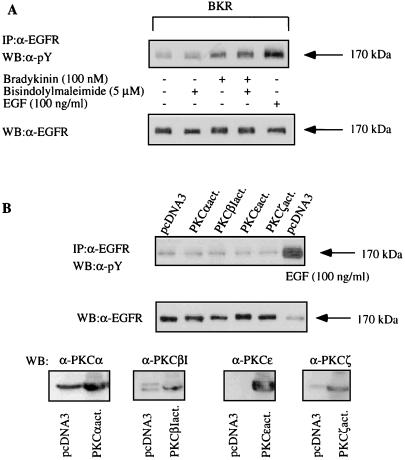
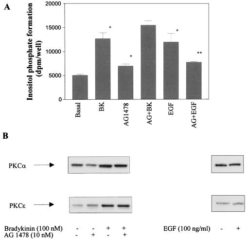
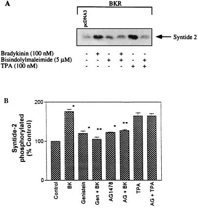
The signaling routes linking G-protein-coupled receptors to mitogen-activated protein kinase (MAPK) may involve tyrosine kinases, phosphoinositide 3-kinase gamma (PI3Kgamma), and protein kinase C (PKC). To characterize the mitogenic pathway of bradykinin (BK), COS-7 cells were transiently cotransfected with the human bradykinin B(2) receptor and hemagglutinin-tagged MAPK. We demonstrate that BK-induced activation of MAPK is mediated via the alpha subunits of a G(q/11) protein. Both activation of Raf-1 and activation of MAPK in response to BK were blocked by inhibitors of PKC as well as of the epidermal growth factor (EGF) receptor. Furthermore, in PKC-depleted COS-7 cells, the effect of BK on MAPK was clearly reduced. Inhibition of PI3-Kgamma or Src kinase failed to diminish MAPK activation by BK. BK-induced translocation and overexpression of PKC isoforms as well as coexpression of inactive or constitutively active mutants of different PKC isozymes provided evidence for a role of the diacylglycerol-sensitive PKCs alpha and epsilon in BK signaling toward MAPK. In addition to PKC activation, BK also induced tyrosine phosphorylation of EGF receptor (transactivation) in COS-7 cells. Inhibition of PKC did not alter BK-induced transactivation, and blockade of EGF receptor did not affect BK-stimulated phosphatidylinositol turnover or BK-induced PKC translocation, suggesting that PKC acts neither upstream nor downstream of the EGF receptor. Comparison of the kinetics of PKC activation and EGF receptor transactivation in response to BK also suggests simultaneous rather than consecutive signaling. We conclude that in COS-7 cells, BK activates MAPK via a permanent dual signaling pathway involving the independent activation of the PKC isoforms alpha and epsilon and transactivation of the EGF receptor. The two branches of this pathway may converge at the level of the Ras-Raf complex.


Figures

FIG. 1
FIG. 1
Activation of MAPK by BK is insensitive to PTX or βγ-scavenging proteins. (A) COS-7 cells were cotransfected with 6 μg of BKR DNA and 0.5 μg of HA-MAPK DNA. After 2 days, cells were preincubated with PTX (200 ng/ml) for 24 h. The effects of PTX treatment on basal and BK-stimulated MAPK activity were determined following a 5-min exposure to BK (100 nM). For control, COS-7 cells were stimulated with 10 μM LPA (5 min). MAPK activity was assessed as phosphorylation by MBP by immunoprecipitated p42HA-MAPK. The amount of immunoprecipitated HA-MAPK was determined by Western blot analysis with HA-specific MAb 12CA5. (IP: α-HA). (B) BKR and HA-MAPK DNAs were cotransfected into COS-7 cells together with plasmids containing the CD8-βARK chimera or the pcDNA vector. Cells were then serum starved and stimulated with isoproterenol (10 μM), BK (100 nM), or EGF (100 ng/ml) for 5 min. MAPK activity was determined in immunoprecipitates with MBP as the substrate, subjected to SDS-PAGE, and autoradiographed. The results shown are representative of at least three experiments. In all relevant figures, IgG denotes immunoglobulin G.
FIG. 2
FIG. 2
Effects of various inhibitors on BK-induced activation of MAPK. COS-7 cells, cotransfected with BKR and HA-MAPK DNAs as described in the text, were serum starved and preincubated with (A) the PI3K inhibitor wortmannin (100 nM), the PKC inhibitor bisindolylmaleimide (5 μM) (B) or Ro 31-8220 (30 μM) (C), the Src inhibitor protein phosphatase (PP1; 10 and 50 nM) (D), or the EGFR tyrosine kinase inhibitor AG1478 (10 nM) (E). R.31-8220 was kindly provided by D. Bradshaw (Roche, Welwyn Garden, United Kingdom). After preincubation for 30 min (A to C), 15 min (D), or 10 min (E), cells were stimulated with BK (100 nM to 1 μM) for 5 min. MAPK activity was assayed in lysates using the MBP phosphorylation assay as described in the text. Shown are blots representative of three independent experiments.
FIG. 3
FIG. 3
BK-induced activation of MAPK is decreased in PKC-depleted cells. COS-7 cells transfected with BKR and HA-MAPK were stimulated with TPA (1 μM) for 24 h to induce down-regulation of PKC and exposed to BK (10 nM) for 5 min, then lysis buffer was added, and MAPK activity was determined. The expression level of HA-MAPK was controlled by Western blotting with HA-specific MAb 12CA5 (WB: α-HA). Results are representative of three experiments.
FIG. 4
FIG. 4
BK induces translocation of the endogenous PKC isoforms α, βI, ɛ, and ζ. COS-7 cells were transfected with 6 μg of BKR DNA. After 2 days, serum-starved cells were stimulated with 100 nM BK for increasing times as indicated. Then a membrane fraction was prepared very quickly. Membranes were lysed, subjected to SDS-PAGE, and blotted onto PVDF membranes. Immunoblots obtained with antisera against the different PKC isoforms (1 μg/ml; Santa Cruz) are shown. Western blots shown are representative of at least two experiments.
FIG. 5
FIG. 5
Expression of inactive mutants of PKC isoforms in COS-7 cells. cDNA (1.5 μg) of inactive (inact.) mutants of PKC isoforms α, βI, ɛ, and ζ were cotransfected with BKR and HA-MAPK DNAs as described in Materials and Methods. After stimulation with BK (100 nM, 5 min), COS-7 cells were lysed, HA-MAPK was immunoprecipitated, and MAPK activity was assayed with MBP as the substrate. For control, the amount of immunoprecipitated HA-MAPK was determined by immunoblotting with a MAb against HA (IP:α-HA). Expression levels of the cotransfected inactive PKC isoforms were analyzed by Western blotting (WB) with antibodies against the respective isoforms (Santa Cruz). For comparison, the basal levels of the corresponding endogenous PKC isoforms are shown (pcDNA3). Results are representative of three experiments.
FIG. 6
FIG. 6
Effects of constitutively active mutants of the PKC isoforms on MAPK activity. In COS-7 cells, cDNA of active PKC isoform α or βI (2 μg) or ɛ or ζ (1 μg) was cotransfected with HA-MAPK DNA (0.5 μg). After 2 days, HA-MAPK was immunoprecipitated and assayed for activity as described in the text. For control, the expression levels of HA-MAPK and of the active PKC mutants determined by Western blotting are shown. Results are representative of three separate experiments. Notation is as for Fig. 5.
FIG. 7
FIG. 7
Time course of BK-induced tyrosine phosphorylation of the EGFR and EGFR-dependent activation of MAPK. (A) COS-7 cells expressing the BKR were serum starved for 12 h and then stimulated with BK (100 nM) for increasing times as indicated. Immunoprecipitates (IP) of the endogenous EGFR (anti-EGFR MAb; E. Merck AG, Darmstadt, Germany) were resolved by SDS-PAGE (7.5% gel) transferred to PVDF membranes, and Western blotted (WB) with either antiphosphotyrosine (α-pY) MAb 4G10 (top panel) or anti-EGFR antibody (polyclonal; Santa Cruz) (bottom panel). The results shown are representative of four separate experiments. (B) COS-7 cells were cotransfected with BKR and HA-MAPK, serum starved for 12 h, and stimulated with 100 nM BK in the absence or presence of AG1478 (10 nM, 10-min preincubation) for the times indicated. Shown is a representative Western blot with a polyclonal anti-phospho-MAPK antibody (Promega), representing two independent experiments.
FIG. 8
FIG. 8
BK-induced tyrosine phosphorylation of the EGFR is independent of PKC (notation is as in Fig. 7). (A) COS-7 cells transfected with pcDNA BKR were serum starved for 12 h, preincubated with the PKC inhibitor bisindolylmaleimide (5 μM) for 30 min, and stimulated with 100 nM BK for 5 min or 100 ng of EGF per ml (for control). The EGFR immunoprecipitates were subjected to SDS-PAGE, blotted, and analyzed by Western blotting with antiphosphotyrosine antibody (top). After stripping, the immunoprecipitates were quantified by using an anti-EGFR antibody (bottom). Results are representative of three independent experiments. (B) COS-7 cells were transfected with 2 μg of cDNA of constitutively active mutants of the four BK-sensitive PKC isoforms. After 2 days, cells were lysed, and the endogenous EGFR was immunoprecipitated and selected by using anti-phosphotyrosine antibody. For control, the EGFR was induced with 100 μg of EGF per ml. Also shown are the control blots with anti-EGFR antibody (middle panel) and with anti-PKC antibodies to detect the expression of the various PKC isoforms (bottom panel). The results are representative of four experiments.
FIG. 9
FIG. 9
Both BK-induced inositol phosphate formation and PKC translocation are independent of EGFR transactivation. (A) COS-7 cells were transfected with BKR DNA, prelabeled with [myo-3H]inositol for 24 h, subjected to serum-starved medium, preincubated for 10 min with 10 nM AG1478, and then stimulated with BK (100 nM, 5 min) or EGF (100 ng/ml, 7 min) as indicated. Inositol phosphate formation was determined in quadruplicate as described in Materials and Methods. ∗, inositol phosphate levels are significantly enhanced in the presence of BK, AG1478, and EGF compared with the control; ∗∗, EGF-induced inositol phosphate formation is significantly inhibited by AG1478 (Student’s t test; P < 0.05). (B) COS-7 cells expressing the BKR were stimulated with BK or EGF as indicated in absence or presence of AG1478 (10 nM, 10-min preincubation). PKC translocation was determined as described in Materials and Methods. Shown are Western blots for PKCs isoforms α and ɛ representative of two separate experiments.
FIG. 10
FIG. 10
Effects of PKC and tyrosine kinase inhibitors on activation of Raf-1 by BK. COS-7 cells transfected with the BKR were subjected to serum-starved medium and preincubated with bisindolylmaleimide (5 μM, 30 min), genistein (50 μM, 2 h), or AG1478 (10 nM, 10 min) followed by stimulation with BK (100 nM, 5 min) or TPA (100 nM, 3 min) as indicated. Raf-1 was immunoprecipitated with anti-Raf-1 polyclonal antibodies (Santa Cruz), and the immunocomplex was incubated with the substrate peptide syntide-2 and [γ-32P]ATP. Shown are results obtained with different experimental approaches to determine Raf-1 activation. (A) Autoradiogram of phosphorylated syntide-2. (B) Measurement of radioactivity (in quadruplicate) incorporated into syntide-2, using Whatman P81 phosphocellulose paper. Results are means ± standard errors of the means of three different experiments. ∗, significantly different compared with the control; ∗∗, significantly different compared with the effect of BK (Student’s t test; P < 0.05). Gen, genistein; AG, AG1478.

References

    1. Akiyama T, Ishida J, Nakagawa S, Ogawara H, Watanabe S, Itoh N, Shibuya M, Fukami Y. Genistein, a specific inhibitor of tyrosine-specific protein kinases. J Biol Chem. 1987;262:5592–5995. - PubMed
    1. Cai H, Smola U, Wixler V, Eisenmann-Trappe I, Diaz-Meco M, Moscat J, Rapp U, Cooper G M. Role of diacylglycerol-regulated protein kinase C isotypes in growth factor activation of the Raf-1 protein kinase. Mol Cell Biol. 1997;17:732–741. - PMC - PubMed
    1. Coutant K D, Corvaia N, Ryder N S. Bradykinin induces tyrosine phosphorylation of epidermal growth factor-receptor and focal adhesion proteins in human keratinocytes. Biochem Biophys Res Commun. 1995;210:774–780. - PubMed
    1. Crespo P, Xu N, Simonds W F, Gutkind J S. Ras-dependent activation of MAP kinase pathway mediated by G-protein beta gamma subunits. Nature. 1994;369:418–420. - PubMed
    1. Crespo P, Cachero T G, Xu N, Gutkind J S. Dual effect of beta-adrenergic receptors on mitogen-activated protein kinase. Evidence for a beta gamma-dependent activation and a G alpha s-cAMP-mediated inhibition. J Biol Chem. 1995;270:25259–25265. - PubMed

Publication types

MeSH terms

Substances