. 1999 Aug;19(8):5308-15.
doi: 10.1128/MCB.19.8.5308.

B-Raf inhibits programmed cell death downstream of cytochrome c release from mitochondria by activating the MEK/Erk pathway

Affiliations

B-Raf inhibits programmed cell death downstream of cytochrome c release from mitochondria by activating the MEK/Erk pathway

P Erhardt et al. Mol Cell Biol. 1999 Aug.

Abstract

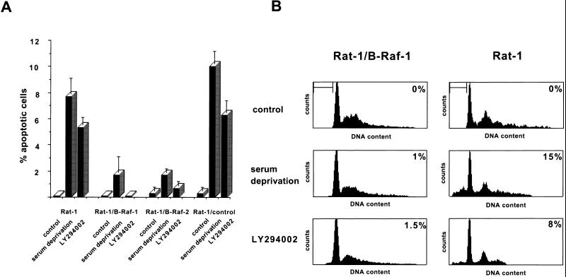
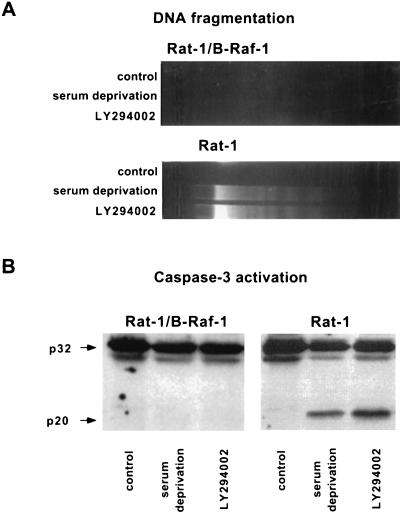
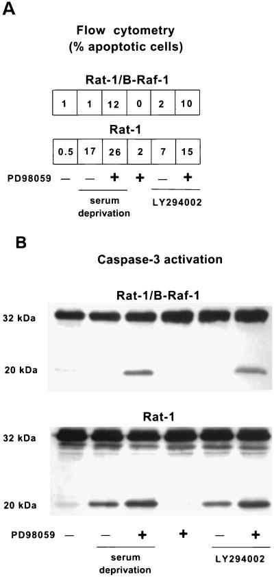
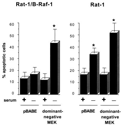
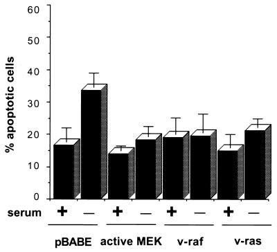
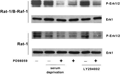
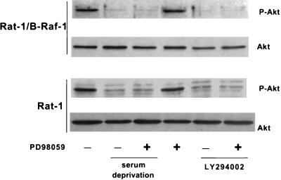
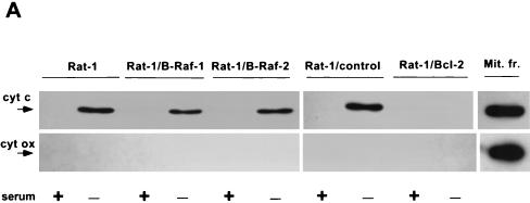
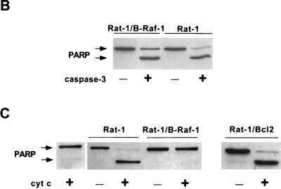
Growth factor-dependent kinases, such as phosphatidylinositol 3-kinase (PI 3-kinase) and Raf kinases, have been implicated in the suppression of apoptosis. We have recently established Rat-1 fibroblast cell lines overexpressing B-Raf, leading to activation of the MEK/Erk mitogen-activated protein kinase pathway. Overexpression of B-Raf confers resistance to apoptosis induced by growth factor withdrawal or PI 3-kinase inhibition. This is accompanied by constitutive activation of Erk without effects on the PI 3-kinase/Akt pathway. The activity of MEK is essential for cell survival mediated by B-Raf overexpression, since either treatment with the specific MEK inhibitor PD98059 or expression of a dominant inhibitory MEK mutant blocks the antiapoptotic activity of B-Raf. Activation of MEK is not only necessary but also sufficient for cell survival because overexpression of constitutively activated MEK, Ras, or Raf-1, like B-Raf, prevents apoptosis after growth factor deprivation. Overexpression of B-Raf did not interfere with the release of cytochrome c from mitochondria after growth factor deprivation. However, the addition of cytochrome c to cytosols of cells overexpressing B-Raf failed to induce caspase activation. It thus appears that the B-Raf/MEK/Erk pathway confers protection against apoptosis at the level of cytosolic caspase activation, downstream of the release of cytochrome c from mitochondria.


Figures

FIG. 1
FIG. 1
Induction of apoptosis by growth factor deprivation and PI 3-kinase inhibition. (A) Wild-type Rat-1 cells (Rat-1), transfected Rat-1 cells overexpressing B-Raf (Rat-1/B-Raf-1 and -2), and transfected Rat-1 cells that fail to overexpress B-Raf (Rat-1/control) were maintained in normal growth medium with or without the addition of 50 μM LY294002 or were incubated in serum-free medium for 16 h. Cells were then fixed, permeabilized, and stained with the DNA dye bisbenzimide (Hoechst 33258), and the apoptotic nuclei were scored on the basis of nuclear morphology. Data were averaged from three experiments, and at least 300 cells were counted per experiment. The error bars are standard errors of the mean. (B) Rat-1/B-Raf-1 and Rat-1 cells were maintained and treated as described in the legend to Fig. 1A. They were then collected and stained with propidium iodide, and the DNA content was analyzed by flow cytometry. The bar indicates cells with sub-G1 DNA content, a characteristic of apoptosis. The percentage of cells with sub-G1 DNA content is indicated in the upper right corner of each panel. The results are representative of at least two similar experiments.
FIG. 2
FIG. 2
Induction of oligonucleosomal DNA fragmentation and processing of caspase-3 by growth factor deprivation and PI 3-kinase inhibition. Rat-1/B-Raf-1 and Rat-1 cells were maintained and treated as described in the legend to Fig. 1. (A) Oligonucleosomal fragmentation of DNA. (B) Immunoblot analysis of caspase-3. The positions of the 32-kDa proenzyme and the 20-kDa active subunit are indicated by arrows.
FIG. 3
FIG. 3
Induction of apoptosis by inhibiting MEK with PD98059. Cells were maintained in normal growth medium or in serum-free medium and treated with 50 μM LY294002 or 50 μM PD98059, as indicated, for 16 h. (A) Fraction of apoptotic cells with sub-G1 DNA content as determined by flow cytometry. (B) Immunoblot analysis of caspase-3. The positions of the 32-kDa proenzyme and the 20-kDa active subunits are indicated. The results are representative of at least two similar experiments.
FIG. 4
FIG. 4
Induction of apoptosis by ectopic expression of dominant-negative MEK. Cells were cotransfected with an expression vector for dominant-negative MEK or an empty vector control (pBABE), together with an expression construct for GFP. Cells were maintained in normal growth medium or in serum-free medium for an additional 24 h. Transfected cells were then identified by fluorescence microscopy and scored for apoptosis on the basis of nuclear morphology. Data are presented as the percentage of GFP-positive cells with apoptotic nuclei. Data were averaged from three experiments, and 100 cells transfected with each vector were counted per experiment. The error bars are standard errors of the mean. Asterisks indicate a significant difference (P < 0.05) from controls maintained in serum (analysis of variance test).
FIG. 5
FIG. 5
Inhibition of apoptosis by expression of activated MEK, Raf, or Ras in Rat-1 cells. Rat-1 cells were cotransfected with the indicated expression vectors and a GFP expression construct. Transfected cells were maintained in normal growth medium or in serum-free medium for an additional 24 h. Data are averaged from three experiments, as in Fig. 4. The extent of apoptosis in pBABE-transfected cells deprived of serum was significantly different (P < 0.05) from controls maintained in serum, whereas apoptosis in serum-deprived cells transfected with MEK, Raf, or Ras expression constructs did not differ significantly from controls.
FIG. 6
FIG. 6
Overexpression of B-Raf results in increased Erk activity. Cells were maintained in normal growth medium or in serum-free medium and were treated with either 50 μM LY294002 or 50 μM PD98059, as indicated, for 16 h. Collected cell lysates were analyzed by immunoblotting with antibodies to phospho-Erk1/2 or Erk1.
FIG. 7
FIG. 7
Overexpression of B-Raf does not affect activation of Akt. Cells were maintained in normal growth medium or in serum-free medium and were treated with either 50 μM LY294002 or 50 μM PD98059, as indicated, for 16 h. Collected cell lysates were analyzed by immunoblotting with antibodies to phospho-Akt or Akt.
FIG. 8
FIG. 8
Effect of B-Raf overexpression on cytochrome c-dependent caspase activation. (A) Wild-type Rat-1 cells (Rat-1), transfected Rat-1 cells overexpressing B-Raf (Rat-1/B-Raf-1 and -2) or Bcl-2 (Rat-1/Bcl-2), and transfected Rat-1 cells that fail to overexpress B-Raf (Rat-1/control) were maintained in the presence of serum or deprived of growth factors for 24 h. Equal amounts of cytosolic proteins were then immunoblotted for cytochrome c (cyt c) and cytochrome oxidase subunit II (cyt ox), along with similar amounts of mitochondrial fraction (Mit. fr.) of Rat-1 cells maintained in serum as a positive control. The positions of cytochrome c and cytochrome oxidase are indicated by arrows. (B) Cytosols from Rat-1 and Rat-1/B-Raf cells were combined with purified, active His6-tagged human caspase-3 and intact HeLa cell nuclei and then incubated for 1 h at 37°C. The reaction mixture was used for immunoblot analysis to measure the cleavage of the caspase-3 substrate PARP. Intact PARP (116 kDa) and the cleaved fragment (85 kDa) are indicated by arrows. (C) Activation of caspase-3 was induced by the addition of cytochrome c to cytosols from nonapoptotic Rat-1, Rat-1/B-Raf, and Rat-1/Bcl-2 cells maintained in the presence of serum. Reaction mixtures were combined with intact HeLa cell nuclei, and the cleavage of PARP was assayed by immunoblot analysis. Lane 1 is a control in which cytochrome c was added to nuclei without cytosolic extract. The results are representative of at least three similar experiments.
FIG. 8
FIG. 8
Effect of B-Raf overexpression on cytochrome c-dependent caspase activation. (A) Wild-type Rat-1 cells (Rat-1), transfected Rat-1 cells overexpressing B-Raf (Rat-1/B-Raf-1 and -2) or Bcl-2 (Rat-1/Bcl-2), and transfected Rat-1 cells that fail to overexpress B-Raf (Rat-1/control) were maintained in the presence of serum or deprived of growth factors for 24 h. Equal amounts of cytosolic proteins were then immunoblotted for cytochrome c (cyt c) and cytochrome oxidase subunit II (cyt ox), along with similar amounts of mitochondrial fraction (Mit. fr.) of Rat-1 cells maintained in serum as a positive control. The positions of cytochrome c and cytochrome oxidase are indicated by arrows. (B) Cytosols from Rat-1 and Rat-1/B-Raf cells were combined with purified, active His6-tagged human caspase-3 and intact HeLa cell nuclei and then incubated for 1 h at 37°C. The reaction mixture was used for immunoblot analysis to measure the cleavage of the caspase-3 substrate PARP. Intact PARP (116 kDa) and the cleaved fragment (85 kDa) are indicated by arrows. (C) Activation of caspase-3 was induced by the addition of cytochrome c to cytosols from nonapoptotic Rat-1, Rat-1/B-Raf, and Rat-1/Bcl-2 cells maintained in the presence of serum. Reaction mixtures were combined with intact HeLa cell nuclei, and the cleavage of PARP was assayed by immunoblot analysis. Lane 1 is a control in which cytochrome c was added to nuclei without cytosolic extract. The results are representative of at least three similar experiments.

Similar articles

Cited by

References

    1. Alessi D R, Andjelkovic M, Caudwell B, Cron P, Morrice N, Cohen P, Hemmings B A. Mechanism of activation of protein kinase B by insulin and IGF-1. EMBO J. 1996;15:6541–6551. - PMC - PubMed
    1. Arends M J, McGregor A H, Toft N J, Brown E J, Wyllie A H. Susceptibility to apoptosis is differentially regulated by c-myc and mutated Ha-ras oncogenes and is associated with endonuclease availability. Br J Cancer. 1993;68:1127–1133. - PMC - PubMed
    1. Armstrong R C, Aja T, Xiang J, Gaur S, Krebs J F, Hoang K, Bai X, Korsmeyer S J, Karanewsky D S, Fritz L F, Tomaselli K J. Fas-induced activation of the cell death-related protease CPP32 is inhibited by Bcl-2 and by ICE family protease inhibitors. J Biol Chem. 1996;271:16850–16855. - PubMed
    1. Bergmann A, Agapite J, McCall K, Steller H. The Drosophila gene hid is a direct molecular target of ras-dependent survival signaling. Cell. 1998;95:331–341. - PubMed
    1. Berra E, Diaz-Meco M T, Moscat J. The activation of p38 and apoptosis by the inhibition of Erk is antagonized by the phosphoinositide 3-kinase/Akt pathway. J Biol Chem. 1998;273:10792–10797. - PubMed

Publication types

MeSH terms

Substances