. 1999 Aug;19(8):5363-72.
doi: 10.1128/MCB.19.8.5363.

Purification and identification of p68 RNA helicase acting as a transcriptional coactivator specific for the activation function 1 of human estrogen receptor alpha

Affiliations

Purification and identification of p68 RNA helicase acting as a transcriptional coactivator specific for the activation function 1 of human estrogen receptor alpha

H Endoh et al. Mol Cell Biol. 1999 Aug.

Retraction in

Abstract

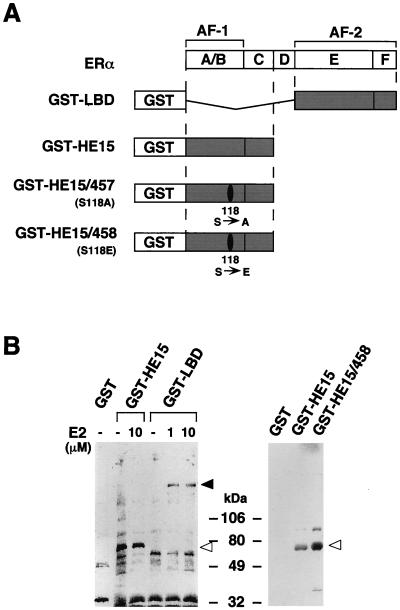
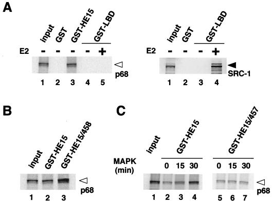
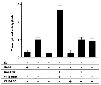
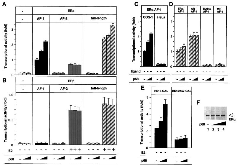
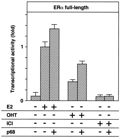
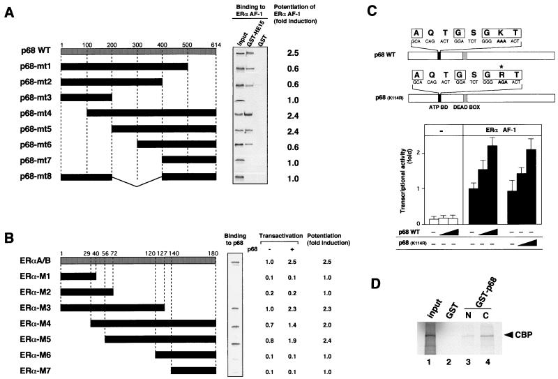
The estrogen receptor (ER) regulates the expression of target genes in a ligand-dependent manner. The ligand-dependent activation function AF-2 of the ER is located in the ligand binding domain (LBD), while the N-terminal A/B domain (AF-1) functions in a ligand-independent manner when isolated from the LBD. AF-1 and AF-2 exhibit cell type and promoter context specificity. Furthermore, the AF-1 activity of the human ERalpha (hERalpha) is enhanced through phosphorylation of the Ser(118) residue by mitogen-activated protein kinase (MAPK). From MCF-7 cells, we purified and cloned a 68-kDa protein (p68) which interacted with the A/B domain but not with the LBD of hERalpha. Phosphorylation of hERalpha Ser(118) potentiated the interaction with p68. We demonstrate that p68 enhanced the activity of AF-1 but not AF-2 and the estrogen-induced as well as the anti-estrogen-induced transcriptional activity of the full-length ERalpha in a cell-type-specific manner. However, it did not potentiate AF-1 or AF-2 of ERbeta, androgen receptor, retinoic acid receptor alpha, or mineralocorticoid receptor. We also show that the RNA helicase activity previously ascribed to p68 is dispensable for the ERalpha AF-1 coactivator activity and that p68 binds to CBP in vitro. Furthermore, the interaction region for p68 in the ERalpha A/B domain was essential for the full activity of hERalpha AF-1. Taken together, these findings show that p68 acts as a coactivator specific for the ERalpha AF-1 and strongly suggest that the interaction between p68 and the hERalpha A/B domain is regulated by MAPK-induced phosphorylation of Ser(118).


Figures

FIG. 1
FIG. 1
Detection of binding proteins to the hERα A/B domain in MCF-7 cells. (A) The hERα (with regions A to F shown) and the GST-ER fusion proteins used. (B) Binding proteins to the hERα A/B domain in MCF-7 cells. Aliquots of the 35S-labeled MCF-7 nuclear extract were incubated with glutathione-Sepharose beads loaded with GST alone, GST-HE15, GST-HE15/458, GST-HE15/457, or GST-LBD in the absence or presence of E2 at 1 and 10 μM. The bound proteins were subjected to SDS-PAGE (5 to 20% polyacrylamide gradient gel) followed by autoradiography. Open arrowheads indicate the position of a protein of 68 kDa. Size markers are indicated in kilodaltons. The solid arrowhead indicates the position of the SRC-1/TIF2 160-kDa family proteins (9, 14).
FIG. 2
FIG. 2
Amino acid sequence of human p68 RNA helicase protein. The five sequences determined by microsequencing are underlined and completely matched to the reported p68 RNA helicase protein (GenBank accession no. X15729 and X52104). The nuclear receptor recognition motif (LXXLL motif [15]) is doubly underlined, and the DEAD box motif is boxed.
FIG. 3
FIG. 3
Expression patterns of p68 RNA helicase transcripts in normal human tissues (A) and cancer cell lines (B). Northern blot analysis was performed as described previously (43). PBL, peripheral blood leukocyte. The cancer cell lines are as follows: HL60, promyelocytic leukemia cell line; HeLa S3, cervical carcinoma cell line; K562, chronic myelogenous leukemia cell line; Raji, Burkitt’s lymphoma cell line; SW480, colorectal adenocarcinoma cell line; A549, lung carcinoma cell line; G361, melanoma cell line; MCF-7, breast cancer cell line; T47D, breast cancer cell line; LNCaP, prostate carcinoma cell line; HOS, osteosarcoma cell line; HTOA, ovarian cancer cell line; Ishikawa, uterine body cancer cell line; PC12, rat pheochromocytoma cell line. The glyceraldehyde-3-phosphate dehydrogenase (G3PDH) transcript was used as an internal control. The relative positions of RNA markers (in kilobases) are shown on the right of panel B.
FIG. 4
FIG. 4
The recombinant p68 RNA helicase specifically binds to the hERα A/B domain in vitro. (A) p68 interacts only with the hERα A/B domain but not with the LBD, irrespective of the presence of E2. In vitro-translated p68 RNA helicase recombinant protein (left panel, lane 1) was analyzed by SDS-PAGE (5 to 20% polyacrylamide gradient gel). GST-ER fusion proteins, immobilized on beads, were mixed with 15 μl of in vitro translation reaction mixtures of p68; 2 μl of translation reaction mixture was loaded on the input lane. SRC-1 interacts with the hERα LBD only in the presence of E2 (right panel; lane 4) as previously reported (39). The open arrowhead indicates the position of a p68 RNA helicase protein, and the solid arrowhead indicates the position of the SRC-1 protein. (B) The p68 interaction is enhanced by replacing the Ser118 residue with Glu (S118E) in the bacterially expressed GST fusion protein [GST-HE15/458]. (C) Phosphorylation of the hERα A/B domain by MAPK increases its binding to p68. GST-HE15 or GST-HE15/457 that was incubated with activated MAPK for 0, 15, or 30 min at 30°C was used as the probe for an in vitro pull-down assay with 35S-labeled p68 protein.
FIG. 5
FIG. 5
p68 interacts with the hERα A/B domain but not with the LBD in vivo. p68 interacts with the hERα A/B domain in the mammalian two-hybrid system. A mammalian two-hybrid system with GAL4-p68 fusion protein and VP16-ER fusion proteins (VP16-HE15 and VP16-LBD) was used in COS-1 cells. COS-1 cells were cotransfected with 1 μg of either GAL4-DBD, GAL4-p68, VP16-HE15, or VP16-LBD in the presence or absence of E2 (10−7 M), along with 2 μg of 17M2G-CAT reporter plasmid. Significant interaction was detected only between p68 and the hERα A/B domain.
FIG. 6
FIG. 6
p68 potentiates the ligand-induced transactivation function of hERα through AF-1. (A) p68 potentiates AF-1 but not AF-2 of hERα. COS-1 cells were cotransfected with 0.40 μg of HE15-GAL(AF-1), LBD-GAL(AF-2), or HEGO (AF-1 plus AF-2) (26) and with either 0, 0.3, or 0.6 μg of pSG5-p68 in the presence or absence of E2 (10−7 M), along with 2 μg of 17M2G-CAT or 2 μg of ERE-G-CAT (for HEGO only). p68 potentiated the ligand-induced transactivation of the full-length hERα and AF-1 but not AF-2 activated by E2. (B) p68 has no effect in the transactivation function (AF-1 and AF-2) of hERβ. (C) p68 potentiates the hERα AF-1 activity in COS-1 cells but not in HeLa cells. (D) p68 has no effect on the AF-1 activities of the other nuclear receptors. (E) p68 potentiates the hERα AF-1 phosphorylated at the Ser118 residue. COS-1 cells were cotransfected with HE15-GAL or HE15/457-GAL along with either 0, 0.3, or 0.6 μg of pSG5-p68 and 2 μg of 17M2G-CAT. (F) p68 does not affect the expression levels of the hERα A/B domain. COS-1 cells were transfected with 0.40 μg of HE15-GAL and with either 0, 0.15, 0.3, or 0.6 μg of pSG5-p68. A Western blot analysis shows that the amount of the expressed chimeric protein is not affected by p68 expression. The open arrowhead indicates the position of the protein.
FIG. 7
FIG. 7
p68 potentiates the transactivation function of hERα induced by OHT but not ICI. COS-1 cells were cotransfected with 0.40 μg of HEGO and 2 μg of ERE-G-CAT, along with either 0, 0.3, or 0.6 μg of pSG5-p68, and treated with 100 nM E2, OHT, or ICI.
FIG. 8
FIG. 8
The RNA helicase activity of p68 is not required for the coactivator activity for the hERα AF-1. (A) The interaction domain of p68 for the hERα A/B domain is essential for the p68 coactivator activity for hERα AF-1. Representations of the p68 deletion mutants used for in vitro GST pull-down assay (middle panel) and the transient-expression assay (right panel) are shown. 35S-labeled p68 mutants were assayed with the GST-HE15 protein as a probe, showing that the region (aa 300 to 400) of the ATP binding domain and the DEAD box motif is required for direct interaction with the hERα A/B domain. The coactivator activities of p68 deletion mutants were determined in the transient-expression assay. COS-1 cells were cotransfected with 0.40 μg of HE15-GAL, 2 μg of 17M2G-CAT, and 0.6 μg of the expression vector of the p68 mutant. The mean fold induction of the AF-1 activity by the p68 mutant from three independent experiments is shown. (B) The interaction region of the hERα A/B domain for p68 is necessary for the potentiation of AF-1 activity by p68. Representations of the hERα A/B domain deletion mutants used for in vitro GST pull-down assay (middle panel) and the transient-expression assay (right panel) are shown. 35S-labeled p68 was assayed with the GST-fused A/B domain deletion mutant proteins as a probe, showing that the region (aa 56 to 127) in the A/B domain is required for direct p68 binding. The coactivator activities of p68 for the A/B domain deletion mutants were determined in the transient-expression assay. COS-1 cells were cotransfected with either 0.40 μg of A/B deletion mutants-plus-GAL (pM-A/B-Ms) with 2 μg of 17M2G-CAT and 0.6 μg of the expression vector of p68. The mean fold induction of the AF-1 activity by p68 from three independent experiments is shown. (C) A p68 mutant with a mutation in the ATP binding domain essential for the RNA helicase activity still potentiates the hERα AF-1. The ATP binding domain and DEAD box motif are indicated by solid and shaded boxes, respectively. The amino acid residues (AXXGXGKT), which are highly conserved among helicases, are boxed. The asterisk shows the replaced amino acid. COS-1 cells were cotransfected with 0.40 μg of pM-HE15 and 2 μg of 17M2G-CAT and with 0, 0.3, or 0.6 μg of the expression vector for the p68 mutant. The activity of hERα AF-1 was enhanced by both the wild-type p68 and the K144R mutant of p68. (D) p68 binds to CBP. To test the binding between p68 and CBP, GST control protein, GST-p68N (aa 1 to 387) fusion protein, or GST-p68C (aa 388 to 614) fusion protein was immobilized on beads and mixed with 15 μl of in vitro CBP translation reaction mixtures. Binding proteins were analyzed by SDS-PAGE. In vitro-translated CBP binds to both GST-fused p68N and p68C but not to the GST control protein.

References

    1. Ali S, Metzger D, Bornert J M, Chambon P. Modulation of transcriptional activation by ligand-dependent phosphorylation of the human oestrogen receptor A/B region. EMBO J. 1993;12:1153–1160. - PMC - PubMed
    1. Anzick S L, Kononen J, Walker R L, Azorsa D O, Tanner M M, Guan X Y, Sauter G, Kallioniemi O P, Trent J M, Meltzer P S. AlB1, a steroid receptor coactivator amplified in breast and ovarian cancer. Science. 1997;277:965–968. - PubMed
    1. Aronica S M, Katzenellenbogen B S. Stimulation of estrogen receptor-mediated transcription and alteration in the phosphorylation state of the rat uterine estrogen receptor by estrogen, cyclic adenosine monophosphate, and insulin-like growth factor-I. Mol Endocrinol. 1993;7:743–752. - PubMed
    1. Baniahmad A, Ha L, Reinberg D, Tsai S, Tsai M-J, O’Malley B. Interaction of human thyroid hormone receptor beta with transcription factor TFIIB may mediate target gene derepression and activation by thyroid hormone. Proc Natl Acad Sci USA. 1993;90:8832–8836. - PMC - PubMed
    1. Beato M, Herrlich P, Schutz G. Steroid hormone receptors: many actors in search of a plot. Cell. 1995;83:851–857. - PubMed

Publication types

MeSH terms

LinkOut - more resources