. 1999 Aug;19(8):5523-34.
doi: 10.1128/MCB.19.8.5523.

Kinase suppressor of Ras forms a multiprotein signaling complex and modulates MEK localization

Affiliations

Kinase suppressor of Ras forms a multiprotein signaling complex and modulates MEK localization

S Stewart et al. Mol Cell Biol. 1999 Aug.

Abstract

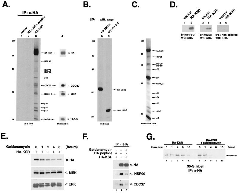
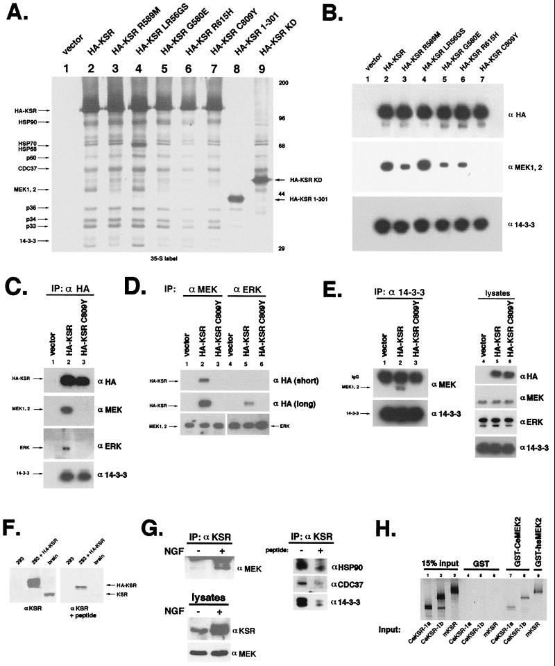
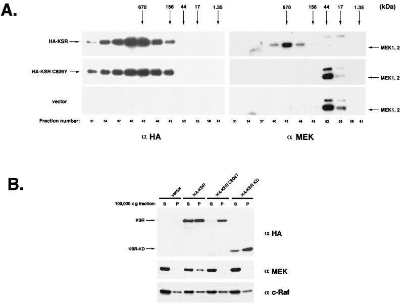
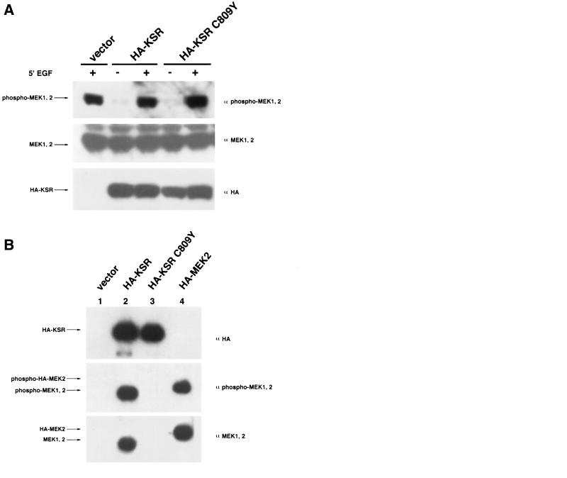
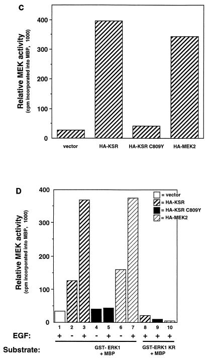
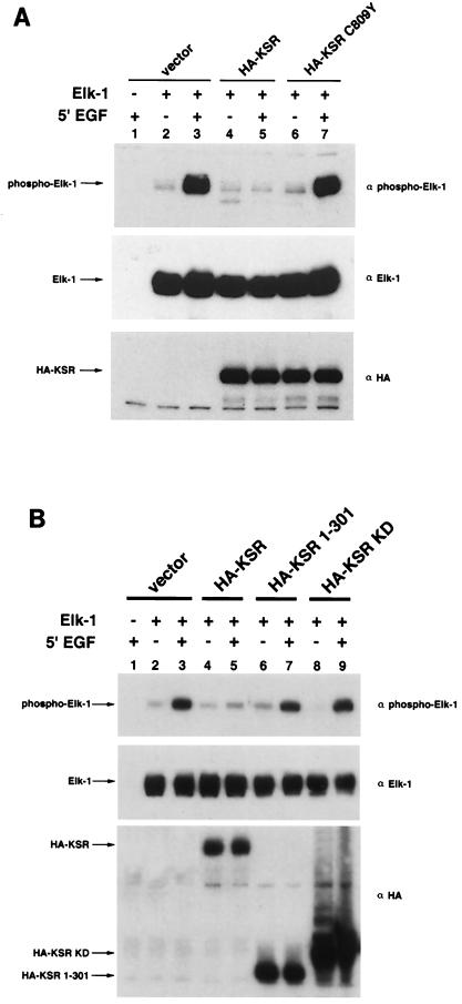
Genetic screens for modifiers of activated Ras phenotypes have identified a novel protein, kinase suppressor of Ras (KSR), which shares significant sequence homology with Raf family protein kinases. Studies using Drosophila melanogaster and Caenorhabditis elegans predict that KSR positively regulates Ras signaling; however, the function of mammalian KSR is not well understood. We show here that two predicted kinase-dead mutants of KSR retain the ability to complement ksr-1 loss-of-function alleles in C. elegans, suggesting that KSR may have physiological, kinase-independent functions. Furthermore, we observe that murine KSR forms a multimolecular signaling complex in human embryonic kidney 293T cells composed of HSP90, HSP70, HSP68, p50(CDC37), MEK1, MEK2, 14-3-3, and several other, unidentified proteins. Treatment of cells with geldanamycin, an inhibitor of HSP90, decreases the half-life of KSR, suggesting that HSPs may serve to stabilize KSR. Both nematode and mammalian KSRs are capable of binding to MEKs, and three-point mutants of KSR, corresponding to C. elegans loss-of-function alleles, are specifically compromised in MEK binding. KSR did not alter MEK activity or activation. However, KSR-MEK binding shifts the apparent molecular mass of MEK from 44 to >700 kDa, and this results in the appearance of MEK in membrane-associated fractions. Together, these results suggest that KSR may act as a scaffolding protein for the Ras-mitogen-activated protein kinase pathway.


Figures

FIG. 1
FIG. 1
KSR specifically associates with a number of proteins in vivo. (A) Metabolic labeling and immunoprecipitation (IP) of transfected mouse KSR. 293T cells were transfected with HA-KSR (lanes 2 and 3) or vector (lane 1), labeled with [35S]Met/Cys, and immunoprecipitated with α-HA (lanes 1 and 3) or with α-HA that had been preincubated with competitor peptide (lane 2). Immunocomplexes were separated by SDS-PAGE and visualized by autoradiography. Lane 4 represents an immunoblot of lane 3 probed with α-HA, α-p50CDC37, α-MEK, and α-14-3-3 as indicated. Positions of size markers are indicated in kilodaltons on the left. (B) Metabolic labeling and immunoprecipitation of transfected MEK and 14-3-3. 293T cells, transfected with HA-MEK2 or Myc-14-3-3 β, were processed as for panel A, separated by SDS-PAGE, and visualized by autoradiography. (C) Coomassie blue staining of HA-KSR complex affinity purified from 293T cells as for panel A. (D) HA-KSR is present in MEK1/2 and 14-3-3 immunoprecipitates. 293T cells transfected with HA-KSR were subjected to IP with α-14-3-3 (lanes 1 and 2), α-MEK1/2 (lanes 3 and 4), or control antibody (lanes 5 and 6) as described above, followed by Western blotting (WB) with α-HA to detect HA-KSR. (E) Reduction of KSR protein levels in response to geldanamycin treatment. HEK 293T cells transfected with HA-KSR were treated with 10 μM geldanamycin for the indicated times before harvesting and immunoblotting of whole-cell lysates with α-HA (top), α-MEK (middle), and α-ERK (bottom). (F) Geldanamycin treatment selectively disrupts the association of KSR with HSP90 and p50CDC37. HEK 293T cells transfected with HA-KSR were treated with 10 μM geldanamycin for 1 h (lane 3) before harvesting and immunoprecipitation with α-HA followed by immunoblotting with α-HA (top), α-HSP90 (middle), and α-p50CDC37 (bottom). HA peptide was included (lane 1) to demonstrate the specificity of the IP. (G) Geldanamycin destabilizes KSR. HA-KSR-transfected 293T cells were labeled for 30 min with [35S]Met/Cys with or without geldanamycin. Cells were chased for the indicated times and then immunoprecipitated with α-HA. Cells that had been treated with geldanamycin (lanes 6 to 10) showed a rapid loss of 35S-labeled HA-KSR. All results in this and subsequent figures are representative examples of at least three independent experiments except for Fig. 3A (performed twice).
FIG. 2
FIG. 2
Mutant KSRs, corresponding to loss-of-function alleles in C. elegans, are compromised in MEK1/2 binding. (A) Metabolic labeling and immunoprecipitation (IP) of mutant KSRs. 293T cells were transfected with vector (lane 1), wild-type KSR (lane 2), or mutant HA-tagged KSR (lanes 3 to 9). Metabolically labeled lysates were subjected to IP with α-HA as for Fig. 1A prior to SDS-PAGE and autoradiography. Positions of size markers are indicated in kilodaltons on the right. (B) Loss-of-function mutant KSR C809Y binds 14-3-3 but not MEK1/2. 293T cells were transfected as for panel A and then immunoprecipitated with α-HA. Immunocomplexes were separated by SDS-PAGE and immunoblotted with α-HA (to detect HA-KSR; top) α-MEK1/2 (middle), and α-14-3-3 (bottom). KSR C809Y showed no detectable MEK binding, while KSR R589M, G580E, and R615H displayed reduced MEK binding. (C) KSR forms a signaling complex in vivo with ERK and MEK. 293T cells were transfected with vector (lane 1), wild-type KSR (lane 2), or HA-KSR C809Y (lane 3) and then immunoprecipitated with α-HA prior to SDS-PAGE and immunoblotting with α-HA, α-MEK, α-ERK, and α-14-3-3 as indicated. Note that we reproducibly detect far more MEK1/2 (>10-fold) than ERK1/2 associated with HA-KSR in these cells. (D) Comparison of MEK-KSR and ERK-KSR association in vivo. 293T cells, transfected as for panel A, were lysed, immunoprecipitated with α-MEK (lanes 1 to 3) or α-ERK (lanes 4 to 6), and then immunoblotted with α-HA (to detect KSR; upper two panels), α-ERK (lower panel, lanes 4 to 6), and α-MEK (lower panel, lanes 1 to 3). Two exposures of the α-HA blot are presented to emphasize the relative amounts of KSR in MEK and ERK immunoprecipitates. (E) MEK-14-3-3 association is mediated by KSR and dependent on cysteine 809. Lysates were immunoprecipitated with α-14-3-3 and then immunoblotted with α-MEK (upper panel, lanes 1 to 3) and α-14-3-3 (lower panel). Representative immunoblot of whole-cell extracts shows expression of transfected HA-KSRs and endogenous MEK, ERK, and 14-3-3 (lanes 4 to 6). IgG, immunoglobulin G. (F) Detection of endogenous KSR in brain and PC12 cells. Extracts from vector- or HA-KSR-transfected 293T cells or mouse brain were probed with affinity-purified α-KSR. Reactive bands were effectively competed by competition by peptide antigen. (G) Association of endogenous KSR with MEK, HSP90, p50CDC37, and 14-3-3 in PC12 cells. Lysates from undifferentiated or day 5 differentiated PC12 cells were blotted with α-KSR or α-MEK as indicated. KSR is induced by NGF treatment. Lysates from differentiated PC12 cells were immunoprecipitated with α-KSR and then immunoblotted with α-MEK, α-HSP90, α-p50CDC37, and α-14-3-3. Where indicated, immunizing peptide was included in the IP. (H) MEK binding is a conserved function of KSR. 35S-labeled in vitro-translated CeKSR-1a and CeKSR-1b (see Materials and Methods) were incubated with either GST- or GST-CeMEK2 coupled to glutathione-Sepharose. After mixing for 1 h, beads were collected by centrifugation and washed several times in buffer before elution with glutathione. Eluted proteins were separated by SDS-PAGE and subjected to autoradiography. Mammalian KSR and GST-MEK were included for comparison. Note that CeMEK2 and CeKSR-1 can associate as effectively as their mammalian counterparts.
FIG. 3
FIG. 3
KSR exists in a high-molecular-weight complex in vivo. (A) HA-KSR recruits MEK to a large complex. HA-KSR-, HA-KSR C809Y-, or vector-transfected 293T cells were homogenized, and the resulting lysates were fractionated on a Superose 6 column (Pharmacia). Equal volumes of each fraction were subjected to SDS-PAGE followed by immunoblotting with α-HA (left) and α-MEK (right). Elution profiles of molecular weight standards are indicated (thyroglobulin, 670 kDa; gamma globulin, 156 kDa; ovalbumin, 44 kDa; myoglobin, 17 kDa; and cobalamin, 1.35 kDa; Bio-Rad Laboratories). Endogenous MEK1/2 exhibits an apparent molecular mass of 700 kDa only in the presence of HA-KSR (top). In contrast, endogenous MEK elutes at approximately 44 kDa in cells transfected with vector or KSR C809Y (bottom and middle). (B) KSR alters the cellular distribution of MEK. HA-KSR-, HA-KSR C809Y-, or HA-KSR KD-transfected 293T cells were Dounce homogenized and then centrifuged at 6,000 × g for 15 min. The resulting supernatant was spun at 100,000 × g for 60 min to separate it into soluble (S) and membrane (P) fractions. Equal volumes of each fraction were subjected to SDS-PAGE and immunoblot analysis with α-HA, α-MEK, and α-c-Raf.
FIG. 4
FIG. 4
KSR-associated MEK is phosphorylated and can activate ERK in response to growth factors. (A) KSR expression does not effect MEK1/2 phosphorylation at serine 218/222. 293T cells were transfected with vector, HA-KSR, or HA-KSR C809Y (1 μg of each). After 24 h of serum deprivation, cells were stimulated for 5 min with EGF (50 ng/ml) as indicated. Whole-cell extracts were prepared and separated by SDS-PAGE followed by immunoblot analysis with α-phospho-MEK1/2, α-MEK, and α-HA, as indicated (lower panel). (B) MEK in the KSR complex is phosphorylated. α-HA immunoprecipitates from vector-, HA-KSR-, HA-KSR C809Y-, or HA-MEK2-transfected 293T cells were subjected to SDS-PAGE and immunoblot analysis with α-phospho-MEK1/2, α-MEK, and α-HA, as indicated. Note that the electrophoretic mobility of HA-MEK2 is slower than that of endogenous MEK due to the presence of two HA epitopes at its amino terminus. (C) KSR-associated MEK is active. Equal amounts of MEK from each of the immunoprecipitates shown in panel B were used to activate recombinant GST-ERK1 in a coupled kinase assay using MBP as a substrate (see Materials and Methods). (D) Binding of KSR to MEK does not alter the ability of MEK to activate ERK in response to growth factor stimulation. 293T cells were transfected with the indicated KSR or MEK expression vector. After 24 h of serum starvation, cells were stimulated with EGF for 5 min as indicated and then immunoprecipitated with α-HA. Equal amounts of MEK from all immunoprecipitates were used to activate recombinant GST-ERK1 or GST-ERK1 KR (kinase-inactive control) in a coupled kinase assay.
FIG. 4
FIG. 4
KSR-associated MEK is phosphorylated and can activate ERK in response to growth factors. (A) KSR expression does not effect MEK1/2 phosphorylation at serine 218/222. 293T cells were transfected with vector, HA-KSR, or HA-KSR C809Y (1 μg of each). After 24 h of serum deprivation, cells were stimulated for 5 min with EGF (50 ng/ml) as indicated. Whole-cell extracts were prepared and separated by SDS-PAGE followed by immunoblot analysis with α-phospho-MEK1/2, α-MEK, and α-HA, as indicated (lower panel). (B) MEK in the KSR complex is phosphorylated. α-HA immunoprecipitates from vector-, HA-KSR-, HA-KSR C809Y-, or HA-MEK2-transfected 293T cells were subjected to SDS-PAGE and immunoblot analysis with α-phospho-MEK1/2, α-MEK, and α-HA, as indicated. Note that the electrophoretic mobility of HA-MEK2 is slower than that of endogenous MEK due to the presence of two HA epitopes at its amino terminus. (C) KSR-associated MEK is active. Equal amounts of MEK from each of the immunoprecipitates shown in panel B were used to activate recombinant GST-ERK1 in a coupled kinase assay using MBP as a substrate (see Materials and Methods). (D) Binding of KSR to MEK does not alter the ability of MEK to activate ERK in response to growth factor stimulation. 293T cells were transfected with the indicated KSR or MEK expression vector. After 24 h of serum starvation, cells were stimulated with EGF for 5 min as indicated and then immunoprecipitated with α-HA. Equal amounts of MEK from all immunoprecipitates were used to activate recombinant GST-ERK1 or GST-ERK1 KR (kinase-inactive control) in a coupled kinase assay.
FIG. 5
FIG. 5
Effects of KSR mutants on Elk-1 phosphorylation. (A) KSR C809Y is deficient in Elk-1 inhibition. 293T cells were transfected with Elk-1 (250 ng) and either vector, HA-KSR, or HA-KSR C809Y (1 μg). After 24 h of serum starvation, cells were stimulated with EGF (50 ng/ml) for 5 min prior to lysis and immunoblotting with the indicated antibodies. α-Phospho-Elk-1 recognizes only phosphorylated, active Elk-1. (B) Neither the isolated amino terminus nor the kinase domain of KSR is sufficient to block Elk-1 phosphorylation. 293T cells were transfected with Elk-1 and the indicated KSR expression vector. After 24 h of serum starvation, cells were stimulated with EGF (50 ng/ml) for 5 min prior to lysis and immunoblotting with α-phospho-Elk-1 (top), α-Elk-1 (middle), and α-HA (bottom).

References

    1. Beitel G J, Clark S G, Horvitz H R. Caenorhabditis elegans ras gene let-60 acts as a switch in the pathway of vulval induction. Nature. 1990;348:503–509. - PubMed
    1. Brenner S. The genetics of Caenorhabditis elegans. Genetics. 1974;77:71–94. - PMC - PubMed
    1. Choi K Y, Satterberg B, Lyons D M, Elion E A. Ste5 tethers multiple protein kinases in the MAP kinase cascade required for mating in S. cerevisiae. Cell. 1994;78:499–512. - PubMed
    1. Cutforth T, Rubin G M. Mutations in Hsp83 and cdc37 impair signaling by the sevenless receptor tyrosine kinase in Drosophila. Cell. 1994;77:1027–1036. - PubMed
    1. Dang A, Frost J A, Cobb M H. The MEK1 proline-rich insert is required for efficient activation of the mitogen-activated protein kinases ERK1 and ERK2 in mammalian cells. J Biol Chem. 1998;273:19909–19913. - PubMed

Publication types

MeSH terms

Substances