. 1999 Aug;19(8):5576-87.
doi: 10.1128/MCB.19.8.5576.

The Wnt/Wg signal transducer beta-catenin controls fibronectin expression

Affiliations

The Wnt/Wg signal transducer beta-catenin controls fibronectin expression

D Gradl et al. Mol Cell Biol. 1999 Aug.

Abstract

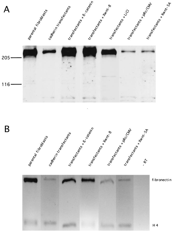
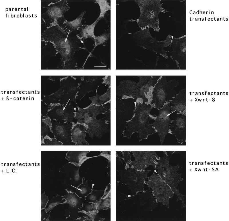
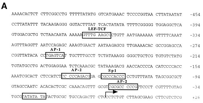
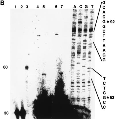
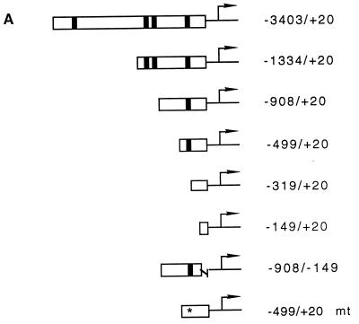
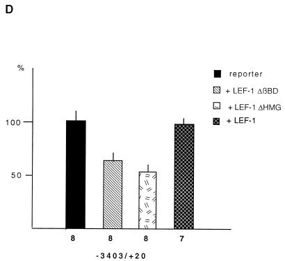
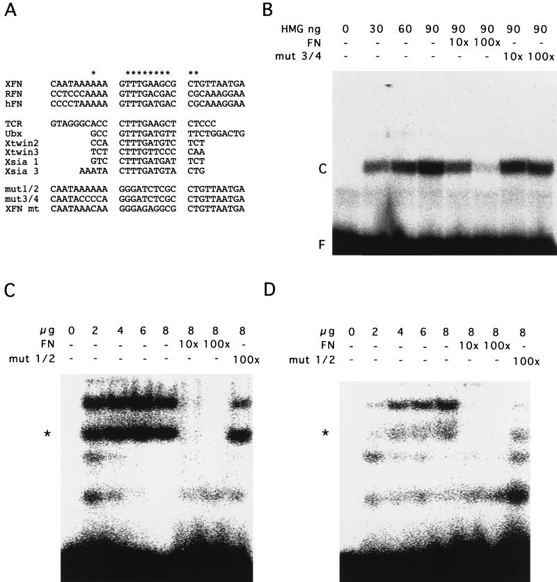
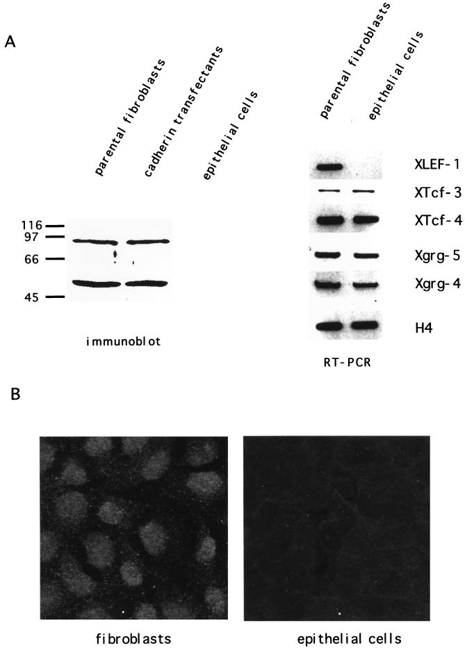
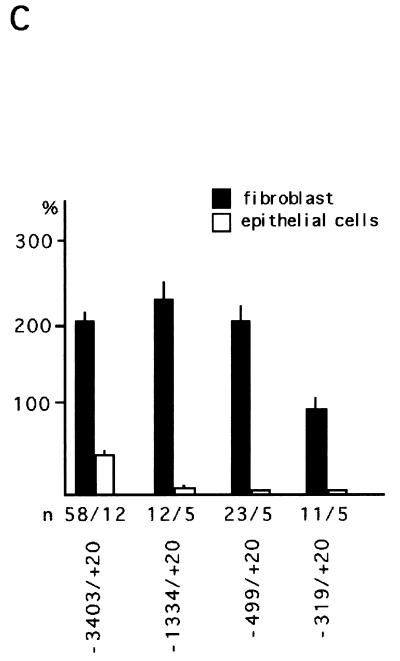
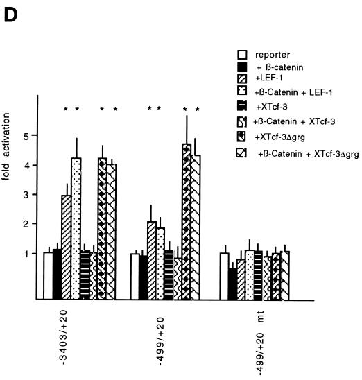
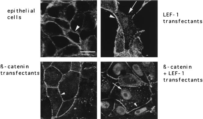
beta-Catenin stabilizes the cadherin cell adhesion complex but, as a component of the Wnt/Wg signaling pathway, also controls gene expression by forming a heterodimer with a transcription factor of the LEF-TCF family. We demonstrate that the substrate adhesion molecule fibronectin is a direct target of Wnt/Wg signaling. Nuclear depletion of beta-catenin following cadherin transfection in Xenopus fibroblasts resulted in downregulation of fibronectin expression which was restored by activating the Wnt/Wg signaling cascade via LiCl treatment or transfection of either Xwnt-8 or beta-catenin. We isolated the Xenopus fibronectin gene (FN) promoter and found four putative LEF-TCF binding sites. By comparing the activities of different fibronectin gene reporter constructs in fibroblasts and cadherin transfectants, the LEF-TCF site at position -368 was identified as a Wnt/Wg response element. LEF-1-related proteins were found in nuclei of the fibroblasts but were absent in a kidney epithelial cell line. Consistent with the lack of these transcription factors, the FN promoter was silent in the epithelial cells but was activated upon transfection of LEF-1. Wild-type Xenopus Tcf-3 (XTcf-3) was unable to activate FN promoter reporter constructs, while a mutant lacking the groucho binding region behaved like LEF-1. In contrast to XTcf-3, LEF-1 does not interact with groucho proteins, which turn TCFs into activators or repressors (J. Roose, M. Molenaar, J. Hurenkamp, J. Peterson, H. Brantjes, P. Moerer, M. van de Wetering, O. Destreé, and H. Clevers, Nature 395:608-612, 1998). Together these data provide evidence that expressing LEF-1 enables fibroblasts, in contrast to epithelial cells, to respond to the Wnt/Wg signal via beta-catenin in stimulating fibronectin gene transcription. Our findings further promote the idea that due to its dual function, beta-catenin regulates the balance between cell-cell and cell-substrate adhesion.


Figures

FIG. 1
FIG. 1
Fibronectin expression is restored in cadherin-transfected cells by transient cotransfection of β-catenin or Xwnt-8 or by lithium treatment. (A) Immunoblot analyses of fibronectin expression with monoclonal antibody 6D9, specific for Xenopus fibronectin. (B) RT-PCR analyses showing the 550-bp fragment of fibronectin and, as an internal standard, the 220-bp fragment of histone H4.
FIG. 2
FIG. 2
Subcellular localization of endogenous β-catenin in fibroblasts and cadherin transfectants, prior to and after stimulation of the Wnt/Wg signaling cascade. In the parental fibroblastic cell line, β-catenin is found in nuclei (arrow) and the cell membrane (arrowhead). In the cadherin transfectants, the β-catenin signal is restricted to the cell membrane (arrowheads). Nuclear staining of β-catenin in the cadherin transfectants (arrows) was observed only following cotransfection with β-catenin or Xwnt-8 or LiCl treatment and not after cotransfection with Xwnt-5A. Bar, 10 μm.
FIG. 3
FIG. 3
Determination of the transcription start site of the FN promoter. (A) Partial nucleotide sequence (−533/+27) of the genomic fibronectin clone XFN30.1. Numbering is with respect to the transcription start site (arrow). Conserved transcription factor binding sites are boxed. (B) Primer extension mapping of the 5′ termini of fibronectin transcripts. For primer extension, oligonucleotide FN-ext.3 (lane 1) or FN-ext.4 (lane 2), each 30 nucleotides in size, was labeled with T4 polynucleotide kinase. For size standards, either an unrelated oligonucleotide of 60 nucleotides was labeled (lane 3) or Sanger sequencing of an unrelated sequence of known composition was performed (lanes A, C, G, and T). Oligonucleotides were annealed to total CsCl-purified RNA from XTC cells and extended with MMLV reverse transcriptase. Two independent experiments are shown in lanes 4 and 5 and in lanes 6 and 7. Extension products of FN-ext.3 are shown in lanes 4 and 6, and those of FN-ext.4 are shown in lanes 5 and 7. RNA from lanes 6 and 7 was digested with DNase I prior to hybridization to exclude contamination with genomic DNA. No differences between the two experiments were detectable. In both cases, the extension product of FN-ext.3 was 92 nucleotides long, and that of FN-ext.4 was 53 nucleotides. None of the oligonucleotides annealed with tRNA (not shown).
FIG. 3
FIG. 3
Determination of the transcription start site of the FN promoter. (A) Partial nucleotide sequence (−533/+27) of the genomic fibronectin clone XFN30.1. Numbering is with respect to the transcription start site (arrow). Conserved transcription factor binding sites are boxed. (B) Primer extension mapping of the 5′ termini of fibronectin transcripts. For primer extension, oligonucleotide FN-ext.3 (lane 1) or FN-ext.4 (lane 2), each 30 nucleotides in size, was labeled with T4 polynucleotide kinase. For size standards, either an unrelated oligonucleotide of 60 nucleotides was labeled (lane 3) or Sanger sequencing of an unrelated sequence of known composition was performed (lanes A, C, G, and T). Oligonucleotides were annealed to total CsCl-purified RNA from XTC cells and extended with MMLV reverse transcriptase. Two independent experiments are shown in lanes 4 and 5 and in lanes 6 and 7. Extension products of FN-ext.3 are shown in lanes 4 and 6, and those of FN-ext.4 are shown in lanes 5 and 7. RNA from lanes 6 and 7 was digested with DNase I prior to hybridization to exclude contamination with genomic DNA. No differences between the two experiments were detectable. In both cases, the extension product of FN-ext.3 was 92 nucleotides long, and that of FN-ext.4 was 53 nucleotides. None of the oligonucleotides annealed with tRNA (not shown).
FIG. 4
FIG. 4
Identification of a Wnt/Wg response element in the Xenopus fibronectin promoter. (A) Luciferase reporter gene constructs used in this study. Putative LEF-TCF binding sites are indicated as filled boxes. The asterisk marks a mutated LEF-TCF site (for sequence comparison, see Fig. 5A). (B) Promoter activities of different constructs in parental fibroblasts (open bars) and cadherin-transfected fibroblasts (filled bars). Promoter activities were normalized to account for differences in transfection efficiency. The activities of the different constructs are shown as percentages relative to that of the CMV promoter. n, number of independent transfections; asterisks, significant differences by the Student t test (P < 0.005). (C) Influence of β-catenin (β-cat), Xwnt-8, and Xwnt-5A on fibronectin promoter activity in cadherin-transfected fibroblasts. n, number of experiments; asterisks, significant differences by the Student t test (P < 0.005). The activity of the corresponding reporter gene construct was set to 100%. (D) Influence of LEF-1 and LEF-1 deletion mutants, lacking either the β-catenin binding site (LEF-1 ΔβBD) or the DNA binding site (LEF-1 ΔHMG), on the fibronectin promoter in parental fibroblasts. The activity of the reporter was set as 100%. The number of experiments is shown below each bar.
FIG. 4
FIG. 4
Identification of a Wnt/Wg response element in the Xenopus fibronectin promoter. (A) Luciferase reporter gene constructs used in this study. Putative LEF-TCF binding sites are indicated as filled boxes. The asterisk marks a mutated LEF-TCF site (for sequence comparison, see Fig. 5A). (B) Promoter activities of different constructs in parental fibroblasts (open bars) and cadherin-transfected fibroblasts (filled bars). Promoter activities were normalized to account for differences in transfection efficiency. The activities of the different constructs are shown as percentages relative to that of the CMV promoter. n, number of independent transfections; asterisks, significant differences by the Student t test (P < 0.005). (C) Influence of β-catenin (β-cat), Xwnt-8, and Xwnt-5A on fibronectin promoter activity in cadherin-transfected fibroblasts. n, number of experiments; asterisks, significant differences by the Student t test (P < 0.005). The activity of the corresponding reporter gene construct was set to 100%. (D) Influence of LEF-1 and LEF-1 deletion mutants, lacking either the β-catenin binding site (LEF-1 ΔβBD) or the DNA binding site (LEF-1 ΔHMG), on the fibronectin promoter in parental fibroblasts. The activity of the reporter was set as 100%. The number of experiments is shown below each bar.
FIG. 5
FIG. 5
Interaction of the identified Wnt/Wg response element with HMG box fusion protein and nuclear extracts. (A) Comparison of putative Wnt/Wg response elements with the LEF-TCF consensus binding sequence in different promoters. XFN, Xenopus fibronectin gene promoter; RFN, rat fibronectin gene promoter (43); HFN, human fibronectin gene promoter (7); TCRα, T-cell receptor α (12), Ubx, ultrabithorax (46); Xtwin2 and -3, Xenopus twin (34); and Xsia 1 and 3, Xenopus siamois (3). mut 1/2 and mut 3/4, two mutated XFN sequences used for competition experiments; mt, mutated LEF-TCF target site in the −499/+20 mt construct. (B) Electrophoretic mobility shift assay with the XFN oligonucleotide and a fusion protein consisting of the LEF-1 HMG box. Amounts of HMG box protein are indicated. For competition studies, oligonucleotide mut 3/4 was used. F, free oligonucleotides; C, complex of oligonucleotide and protein. (C) Electrophoretic mobility shift assay with the XFN oligonucleotide and nuclear extracts of Xenopus fibroblasts (XTC). Amounts of nuclear extracts are indicated. For competition studies, oligonucleotide mut 1/2 was used. Two slower-migrating bands were identified. (D) Electrophoretic mobility shift assay with the XFN oligonucleotide and nuclear extracts of a Xenopus kidney epithelial cell line (A6). Amounts of nuclear extracts are indicated. For competition studies, oligonucleotide mut 1/2 was used. The band marked with an asterisk was specific in both XTC and A6 cells, whereas the slower-migrating band was specific only in XTC cells.
FIG. 6
FIG. 6
Correlation between fibronectin promoter activity and presence of LEF-1-related proteins in kidney epithelial cells (A6). (A) LEF-1 immunoblot and RT-PCR of fibroblasts, cadherin-transfected fibroblasts, and epithelial cells. In transfected and untransfected fibroblasts a protein band of approximately 55 kDa which corresponds to the size of murine LEF-1 was detectable, while lysates of epithelial cells gave no signal. The protein of 90 kDa might represent a LEF-1-related HMG box-containing protein. The RT-PCR showed XTcf-3 and XTcf-4 expression in both fibroblasts and epithelial cells, whereas XLEF-1 was detected only in the fibroblasts. Both cell lines also express XGrg4 and XGrg5. As an internal standard, histone H4 was used. (B) Subcellular localization of LEF-TCF-related proteins in immunostaining with LEF-1 antiserum. In the fibroblasts the signal was found concentrated in the nuclei, while epithelial cells were negative in staining. (C) The fibronectin promoter was inactive in epithelial cells compared to fibroblasts. The activity of the promoter is shown in relation to that of the CMV promoter. The reporter gene constructs used are indicated. n, number of independent transfections. (D) The fibronectin promoter containing the Wnt/Wg response element was activated by LEF-1 alone or by the combination of LEF-1 and β-catenin, while β-catenin alone was not able to enhance promoter activity. Wild-type XTcf-3 does not influence FN promoter activity, either alone or in combination with β-catenin, while the mutant XTcf-3Δgrg upregulates the promoter activity. n, number of independent transfections; asterisks, significant differences by the Student t test (P < 0.005). Activity of the corresponding promoter construct was set at 100%.
FIG. 6
FIG. 6
Correlation between fibronectin promoter activity and presence of LEF-1-related proteins in kidney epithelial cells (A6). (A) LEF-1 immunoblot and RT-PCR of fibroblasts, cadherin-transfected fibroblasts, and epithelial cells. In transfected and untransfected fibroblasts a protein band of approximately 55 kDa which corresponds to the size of murine LEF-1 was detectable, while lysates of epithelial cells gave no signal. The protein of 90 kDa might represent a LEF-1-related HMG box-containing protein. The RT-PCR showed XTcf-3 and XTcf-4 expression in both fibroblasts and epithelial cells, whereas XLEF-1 was detected only in the fibroblasts. Both cell lines also express XGrg4 and XGrg5. As an internal standard, histone H4 was used. (B) Subcellular localization of LEF-TCF-related proteins in immunostaining with LEF-1 antiserum. In the fibroblasts the signal was found concentrated in the nuclei, while epithelial cells were negative in staining. (C) The fibronectin promoter was inactive in epithelial cells compared to fibroblasts. The activity of the promoter is shown in relation to that of the CMV promoter. The reporter gene constructs used are indicated. n, number of independent transfections. (D) The fibronectin promoter containing the Wnt/Wg response element was activated by LEF-1 alone or by the combination of LEF-1 and β-catenin, while β-catenin alone was not able to enhance promoter activity. Wild-type XTcf-3 does not influence FN promoter activity, either alone or in combination with β-catenin, while the mutant XTcf-3Δgrg upregulates the promoter activity. n, number of independent transfections; asterisks, significant differences by the Student t test (P < 0.005). Activity of the corresponding promoter construct was set at 100%.
FIG. 7
FIG. 7
Nuclear localization of β-catenin in epithelial cells (A6) upon transfection with LEF-1 alone or LEF-1 combined with β-catenin. In epithelial cells β-catenin was detected exclusively at the plasma membrane (arrowheads). After introduction of LEF-1, either alone or in combination with β-catenin, a subpopulation of the cells revealed nuclear staining (arrows), whereas transfection with β-catenin gave no additional signal in the nuclei. Bar, 10 μm.

Similar articles

Cited by

References

    1. Behrens J, von der Kries J P, Kühl M, Bruhn L, Wedlich D, Grosschedl R, Birchmeier W. Functional interaction of β-catenin with the transcription factor LEF-1. Nature. 1996;382:638–642. - PubMed
    1. Bradley R S, Cowin P, Brown A M C. Expression of Wnt-1 in PC12 cells results in modulation of plakoglobin and E-cadherin and increased cellular adhesion. J Cell Biol. 1993;123:1857–1865. - PMC - PubMed
    1. Brannon M, Gomperts M, Sumoy L, Moon R T, Kimelman D. A β-catenin/XTcf-3 complex binds to the siamois promoter to regulate dorsal axis specification in Xenopus. Genes Dev. 1997;11:2359–2370. - PMC - PubMed
    1. Cavallo R, Rubinstein D, Peifer M. Armadillo and dTCF: a marriage made in the nucleus. Curr Opin Genet Dev. 1997;7:459–466. - PubMed
    1. Chirgwin J M, Przybyia A E, MacDonald R J, Rutter W J. Isolation of biologically active ribonucleic acid from sources enriched in ribonuclease. Biochemistry. 1979;18:5294–5299. - PubMed

Publication types

MeSH terms

LinkOut - more resources