. 1999 Aug;19(8):5696-706.
doi: 10.1128/MCB.19.8.5696.

Neoplastic transformation of RK3E by mutant beta-catenin requires deregulation of Tcf/Lef transcription but not activation of c-myc expression

Affiliations

Neoplastic transformation of RK3E by mutant beta-catenin requires deregulation of Tcf/Lef transcription but not activation of c-myc expression

F T Kolligs et al. Mol Cell Biol. 1999 Aug.

Abstract

Current models predict that beta-catenin (beta-cat) functions in Wnt signaling via activation of Tcf/Lef target genes and that its abundance is regulated by the adenomatous polyposis coli (APC) and glycogen synthase kinase 3beta (GSK3beta) proteins. In colon and other cancers, mutations in APC or presumptive GSK3beta phosphorylation sites of beta-cat are associated with constitutive activation of Tcf/Lef transcription. In spite of assumptions about its oncogenic potential, prior efforts to demonstrate that mutated beta-cat will induce neoplastic transformation have yielded equivocal results. We report here that mutated, but not wild-type, beta-cat proteins induced neoplastic transformation of RK3E, an adenovirus E1A-immortalized epithelial cell line. Analysis of the properties of mutant beta-cat proteins and studies with a dominant negative Tcf-4 mutant indicated that the ability of beta-cat to bind and activate Tcf/Lef factors is crucial for transformation. c-myc has recently been implicated as a critical Tcf-regulated target gene. However, c-myc was not consistently activated in beta-cat-transformed RK3E cells, and a dominant negative c-Myc mutant protein failed to inhibit beta-cat transformation. Our findings underscore the role of beta-cat mutations and Tcf/Lef activation in cancer and illustrate a useful system for defining critical factors in beta-cat transformation.


Figures

FIG. 1
FIG. 1
Mutated β-cat proteins activate Tcf transcription and accumulate in the cytosol. (A) Schematic outline of β-cat domains and proteins encoded by the retroviral expression constructs. Shown are presumptive GSK3β phosphorylation (phos.) sites in the β-cat N-terminal region; armadillo repeats in the central region; the C-terminal transcriptional activation domain; and the regions required for interaction with α-cat, E-cad, APC, Tcf/Lef factors, and conductin/axin. In addition to wild-type (WT) β-cat, the structures of mutated proteins are indicated. The star indicates the position of the S33Y substitution, the solid boxes represent the β-cat sequences present, and the thin line indicates the in-frame deletions in the constructs. (B) Activation of Tcf transcription by wild-type (WT) and mutated forms of β-cat in RK3E cells following transient transfection of pcDNA3 expression constructs. The ratio of luciferase activities from a Tcf-responsive reporter (pTOPFLASH) and a control luciferase reporter gene construct (pFOPFLASH) was determined 48 h after transfection. The means and standard deviations of three independent experiments are shown. (C) Mutated β-cat proteins accumulate to higher levels than wild-type β-cat (WT). ECL-Western blot analysis with an anti-Flag antibody was carried out on whole-cell lysates prepared 2 days after transient transfection of 293 cells with pcDNA3 constructs encoding wild-type β-cat and the indicated mutant forms. To demonstrate equivalent loading of the lanes, the blot was stripped and ECL-Western blotting with an anti-actin antibody was performed. (D) Mutant forms of β-cat accumulate to higher levels than wild-type β-cat in RK3E cells following infection with retroviruses encoding wild-type (WT) β-cat or mutated forms (S33Y or ΔN132). ECL-Western blot studies with an anti-Flag antibody were carried out on whole-cell or cytosolic lysates. The blot was stripped, and analysis with an anti-actin antibody was performed.
FIG. 2
FIG. 2
Induction of macroscopic foci in RK3E cells following infection with retroviruses encoding mutated forms of β-cat but not wild-type (WT) β-cat. The specific proteins encoded by the retroviruses used for infection, including a control LacZ virus, are indicated in each panel. Schematic representations of the β-cat proteins are shown in Fig. 1A. Four weeks after infection with the retroviruses, the plates were fixed, stained, and photographed, as described in the text.
FIG. 3
FIG. 3
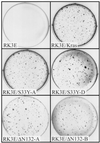
Altered phenotypic properties of stable RK3E cells transformed by mutated β-cat proteins. (A) Morphology of parental RK3E cells and a polyclonal RK3E line with overexpression of wild-type β-cat are shown in the top left and top center panels, respectively. A polyclonal RK3E line transformed by K-Ras (RK3E/Kras) is shown at the top right. Three different RK3E lines transformed by mutant β-cat are shown at the bottom. Magnification for all panels, ×200. (B) ECL-Western blot analysis of Flag-epitope tagged β-cat proteins in stable RK3E cell lines and the negative control RK3E parental line. The blot was stripped, and ECL-Western blot analysis with anti-actin antibody was carried out to control for loading of the lanes. (C) RK3E lines stably transformed by mutated β-cat proteins have markedly elevated Tcf transcription activity compared to control lines (parental RK3E, RK3E/Kras, or RK3E/WTβ1). The ratio of luciferase activities from a Tcf-responsive reporter (pTOPFLASH) and a control luciferase reporter gene construct (pFOPFLASH) was measured for each cell line 48 h after transfection. Mean values and standard deviations from three independent experiments are shown. (D) RK3E lines transformed by mutated β-cat proliferate in 0.5% serum. A total of 2 × 10⁴ cells were seeded in 35-mm dishes in the presence of growth medium containing 10% fetal bovine serum. The following day, the medium was exchanged for medium containing 0.5% fetal bovine serum. Cell numbers were determined at specific time points after the switch to 0.5% serum. Values shown represent the means and standard deviations of triplicate experiments.
FIG. 3
FIG. 3
Altered phenotypic properties of stable RK3E cells transformed by mutated β-cat proteins. (A) Morphology of parental RK3E cells and a polyclonal RK3E line with overexpression of wild-type β-cat are shown in the top left and top center panels, respectively. A polyclonal RK3E line transformed by K-Ras (RK3E/Kras) is shown at the top right. Three different RK3E lines transformed by mutant β-cat are shown at the bottom. Magnification for all panels, ×200. (B) ECL-Western blot analysis of Flag-epitope tagged β-cat proteins in stable RK3E cell lines and the negative control RK3E parental line. The blot was stripped, and ECL-Western blot analysis with anti-actin antibody was carried out to control for loading of the lanes. (C) RK3E lines stably transformed by mutated β-cat proteins have markedly elevated Tcf transcription activity compared to control lines (parental RK3E, RK3E/Kras, or RK3E/WTβ1). The ratio of luciferase activities from a Tcf-responsive reporter (pTOPFLASH) and a control luciferase reporter gene construct (pFOPFLASH) was measured for each cell line 48 h after transfection. Mean values and standard deviations from three independent experiments are shown. (D) RK3E lines transformed by mutated β-cat proliferate in 0.5% serum. A total of 2 × 10⁴ cells were seeded in 35-mm dishes in the presence of growth medium containing 10% fetal bovine serum. The following day, the medium was exchanged for medium containing 0.5% fetal bovine serum. Cell numbers were determined at specific time points after the switch to 0.5% serum. Values shown represent the means and standard deviations of triplicate experiments.
FIG. 4
FIG. 4
RK3E lines transformed by mutated β-cat form colonies in agar. Colony formation in soft agar was assessed for parental RK3E cells, a polyclonal RK3E line transformed by mutant K-Ras, and four representative β-cat-transformed lines. A total of 104 cells of each line were plated in 0.3% agar medium over agar underlayers. After 3 weeks, the dishes were stained with methylene blue and the colonies were photographed.
FIG. 5
FIG. 5
Expression of a dominant negative Tcf-4 mutant protein, lacking the N-terminal 31 aa of Tcf-4 (i.e., Tcf-4ΔN31), inhibits transformation of RK3E by mutated β-cat but not mutated K-Ras. (A) Activation of Tcf transcription by the S33Y mutated form of β-cat is substantially inhibited in a G418-resistant polyclonal RK3E line expressing Tcf-4ΔN31 (RK3E/Tcf-4DN31) compared to a control G418-resistant RK3E line (RK3E/Neo). (B) K-Ras-mediated colony formation in soft agar is not inhibited by expression of the dominant negative Tcf-4 mutant. Following infection with the K-Ras retrovirus, colony formation assays in RK3E/Neo control and RK3E/Tcf-4DN31 cells were carried out as described in the legend to Fig. 4 and the text. (C) β-cat-induced focus formation in RK3E is inhibited by expression of a dominant negative Tcf-4 mutant. Focus formation assays in the control RK3E/Neo and the RK3E/Tcf-4DN31 lines was carried out with retroviruses expressing the S33Y and ΔN132 β-cat mutants.
FIG. 6
FIG. 6
c-myc expression is not uniformly activated in stable RK3E lines transformed by mutated β-cat. Northern blot studies of c-myc, candidate c-Myc-regulated genes (lactate dehydrogenase A [LDH-A] and ornithine decarboxylase [ODC]), and a loading control (glyceraldehyde-3-phosphate dehydrogenase [GAPDH]) were carried out on total RNA from parental RK3E cells, a polyclonal RK3E line transformed by activated K-Ras (RK3E/Kras), and 10 different RK3E lines transformed by mutated β-cat. All β-cat-transformed lines form colonies in soft agar, and several of the lines were tested and found to grow in nude mice (see the text for details).
FIG. 7
FIG. 7
c-myc activation is not required for transformation by mutated β-cat, but increased c-myc expression can contribute to a more aggressive phenotype. (A) A polyclonal RK3E line with stable expression of a dominant negative c-Myc mutant protein (i.e., MycΔ106–143) was generated and termed RK3E/MycΔ106–143. Activation of a c-Myc-responsive reporter (pGLDH637Luc) is substantially inhibited in the RK3E/MycΔ106–143 line. (B) Mutated β-cat proteins induce focus formation in RK3E/MycΔ106–143, but the size of the foci is reduced compared to those induced by mutated β-cat in the control RK3E/Neo line. (C) Colony formation in soft agar following infection with a K-Ras retrovirus is not inhibited in RK3E/MycΔ106–143 cells.

Similar articles

Cited by

References

    1. Aberle H, Butz S, Stappert J, Weissig H, Kemler R, Hoschuetzky H. Assembly of the cadherin-catenin complex in vitro with recombinant proteins. J Cell Sci. 1994;107:3655–3663. - PubMed
    1. Aberle H, Bauer A, Stappert J, Kispert A, Kemler R. β-catenin is a target for the ubiquitin-proteasome pathway. EMBO J. 1997;16:3797–3804. - PMC - PubMed
    1. Aoki M, Hecht A, Kruse U, Kemler R, Vogt P K. Nuclear endpoint of Wnt signaling: neoplastic transformation induced by transactivating lymphoid-enhancing factor 1. Proc Natl Acad Sci USA. 1999;96:139–144. - PMC - PubMed
    1. Barth A I, Nathke I S, Nelson W J. Cadherins, catenins and APC protein: interplay between cytoskeletal complexes and signaling pathways. Curr Opin Cell Biol. 1997;9:683–690. - PubMed
    1. Behrens J, von Kries J P, Kuhl M, Bruhn L, Wedlich D, Grosschedl R, Birchmeier W. Functional interaction of β-catenin with the transcription factor LEF-1. Nature. 1996;382:638–642. - PubMed

Publication types

MeSH terms