. 1999 Aug;19(8):5743-58.
doi: 10.1128/MCB.19.8.5743.

Notch and wingless regulate expression of cuticle patterning genes

Affiliations

Notch and wingless regulate expression of cuticle patterning genes

C S Wesley. Mol Cell Biol. 1999 Aug.

Abstract

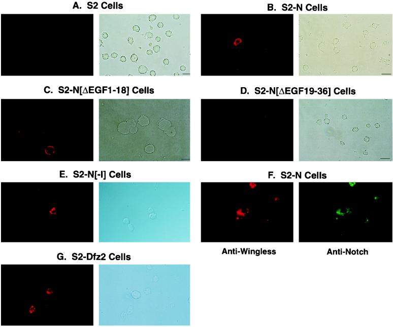
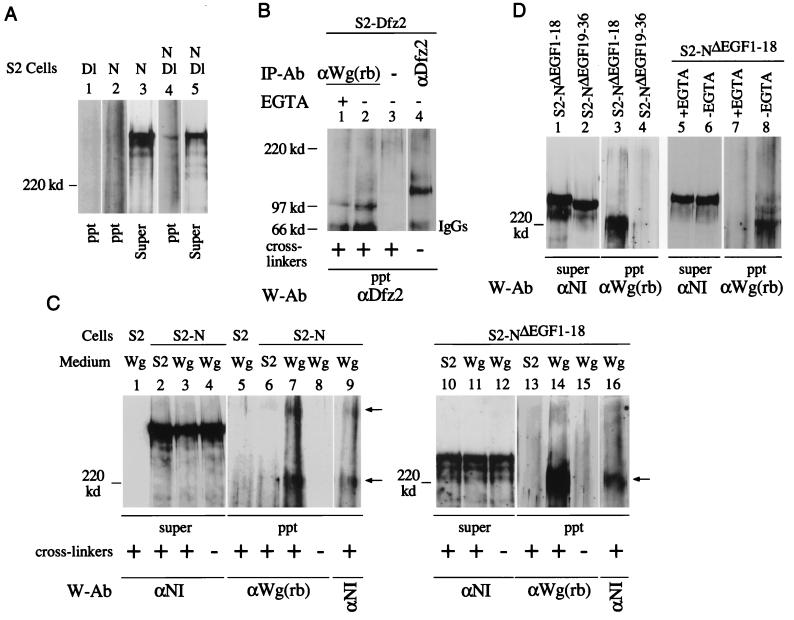
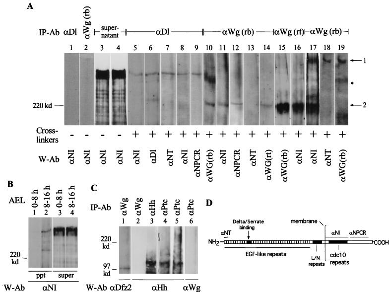
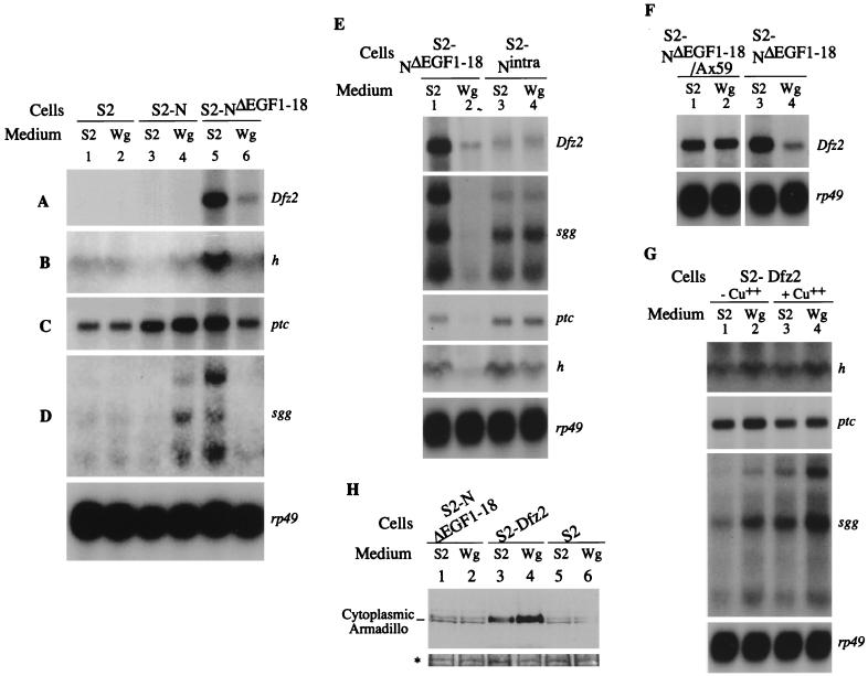
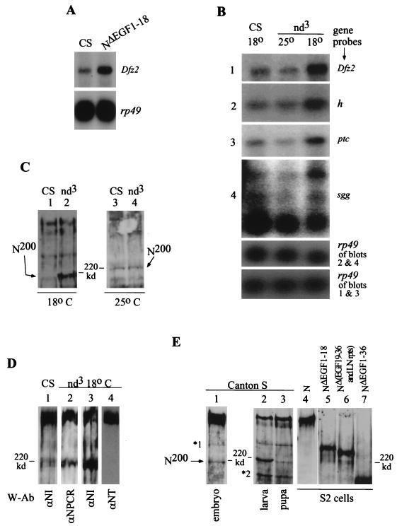
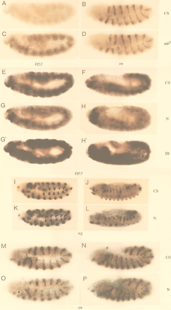
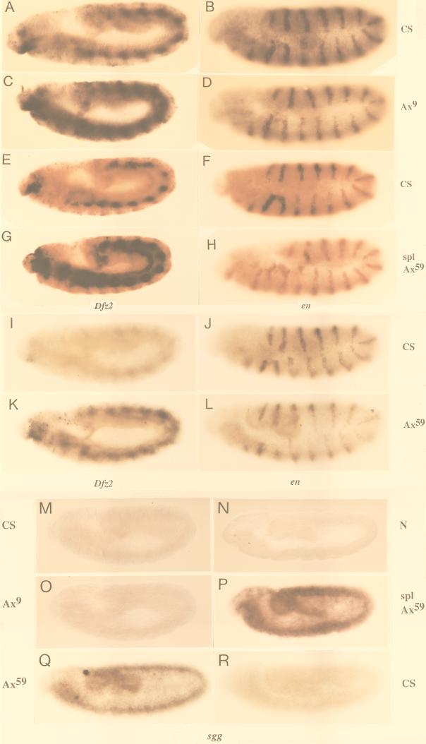
The cell surface receptor Notch is required during Drosophila embryogenesis for production of epidermal precursor cells. The secreted factor Wingless is required for specifying different types of cells during differentiation of tissues from these epidermal precursor cells. The results reported here show that the full-length Notch and a form of Notch truncated in the amino terminus associate with Wingless in S2 cells and in embryos. In S2 cells, Wingless and the two different forms of Notch regulate expression of Dfrizzled 2, a receptor of Wg; hairy, a negative regulator of achaete expression; shaggy, a negative regulator of engrailed expression; and patched, a negative regulator of wingless expression. Analyses of expression of the same genes in mutant N embryos indicate that the pattern of gene regulations observed in vitro reflects regulations in vivo. These results suggest that the strong genetic interactions observed between Notch and wingless genes during development of Drosophila is at least partly due to regulation of expression of cuticle patterning genes by Wingless and the two forms of Notch.


Figures

FIG. 1
FIG. 1
Search for new N ligands. (A) Interspecific sequence comparisons reveal possible ligand binding sites in the extracellular domain of N. Each plot represents a running average of sequence conservation between two homologs, over sliding blocks of 40 amino acids (aa). At the top, the corresponding EGF-like repeats of N are graphically represented. lin12 = lin12/N repeats (42, 88). The line in the middle of each plot represents average similarity between the two sequences compared (the maximum value is 1.5). This average would include sequence conservation due to sequence elements common to all EGF-like repeats. The plot of D. melanogaster N and its D. pseudoobscura homolog is similar to that of N and the D. virilis homolog (differing only in the level of overall conservation). The between-lineage comparison identifies evolutionarily stable conserved regions. dmN, D. melanogaster N; hN1, human homolog of N (23); dvN, D. virilis N. Sites of mutations in nd3 and l(1)NB are from reference , NM1 are from reference , spl and the Ax alleles are from reference , and Nts1 are from . Lethal alleles are in bold letters. (B) Schematic representation of the biopanning screen used for identification of potential N ligands (see Materials and Methods).
FIG. 1
FIG. 1
Search for new N ligands. (A) Interspecific sequence comparisons reveal possible ligand binding sites in the extracellular domain of N. Each plot represents a running average of sequence conservation between two homologs, over sliding blocks of 40 amino acids (aa). At the top, the corresponding EGF-like repeats of N are graphically represented. lin12 = lin12/N repeats (42, 88). The line in the middle of each plot represents average similarity between the two sequences compared (the maximum value is 1.5). This average would include sequence conservation due to sequence elements common to all EGF-like repeats. The plot of D. melanogaster N and its D. pseudoobscura homolog is similar to that of N and the D. virilis homolog (differing only in the level of overall conservation). The between-lineage comparison identifies evolutionarily stable conserved regions. dmN, D. melanogaster N; hN1, human homolog of N (23); dvN, D. virilis N. Sites of mutations in nd3 and l(1)NB are from reference , NM1 are from reference , spl and the Ax alleles are from reference , and Nts1 are from . Lethal alleles are in bold letters. (B) Schematic representation of the biopanning screen used for identification of potential N ligands (see Materials and Methods).
FIG. 2
FIG. 2
Soluble Wg binds N in the region containing EGF-like repeats 19 to 36. Wg binds surfaces of S2-N (B), S2-NΔEGF1–18 (C), and S2-NΔI cells (E) but not surfaces of S2 (A) and S2-NΔEGF19–36 cells (D). Only N-expressing cells bind Wg (F), and the frequency of N-expressing cells binding Wg is comparable to the frequency of S2-Dfz2 cells binding Wg (G). (A to E and G) The photograph on the left shows anti-Wg immunofluorescence in a microscopic field of cells, and the photograph on the right shows Nomarski illumination of the same microscopic field of cells. (F) The photograph on the left shows immunofluorescence generated by the anti-Wg antibody, and the photograph on the right shows immunofluorescence generated in the same microscopic field of cells by an anti-N antibody. Cells were treated with unconcentrated culture medium conditioned by growth of S2-Wg cells. Wg on cell surfaces was detected immunocytochemically with anti-Wg (rb), an antibody made in rabbit (69), and a rhodamine-conjugated secondary antibody. N on cell surfaces was detected with αNI (52) and a fluorescein-conjugated secondary antibody. None of the cells incubated with culture medium conditioned by growth of S2 cells showed any detectable signals. Only cells expressing high levels of N are apparent in the photographs. An actual count of all immunofluorescent cells indicates a Wg-positive frequency of ∼40% (N is expressed by only 50% of the cells stably cotransfected with the hygromycin gene). A short binding period was used because N was found to be lost from the cell surfaces within minutes of treatment with Wg.
FIG. 3
FIG. 3
Two different Wg- and N-containing complexes are recovered from N-expressing S2 cell surfaces. (A) N- and Dl-containing complexes are recovered from S2-N and S2-Dl cell aggregates in the presence of cross-linkers. Dl-containing cross-linked complexes were immunoprecipitated by the monoclonal anti-Dl antibody, MAb 202 (24) and analyzed by Western blotting with anti-NI antibody. (B) Wg- and Dfz2-containing cross-linked complexes are recovered from S2-Dfz2 cells treated with Wg medium containing cross-linkers, in the presence or absence of EGTA (lanes 1 and 2). A mouse monoclonal anti-Dfz2 antibody (kindly provided by R. Nusse) was used for immunoprecipitation (lane 4) and for detection of Wg-Dfz2 complexes by Western blotting. Wg-Dfz2 complexes were not recovered from S2-Dfz2 cells treated with medium conditioned by growth of S2 cells (not shown). (C) Two Wg- and N-containing cross-linked complexes (arrows) are immunoprecipitated from S2-N cells (lanes 7 and 9), and only one is immunoprecipitated from S2-NΔEGF1–18 cells (lanes 14 and 16), treated with Wg-containing medium. N- and Wg-containing complexes were immunoprecipitated with anti-Wg(rb) and detected by Western blotting with the indicated antibodies (W-Ab). Lanes 7 and 9 and lanes 14 and 16 show reaction of the same blots with anti-Wg(rb) and anti-NI antibodies. (D) Wg and N containing cross-linked complexes are recovered from S2-NΔEGF1–18 cells (lane 3) in the absence of EGTA (lane 8) but not from S2-NΔEGF19–36 cells (lane 4). S2-NΔEGF1–18 or S2-NΔEGF19–36 cells were treated with Wg medium containing cross-linkers, immunoprecipitation was performed with anti-Wg(rb) antibody, and the Western blots were probed with the indicated antibodies. ppt, immunoprecipitated complexes eluted from GammaBind beads; Super, an aliquot of the protein extract after the last pelleting of the GammaBind beads (see Materials and Methods); IP-Ab, immunoprecipitation antibody; W-Ab, Western blotting antibody; cross-linker, BS3. Wg, medium conditioned by growth of S2-Wg cells; S2, medium conditioned by growth of S2 cells. For panels A, C, and D, 4% polyacrylamide gels were used; for panel B, 6% polyacrylamide gels were used. Only proteins or protein complexes migrating slower than a 120-kDa marker protein are resolved in panels A, C, and D. The tops of all the blots shown coincide with the top of the resolving gel of the discontinuous SDS-PAGE gels.
FIG. 4
FIG. 4
Wg and N form complexes during embryogenesis. (A) Two Wg- and N-containing cross-linked complexes, similar to those recovered from cultured cells, are immunoprecipitated from Canton S embryonic extracts. Anti-Dl, is monoclonal antibody MAb 202; anti-NT is described in reference ; anti-NPCR is described in reference ; and anti-Wg(rt) was kindly provided by A. Martinez-Arias (See panel D for epitope regions for N antibodies). I used 0- to 3-h embryos for lanes 1 to 16, 6- to 12-h embryos for lane 17, and 10- to 16-h embryos for lanes 18 and 19. Arrow 1 shows Wg complexed with full-length N (lanes 17, 18, and 19); arrow 2 shows Wg complexed with a truncated N (lanes 10 to 17). The asterisk marks the Wg complex not containing N (lanes 10 and 19). A single blot was probed sequentially with the indicated antibodies to form lanes 10 and 11; 12, 13, and 14; 15 and 16; and 19 and 18 (numbers also indicate the sequence of probing). The same embryonic extract was used for lanes 1 to 4 and 7 to 14; lanes 5 to 6, 15, 17, and 18 are derived from different embryonic extracts. (B) Ser-N cross-linked complexes are also recovered from cross-linked embryonic extracts. Complexes were immunoprecipitated with anti-Ser antibody (kindly provided by Elizabeth Knust). Complexes migrating faster than a ∼120-kDa marker protein were not analyzed in panels A and B. (C) The procedure recovering Wg-N, Dl-N, and Ser-N complexes also recovers Wg-Dfz2 (lane 1) and Hh-Ptc complexes from cross-linked embryonic extracts (lanes 3 to 6). For lanes 5 and 6, equal volumes of anti-ptc immunoprecipitate was separated in two different lanes and probed with the indicated antibodies. IP-Ab, immunoprecipitation antibody; cross-linker, BS3; W-Ab, Western blotting antibody; AEL, after egg laying. For panels A and B, 4% polyacrylamide gels were used; for panel C, 6% polyacrylamide gels were used. The tops of all the blots shown in panels A, B, and C coincide with the top of the resolving gel of the discontinuous SDS-PAGE gels. (D) Diagram showing the N epitopes used to produce the N antibodies used in the study.
FIG. 5
FIG. 5
N and NΔEGF1–18 have different ligand-independent activities and respond differently to Wg. (A to D) Expression of Dfz2, h, ptc, and sgg in S2-N and S2-NΔEGF1–18 cells are regulated by Wg. en, wg, ac, hh, and m5 and m8 of E(spl)C were not detected in any of the experiments. The sizes of transcripts of all genes were similar to published reports. Dfz2 (8), ptc (35), h (37), rp49 (61), and sgg (77). The largest sgg RNA corresponds in size to the embryonic transcript, while the smallest sgg RNA has a size expected for the ovarian transcript (77). sgg, wg, ac, and m5 and m8 of E(spl)C are known to genetically interact with N (15, 33, 71, 72, 85); Dfz2, ptc, wg, and sgg, are involved in epidermal patterning (4, 7, 8, 20, 35, 57, 65); h is a negative regulator of ac (38, 79, 84). (E) The extracellular domain of NΔEGF1–18 is required for regulation of Dfz2, sgg, ptc, and, to a lesser extent, h expression. (F) Ax59d mutation in NΔEGF1–18 abolishes Wg-mediated down regulation of Dfz2 expression in S2-NΔEGF1–18 cells. The two autoradiographs were derived from the same blot with different exposure times. (G) S2-Dfz2 cells (S2-pMK 33 cells [8]) do not down regulate expressions of ptc, sgg, and h in response to Wg, with or without copper induction. (A to G) Total RNAs were extracted from the indicated cells treated with M3 medium conditioned by growth of S2 cells (S2 medium) or S2-Wg cells (Wg medium) and analyzed by Northern blotting. The same batch of S2 or Wg medium was used for all of the studies. Gene sequences used as probes are indicated on the right of each panel. rp49 was used to indicate the relative levels of RNA in different lanes. The individual blots are exposed to film for different periods. Exposure times: rp49 < Dfz2 < ptc < sgg < h. (H) S2-NΔEGF1–18 cells do not accumulate Arm in the cytoplasm in response to Wg. The bottom panel (∗) shows a India ink-stained protein band visible in all lanes of the blot to indicate the amount of samples loaded. An anti-Arm antibody made in rabbits (kindly provided by Laurent Ruel) was used to detect Arm.
FIG. 6
FIG. 6
NΔEGF1–18 and nd3 embryos show increased expression of genes regulated by NΔEGF1–18 in vitro, and an endogenous form of N resembling NΔEGF1–18 is overproduced in nd3 embryos. (A) Embryos carrying the heat shock-inducible NΔEGF1–18 transgene also overexpress Dfz2. We heat shocked 0- to 20-h Canton S (CS) (yw strain) and NΔEGF1–18 (in Canton S, yw strain) embryos for 30 min, allowed them to recover at room temperature for 45 min, and extracted total RNAs for Northern blot analysis. rp49 indicates relative levels of RNA in different lanes. (B) Dfz2, h, ptc, and sgg are overexpressed in nd3 embryos at 18°C, the temperature at which the overt mutant phenotype is observed. At 25°C, expression of these genes in nd3 does not differ from that in Canton S. Levels of expression in Canton S at 25 and 18°C are indistinguishable (expression at 18°C is shown). Total RNAs were extracted from 0- to 20-h Canton S and nd3 embryos, reared at 25 or 18°C (with appropriate correction for developmental times), and analyzed by Northern blotting. Gene sequences used as probes are shown at the right. Exposure times: rp49 < Dfz2 < ptc < sgg < h. (C) nd3 embryos at 18°C overproduce a ∼200-kDa form of N, N200. Because signals from high-molecular-weight forms of N interfere with assessment of levels of the less abundant N200, total embryonic proteins extracted from 0- to 12-h Canton S and nd3 embryos (at 18 or 25°C) were incubated with anti-NT and cleared prior to SDS-PAGE. Anti-NT does not react with N200 (see below). Extracts containing equivalent levels of ∼350-kDa N were used for lanes 1 and 2. N is detected with anti-NI. (D) N200 is truncated in the amino terminus. N was immunoprecipitated from 0- to 12-h Canton S or nd3 embryos (reared at 18°C) by using anti-NI and separated by SDS-PAGE (4% polyacrylamide), and the Western blots were probed with the indicated antibodies (W-Ab). See Fig. 4D for epitopes for N antibodies. nd3 embryos were used to determine missing epitopes because they produce higher levels of N200 than do Canton S embryos (compare lanes 1 and 3). (E) Different developmental stages of Canton S flies express N200, and N200 lacks more than 18 of the amino-terminal EGF-like repeats. Aliquots of total proteins extracted from Canton S embryos (0 to 3 h), one Canton S larva, one Canton S pupa, S2-N cells, S2-NΔEGF1–18 cells, S2-NΔEGF19–36 and LN rpts cells, and S2-NΔEGF1–36 cells were separated by SDS-PAGE (4% polyacrylamide) and analyzed by Western blotting with anti-NI. ∗1 is recognized by all of the N antibodies studied and is therefore considered to be the partially denatured form of N (43); ∗2 is not recognized by anti-NT and anti-NPCR (not shown) but is recognized by anti-NI.
FIG. 7
FIG. 7
Expression of Dfz2 is reduced in zygotic N embryos, while expression of en and wg are unaffected. (A to D) In situ hybridization of Canton S and nd3 embryos corroborates results from Northern blotting that Dfz2 is overexpressed in nd3 embryos but en is not (E to H, G′, H′) Dfz2 expression is lost in N264-47/Y embryos but not in comparable stages of Canton S or DlX/DlX embryos. (I to L) wg expression is not lost in N264-47/Y. (M to P) en expression is similar in Canton S and N264-47/Y embryos. CS, Canton S; nd3, nd3; N, N264-47/Y; Dl, DlX/DlX embryos; genes used as probes are indicated below the appropriate sets of embryos. Anterior is to the left of each embryo. N264-47/Y and DlX/DlX embryos were identified by lack of β-galactosidase staining associated with the FM7 or TM6 balancer chromosomes (see Materials and Methods). Embryos A to D were processed simultaneously, and so were embryos E to P.
FIG. 8
FIG. 8
Ax embryos overproduce Dfz2 and sgg RNA. (A to L) Ax embryos overproduce Dfz2 RNA (A, C, E, G, I, and K) but not en RNA (B, D, F, H, J, and L). (M to R) Ax embryos overproduce sgg RNA (O to Q) but not Canton S (M and R) and N264-47/Y embryos (N). Due to low-level of expression in a general pattern, reduced sgg expression (as in panel N) and weak sgg overexpression (as in panel O) are more obvious in pools of embryos than in individual embryos. CS, Canton S; Ax9, Ax9B/Y; spl Ax59, spl Ax59d/Y; Ax59, Ax59d/Y; N, N264-47/Y. Anterior is to the left of each embryo. Homozygous Ax or N embryos were identified by the lack of β-galactosidase staining associated with the FM7 balancer chromosomes (see Materials and Methods). Embryos A to H and M to P were processed simultaneously, and so were embryos I to L and Q to R.

Similar articles

Cited by

References

    1. Alton A K, Fechtel K, Kopczynski C C, Shepard S B, Kooh P J, Muskavitch M A T. Molecular genetics of Delta, a locus required for ectodermal differentiation in Drosophila. Dev Genet. 1989;10:261–272. - PubMed
    1. Artavanis-Tsakonas S, Matsuno K, Fortini M E. Notch signaling. Science. 1995;268:225–232. - PubMed
    1. Axelrod J D, Matsuno K, Artavanis-Tsakonas S, Perrimon N. Interaction between Wingless and Notch signaling pathways mediated by Dishevelled. Science. 1996;271:1826–1832. - PubMed
    1. Baker N E. Molecular cloning of sequences from wingless, a segment polarity gene in Drosophila: the spatial distribution of a transcript in embryos. EMBO J. 1987;6:1765–1773. - PMC - PubMed
    1. Baker N E, Mlodzik M, Rubin G M. Spacing differentiation in the developing Drosophila eye: a fibrinogen-related lateral inhibitor encoded by scabrous. Science. 1990;250:1370–1377. - PubMed

Publication types

MeSH terms

Substances

LinkOut - more resources