. 1999 Aug;19(8):5768-84.
doi: 10.1128/MCB.19.8.5768.

Nup124p is a nuclear pore factor of Schizosaccharomyces pombe that is important for nuclear import and activity of retrotransposon Tf1

Affiliations

Nup124p is a nuclear pore factor of Schizosaccharomyces pombe that is important for nuclear import and activity of retrotransposon Tf1

D Balasundaram et al. Mol Cell Biol. 1999 Aug.

Abstract

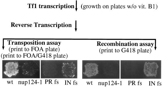
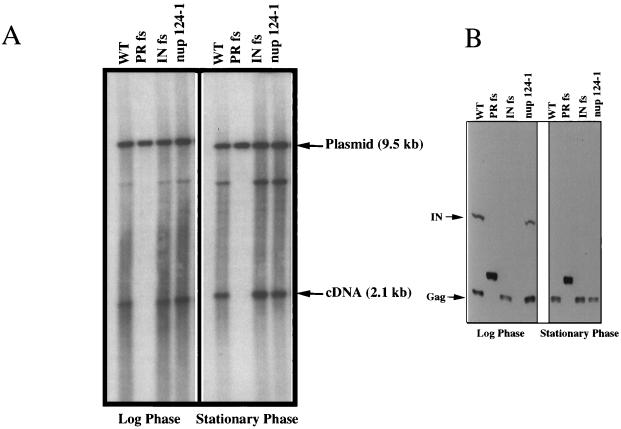
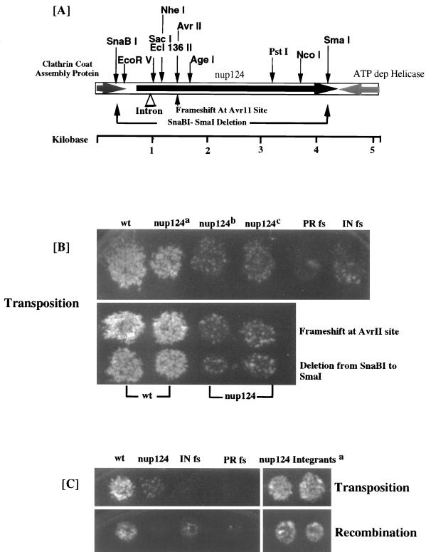
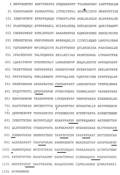
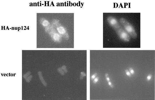
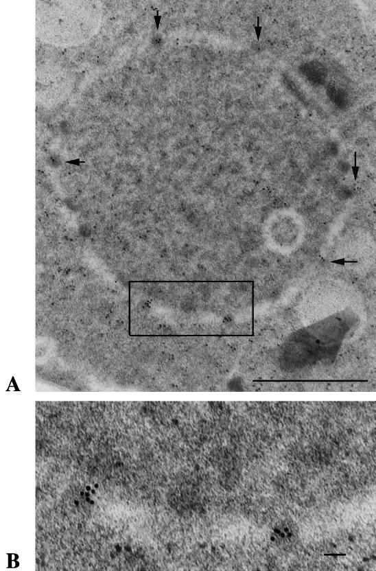
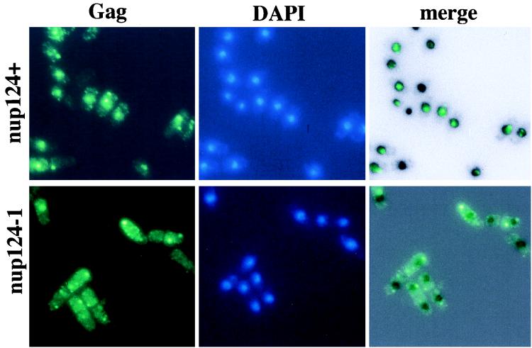
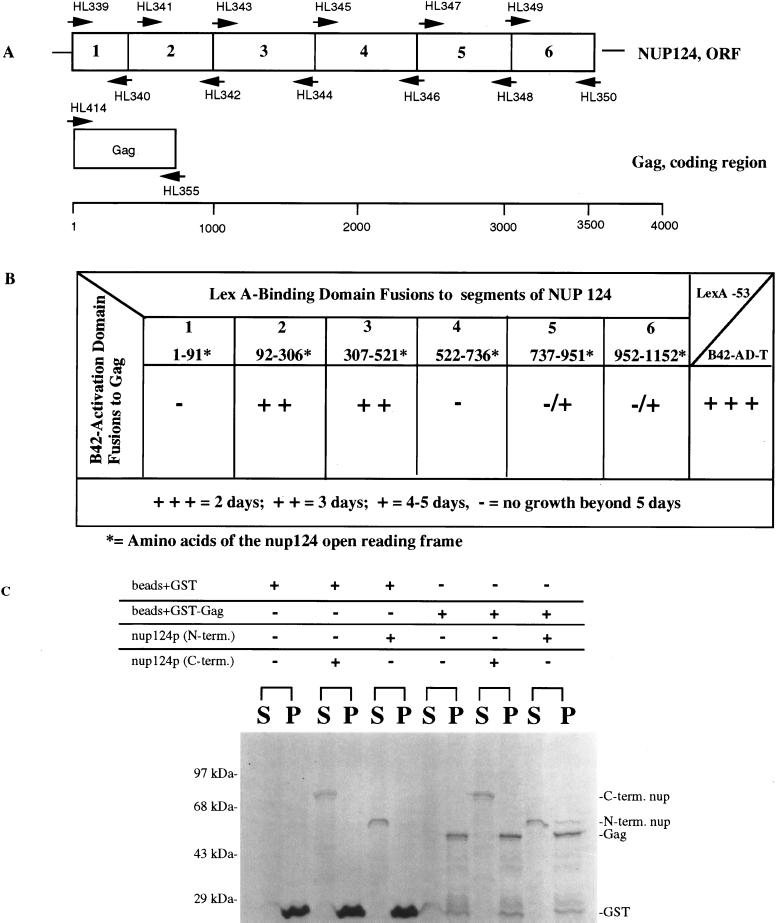
The long terminal repeat (LTR)-containing retrotransposon Tf1 propagates within the fission yeast Schizosaccharomyces pombe as the result of several mechanisms that are typical of both retrotransposons and retroviruses. To identify host factors that contribute to the transposition process, we mutagenized cultures of S. pombe and screened them for strains that were unable to support Tf1 transposition. One such strain contained a mutation in a gene we named nup124. The product of this gene contains 11 FXFG repeats and is a component of the nuclear pore complex. In addition to the reduced levels of Tf1 transposition, the nup124-1 allele caused a significant reduction in the nuclear localization of Tf1 Gag. Surprisingly, the mutation in nup124-1 did not cause any reduction in the growth rate, the nuclear localization of specific nuclear localization signal-containing proteins, or the cytoplasmic localization of poly(A) mRNA. A two-hybrid analysis and an in vitro precipitation assay both identified an interaction between Tf1 Gag and the N terminus of Nup124p. These results provide evidence for an unusual mechanism of nuclear import that relies on a direct interaction between a nuclear pore factor and Tf1 Gag.


Figures

FIG. 1
FIG. 1
Transposition and cDNA recombination assays of Tf1. The genetic manipulations and replica printing required for the transposition and recombination assays are indicated in parentheses. The ability of the reverse transcripts in both assays to produce G418 resistance is shown. Although wild-type (wt) Tf1-neoAI produced G418 resistance in both assays, a mutation that blocked integrase expression, IN fs, greatly reduced growth on the transposition plates without significantly reducing growth on the recombination plates. PR fs is a strain with a frameshift mutation in Tf1 that blocks the expression of PR, RT, and IN. FOA- 5-fluoroorotic acid.
FIG. 2
FIG. 2
Levels of Tf1 cDNA and proteins are not altered by the mutation in nup124-1. (A) Tf1 cDNA production in wild-type and nup124-1 cells at the logarithmic and stationary phases of growth. A DNA blot containing nucleic acid extracted from wild-type (WT) (YHL1282), PR frameshift (YHL1836), INT frameshift (YHL1554), and the nup124-1 mutant (YHL5754), is shown. Genomic DNA from the logarithmic and stationary phases of growth on medium without vitamin B1 was digested with BstXI and loaded onto a 0.6% agarose gel, which was transferred to a filter and probed with a 1.0-kb neo fragment. (B) Tf1 proteins in wild-type and nup124-1 cells at the logarithmic and stationary phases of growth. An immunoblot of extracts from the cells used for the Tf1 cDNA determination in panel A is shown. The filter was probed with both anti-Gag and anti-IN antisera. The arrows marked IN and Gag show the positions of the IN and Gag proteins.
FIG. 3
FIG. 3
Isolation and sequence of the nup124 gene. (A) Restriction fragment of the genomic sequence that complemented the nup124-1 defect, with the locations of the ORFs indicated by large arrows. The C-terminal sections of the ATP-dependent helicase and the clathrin assembly protein are shown with shaded arrows, and the nup124 ORF is shown with a black arrow. Also shown are the positions of restriction sites including AvrII, SnaBI, and SmaI, the sites used to disrupt the nup124 ORF. (B) All strains were assayed for transposition activity. The strains represented in the upper panel are as follows (from left to right); wild type (wt) (YHL5533), nup124a (YHL6106, a nup124-1 strain expressing the entire complementing fragment, nup124b (YHL6061, a strain with an empty vector, pSP1), nup124c (YHL6110, a strain with the empty library vector pHL1288); a wild-type strain containing Tf1 PR fs (YHL4990); and a wild-type strain containing Tf1 IN fs (YHL4992). The strains in the top row of the lower panel are, from left to right, two transformants of a wild-type strain with the complementing fragment that contained the frameshift mutation at the AvrII site (YHL6404) and two transformants of a strain with the nup124-1 mutation and the plasmid with the complementing fragment and the frameshift mutation at the AvrII site (YHL6406). The strains in the lower row of the bottom panel are two transformants of a wild-type strain with the plasmid copy of the complementing fragment that contained the SnaBI-SmaI deletion (YHL6405) and two transformants of a strain with the nup124-1 mutation and the plasmid that contained the SnaBI-SmaI deletion (YHL6407). (C) The strain with the nup124-1 allele was transformed with an integrating plasmid containing the entire complementing sequence. A stable transformant (YHL6136), shown to contain an integration of the suppressor sequence into the nup124-1 loci, was transformed with the Tf1-neoAI plasmid pHL449 (YHL6620) and assayed for transposition (upper panel) and recombination (lower panel). aTwo independent transformants are shown. Also represented in both top and bottom sections are (from left to right) strains with wild-type (wt) and nup124-1 alleles (YHL1282 and YHL5754, respectively) and the standard control strains consisting of Tf1 IN fs (YHL1554) and Tf1 PR fs (YHL1836).
FIG. 4
FIG. 4
Amino acid sequence of the Nup124p (Q09904) protein as predicted with annotation software that identified a small intron marked here with an asterisk. The 11 FXFG repeats at the C-terminal end are underlined. The mutation site of the nup124-1 allele is indicated by a short vertical line before the Q where the codon CAG coding for Q is mutated to a Stop codon, TAG.
FIG. 5
FIG. 5
Cellular location of Nup124p. Two strains, YHL965 (bottom, vector without nup124) and YHL6576 (top, plasmid with HA-tagged nup124) were grown in EMM − leu dropout medium and prepared for immunofluorescence microscopy. The left two panels show the FITC signal produced by the anti-HA antibody, and the right two panels contain images of the DAPI signals.
FIG. 6
FIG. 6
Immunoelectron microscopy demonstrating the presence of a Nup124-GFP fusion protein within nuclear pores. Strain YHL6876 expressed a single-copy allele of GFP fused to the end of Nup124p. Cells were grown in EMM and processed for immunoelectron microscopy with an antibody specific for GFP. (A) A thin section of a cell shows a ring of reduced density that indicates the position of the nuclear envelope. The dark structures embedded in the ring are the nuclear pore. A high concentration of gold particles are associated with the nuclear pores (arrows). The square indicated the region shown at higher magnification in panel B. Bar, 1.0 μm. (B) The inset from panel A was enlarged to allow inspection of the gold particles associated with two nuclear pores. Bar, 0.1 μm.
FIG. 7
FIG. 7
Immunofluorescence of Tf1 FLAG-Gag in wild-type cells. (Top) Strain YHL5895 contained a wild-type allele of nup124 and the Tf1-neoAI FLAG-Gag plasmid, pHL1276. The green signal is specific for the FLAG-Gag protein, and the blue signal is produced by DAPI and indicates the position of the nucleus. The panel on the right is a merge of the FLAG-Gag signal produced by YHL5895 with an inverted black-and-white image of its DAPI stain. The merge was generated with Adobe Photoshop 4.0 with the screen function set at 65% opacity. (Bottom) Same experiment as in the top three panels, except that strain YHL6565 (bottom) contained the nup124-1 allele.
FIG. 8
FIG. 8
Analysis of interactions between Gag and Nup124p. (A) Segments 1 through 6 of Nup124p correspond to the following amino acids of Nup124p: 1, 1 to 91; 2, 92 to 306; 3, 307 to 521; 4, 522 to 736; 5, 737 to 951; 6, 952 to 1152. Corresponding nucleotide sequence were fused to the transcription activation domain B42. Numbers above and below the arrowheads indicate the primers used to generate the PCR products that were inserted into the two-hybrid vectors. (B) Summary of two-hybrid interactions between segments 1 to 6 of Nup124p expressed as fusions to the LexA binding domain and the Tf1 Gag expressed as a fusion to the B42 activation domain. Potential interactions were scored as growth on SC medium containing galactose and lacking leucine. All fusions were tested for intrinsic or nonspecific activation. The AD plasmids were cotransformed with LexA fused to the Bicoid protein to test for specificity of interaction with each of the BD fusions, while all the BD plasmids were cotransformed with an empty AD to test for intrinsic activation. The BD fusion with the Gag protein resulted in significant intrinsic activation and therefore could not be used in this study. All other fusions used did not intrinsically or nonspecifically activate expression of the LEU2 reporter. Additionally, a positive control was used in these two-hybrid experiments based on the strong interaction of murine p53 and SV40 large T antigen. Plasmids containing p53 fused to LexA and SV40 large T antigen fused to B42 (Clontech) were cotransformed into EGY48 and tested for growth on medium lacking leucine. Strains containing these fusion proteins showed visible growth in approximately 1 to 2 days. (C) GST precipitation analysis of interactions between purified samples of Gag and two portions of Nup124p. The N-terminal and C-terminal portions of Nup124p as well as GST and GST-Gag were purified from bacteria. The GST and GST-Gag proteins were coupled to glutathione-Sepharose and combined with either the C-terminal or N-terminal fragments of Nup124p. The samples were incubated at room temperature for 45 min and washed three times in binding buffer. The beads and the supernatant were combined with 2× sample-loading buffer and loaded in equal proportions onto an SDS–10% polyacrylamide gel that was stained with Coomassie blue. The brackets over S and P indicate the pairs of supernatant and pellet fractions from separate binding reactions. The components of each binding reaction are indicated by plus signs above each bracket. The positions of molecular mass standards are indicated on the left of the panel.

Similar articles

Cited by

References

    1. Aitchison J D, Blobel G, Route M P. Kap104p: a karyopherin involved in the nuclear transport of messenger RNA binding proteins. Science. 1996;274:624–627. - PubMed
    1. Atwood A, Choi J, Levin H L. The application of a homologous recombination assay revealed amino acid residues in a long terminal repeat retrotransposon that were critical for integration. J Virol. 1998;72:1324–1333. - PMC - PubMed
    1. Atwood A, Lin J, Levin H. The retrotransposon Tf1 assembles virus-like particles with excess Gag relative to integrase because of a regulated degradation process. Mol Cell Biol. 1996;16:338–346. - PMC - PubMed
    1. Azad A K, Tani T, Shiki N, Tsuneyoshi S, Urushiyama S, Ohshima Y. Isolation and molecular characterization of mRNA transport mutants in Schizosaccharomyces pombe. Mol Biol Cell. 1997;8:825–841. - PMC - PubMed
    1. Berkower C, Loayza D, Michaelis S. Metabolic instability and constitutive endocytosis of STE6, the a-factor transporter of Saccharomyces cerevisiae. Mol Biol Cell. 1994;5:1185–1198. - PMC - PubMed

Publication types

MeSH terms

LinkOut - more resources