. 1999 Aug;19(8):5785-99.
doi: 10.1128/MCB.19.8.5785.

NF-kappaB controls cell growth and differentiation through transcriptional regulation of cyclin D1

Affiliations

NF-kappaB controls cell growth and differentiation through transcriptional regulation of cyclin D1

D C Guttridge et al. Mol Cell Biol. 1999 Aug.

Abstract

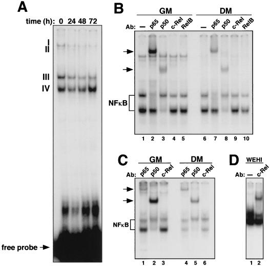
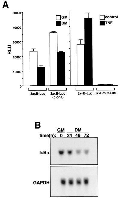
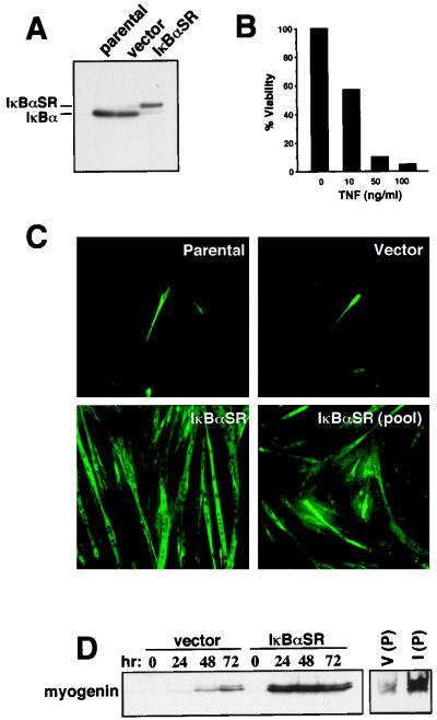
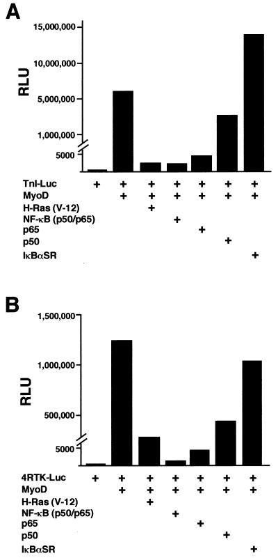
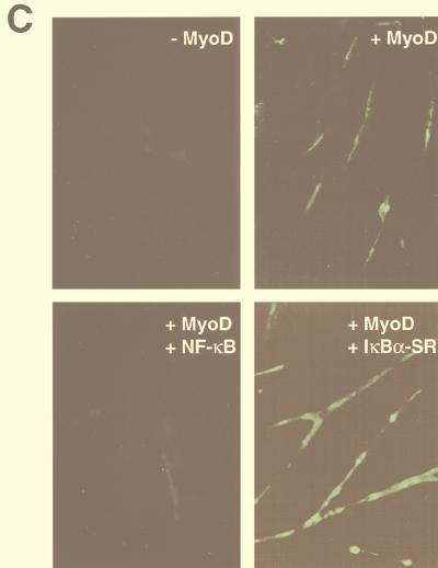
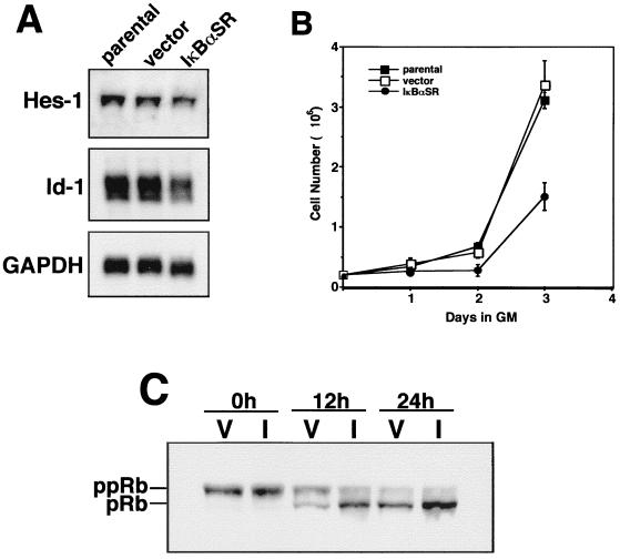
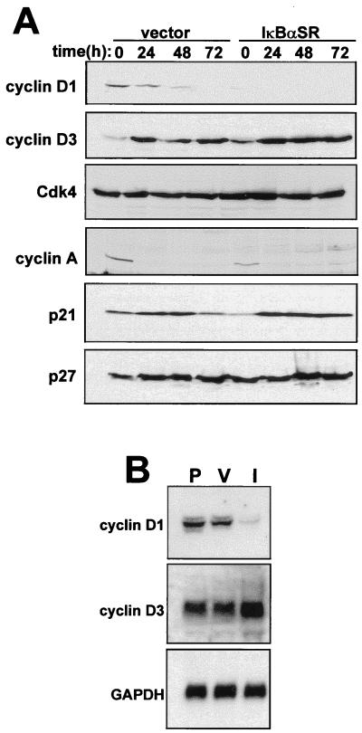
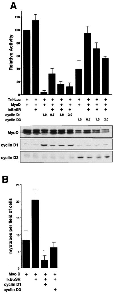
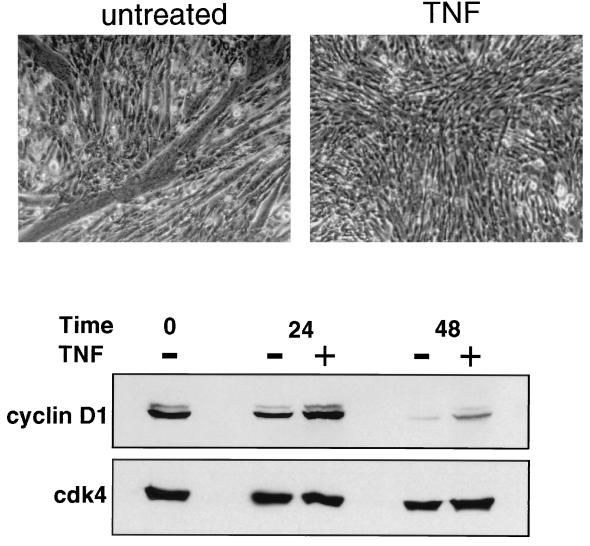
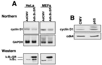
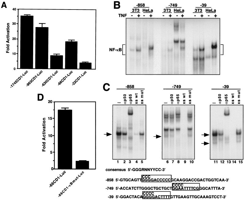
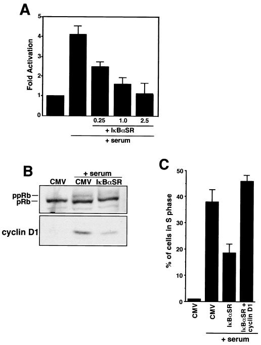
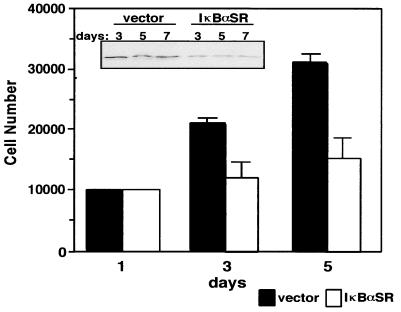
Accumulating evidence implicates the transcription factor NF-kappaB as a positive mediator of cell growth, but the molecular mechanism(s) involved in this process remains largely unknown. Here we use both a skeletal muscle differentiation model and normal diploid fibroblasts to gain insight into how NF-kappaB regulates cell growth and differentiation. Results obtained with the C2C12 myoblast cell line demonstrate that NF-kappaB functions as an inhibitor of myogenic differentiation. Myoblasts generated to lack NF-kappaB activity displayed defects in cellular proliferation and cell cycle exit upon differentiation. An analysis of cell cycle markers revealed that NF-kappaB activates cyclin D1 expression, and the results showed that this regulatory pathway is one mechanism by which NF-kappaB inhibits myogenesis. NF-kappaB regulation of cyclin D1 occurs at the transcriptional level and is mediated by direct binding of NF-kappaB to multiple sites in the cyclin D1 promoter. Using diploid fibroblasts, we demonstrate that NF-kappaB is required to induce cyclin D1 expression and pRb hyperphosphorylation and promote G(1)-to-S progression. Consistent with results obtained with the C2C12 differentiation model, we show that NF-kappaB also promotes cell growth in embryonic fibroblasts, correlating with its regulation of cyclin D1. These data therefore identify cyclin D1 as an important transcriptional target of NF-kappaB and reveal a mechanism to explain how NF-kappaB is involved in the early phases of the cell cycle to regulate cell growth and differentiation.


Figures

FIG. 1
FIG. 1
Loss of NF-κB binding activity during myogenic differentiation. (A) Proliferating C2C12 myoblasts (GM) were induced to differentiate (DM), and at the indicated times nuclear extracts were prepared and EMSA was performed with a radiolabeled oligonucleotide containing an NF-κB-binding site. (B) C2C12 cells were maintained in GM or switched to DM for 72 h. Supershift EMSA was performed with nuclear extracts preincubated with either no antibody (lanes 1 and 6) or antisera specific for p65 (lanes 2 and 7), p50 (lanes 3 and 8), c-Rel (lanes 4 and 9), or RelB (lanes 5 and 10). NF-κB complexes containing p50 and p65 subunits are shown. (C) EMSA and supershift EMSA were performed as described above with nuclear extracts prepared from primary myoblasts undergoing differentiation. (D) Supershift EMSA was performed with nuclear extract prepared from WEHI-231 cells, preincubated either with no antibody or with an antiserum specific to the c-Rel subunit of NF-κB.
FIG. 2
FIG. 2
Myogenic differentiation correlates with loss of NF-κB transactivation function. (A) C2C12 myoblasts stably containing a 3xκB-Luc reporter plasmid were propagated as either a mixed population or a clonal isolate. Cells were plated in triplicate overnight in 6-cm culture dishes, and on the following day they were maintained in GM or switched to DM for 48 h. At that time, cell extracts were prepared, and relative luciferase units were determined by normalizing to total protein (RLU). Promoter activities were also determined for 3xκB-Luc and 3xκBmut-Luc populations that were treated or not treated with 10 ng of TNF-α per ml for 24 h. (B) C2C12 cells were maintained in GM or differentiated in DM for up to 72 h. At the indicated times, total RNA was prepared and 10 μg of sample was used for Northern blot analysis. The blot was hybridized with an IκBα-specific probe, and RNA loading was normalized by stripping the blot and reprobing for GAPDH mRNA.
FIG. 3
FIG. 3
C2C12 cells lacking NF-κB activity have an accelerated rate of differentiation. (A) Whole-cell lysates were prepared from C2C12 parental, vector control, or IκBαSR proliferating myoblasts, and 50 μg of sample was used for Western blot analysis. IκBα and IκBαSR proteins were detected with an IκBα polyclonal antibody (C-19; Santa Cruz Biotechnology) at a 1:1,500 dilution. The IκBαSR protein is FLAG tagged and therefore migrates at a slightly higher mobility compared to the endogenous protein. (B) IκBαSR-expressing myoblasts were seeded in triplicate overnight in 12-well plates, and the following day cells were treated with increasing concentrations of TNF-α for 48 h. Cell viability was scored by trypsinization and the trypan blue exclusion method. Cells not treated with TNF-α were designated 100% viable. (C) C2C12 parental cells, vector control, an IκBαSR clone, or five pooled IκBαSR clones were differentiated in DM for 48 h, at which time the cells were prepared for immunofluorescence to detect for the myosin heavy chain. (D) C2C12 vector control or IκBαSR cells were induced to differentiate in DM for up to 72 h. At the indicated times, lysates were prepared and Western blot analysis was performed probing for myogenin expression. V (P) and I (P) denote myogenin expression from five pooled vector control or IκBαSR clones, respectively, that were differentiated for 48 h.
FIG. 4
FIG. 4
NF-κB inhibits MyoD-induced myogenesis in 10T1/2 fibroblasts. Cells were maintained in growth medium containing 15% FBS and differentiated in DM. The cells were seeded in triplicate overnight in 6-cm dishes, and the following day, cotransfections were performed with Superfect (Qiagen). DNA consisted of 1 μg of a troponin-I-Luc reporter plasmid (TnI-Luc) (A) or 1 μg of the 4RTK-Luc plasmid (B), along with 0.25 μg of an expression plasmid for MyoD alone or in combination with 0.5 μg of expression plasmids for either the activated form of oncogenic ras (H-ras V-12), p65, p50, or 1 μg of IκBαSR. DNA was standardized to 2.5 μg by the addition of Bluescript plasmid (Stratagene). Cells were maintained in growth medium for 24 h following transfections and then switched to DM for 48 h, at which time cell extracts were prepared and relative light units (RLU) were determined, by normalizing values to total protein. (C) For immunofluorescence analysis, 10T1/2 cells were seeded overnight and the next day similar transfections were performed as described above, except that one-third of the amount of DNA was used. At 24 h following transfections, the cells were switched to DM for 72 h, at which time the cells were fixed and probed for the myosin heavy chain. To score for the number of myotubes formed, cells expressing myosin were counted and averaged from a minimum of 10 randomly selected fields.
FIG. 4
FIG. 4
NF-κB inhibits MyoD-induced myogenesis in 10T1/2 fibroblasts. Cells were maintained in growth medium containing 15% FBS and differentiated in DM. The cells were seeded in triplicate overnight in 6-cm dishes, and the following day, cotransfections were performed with Superfect (Qiagen). DNA consisted of 1 μg of a troponin-I-Luc reporter plasmid (TnI-Luc) (A) or 1 μg of the 4RTK-Luc plasmid (B), along with 0.25 μg of an expression plasmid for MyoD alone or in combination with 0.5 μg of expression plasmids for either the activated form of oncogenic ras (H-ras V-12), p65, p50, or 1 μg of IκBαSR. DNA was standardized to 2.5 μg by the addition of Bluescript plasmid (Stratagene). Cells were maintained in growth medium for 24 h following transfections and then switched to DM for 48 h, at which time cell extracts were prepared and relative light units (RLU) were determined, by normalizing values to total protein. (C) For immunofluorescence analysis, 10T1/2 cells were seeded overnight and the next day similar transfections were performed as described above, except that one-third of the amount of DNA was used. At 24 h following transfections, the cells were switched to DM for 72 h, at which time the cells were fixed and probed for the myosin heavy chain. To score for the number of myotubes formed, cells expressing myosin were counted and averaged from a minimum of 10 randomly selected fields.
FIG. 5
FIG. 5
C2C12 cells lacking NF-κB exhibit a growth defect and changes in pRb phosphorylation. (A) Total RNA was prepared from C2C12 parental, vector control, or IκBαSR proliferating myoblasts, and Northern analysis was performed to detect the expression of Hes-1 or Id-1 genes. (B) C2C12 parental, vector control, or IκBαSR myoblasts were plated in triplicate in 10-cm plates and maintained in GM for 3 days. Every 24 h, the total cell number was determined by trypsinization and trypan blue exclusion. (C) C2C12 vector control (V) or IκBαSR cells (I) were induced to differentiate for up to 24 h. At the indicated times, cell lysates were prepared with lysis buffer containing phosphatase inhibitors and Western blotting was performed to probe for the hypo- and hyperphosphorylated forms of Rb with a monoclonal antibody (14001A; Pharmigen).
FIG. 6
FIG. 6
C2C12 cells lacking NF-κB are downregulated for cyclin D1 protein and mRNA. (A) C2C12 vector control or IκBαSR cells were differentiated in DM for up to 72 h. At the indicated times, whole-cell lysates were prepared and 50 μg was used in Western blot analyses to probe for various cell cycle proteins. (B) Total RNA was prepared from proliferating parental (P), vector control (V), or IκBαSR (I) myoblasts, and 10 μg of sample was used in Northern blotting to probe for cyclin D1 or cyclin D3 mRNA.
FIG. 7
FIG. 7
NF-κB inhibits MyoD transactivation function through the regulation of cyclin D1. (A) 10T1/2 fibroblasts were seeded in triplicate overnight in 6-cm culture dishes, and the next day cotransfection was performed with DNA consisting of 1 μg of the troponin-I reporter plasmid (TnI-Luc) and 0.25 μg of a MyoD expression plasmid, along with either 0.25 μg of an IκBαSR expression plasmid or the indicated amounts of cyclin D1 or cyclin D3 expression plasmid. DNA was normalized by the addition of Bluescript plasmid (Stratagene). Transfected cells were maintained in growth medium overnight and on the following day were switched to DM for 48 h, at which time extracts were prepared and a luciferase assay was performed. The level of troponin-I activation by MyoD alone was set to a value of 100. (Below) To verify the expression of proteins, parallel transfections were performed and immunoblot analysis was performed to probe for MyoD, cyclin D1, and cyclin D3. (B) Similar transfections were performed in 10T1/2 fibroblasts with the following amounts of expression plasmids: 0.25 μg of MyoD, 1 μg of IκBαSR, and 2 μg of cyclin D1 or cyclin D3. The cells were differentiated as described above for 72 h, at which point myogenesis was quantitated by counting myotubes from a minimum of 10 fields of cells.
FIG. 8
FIG. 8
TNF-α inhibits C2C12 myogenesis and stabilizes cyclin D1. C2C12 cells were induced to differentiate in the absence or presence of 20 ng of TNF-α per ml. Cytokine addition was repeated at 6 h and every additional 12 h after the induction of differentiation. At 72 h, the cells were washed with PBS, fixed for 10 min at room temperature with 4% paraformaldehyde, and photographed by phase-contrast microscopy. In parallel treatment cultures, cells were harvested at 24 and 48 h and whole-cell lysates were prepared for Western blot analysis. A 50-μg portion of total protein was fractionated by SDS-polyacrylamide gel electrophoresis, and immunoblotting was performed to probe for cyclin D1. The blot was subsequently stripped and reprobed for cdk4, used as an internal control.
FIG. 9
FIG. 9
Regulation of cyclin D1 is specific to NF-κB occurring in multiple cell types. (A) HeLa cells or MEFs were infected with adenovirus containing either the IκBαSR or empty vector (AdCMV) at a multiplicity of infection of 50 or 200, respectively. Total RNA was prepared 48 h postinfection, and Northern analysis was performed to detect cyclin D1 mRNA expression. Western blot analysis with an IκBα-specific antibody is shown to demonstrate the expression of the IκBαSR in HeLa cells and MEFs by using the adenovirus delivery system. (B) Fibroblasts were transfected with either empty vector or an expression plasmid expressing the p65 subunit of NF-κB. The following day, cells were switched to serum-deprived conditions for a 48-h period. Subsequently, whole-cell extracts were prepared and 50 μg was used in immunoblot analysis probing for cyclin D1. The loading efficiency was normalized by reprobing the blot for cdk4 expression.
FIG. 10
FIG. 10
NF-κB regulation of cyclin D1 occurs at the transcriptional level. (A) NIH 3T3 cells were plated in triplicate in 12-well plates. Transfections were performed on the following day with Lipofectamine reagent (Life Technologies) mixed with DNA consisting of 0.2 μg of reporter plasmids containing various 5′ deletions of the human cyclin D1 promoter, along with 0.15 μg of a p65 expression plasmid. DNA remained on the cells for 3 h in serum-free medium, and the cells were then switched for 48 h to complete medium containing 10% calf serum, at which time the luciferase activity was determined. Values were normalized to basal levels of promoter activity obtained by transfecting cyclin D1 promoter constructs in the absence of p65. (B) EMSAs were performed with nuclear extracts prepared from either NIH 3T3 cells or HeLa cells treated (+) or not treated (−) with TNF-α for 30 min. Putative NF-κB-binding sites, located at positions −858, −749, and −39 in the human cyclin D1 promoter, are indicated within the oligonucleotide sequences used for EMSAs. (C) Supershift EMSAs were performed with nuclear extracts prepared from HeLa cells treated with TNF, which were preincubated either with no addition (lanes 1, 6, and 11) or with addition of antisera specific for p50 (lanes 2, 7, and 12), or the p65 subunit (lanes 3, 8, and 13). Arrows denote p65-containing complexes. For competition EMSAs, extracts were preincubated with either a 100-fold molar excess of unlabeled oligonucleotides containing wild-type (xs wt) NF-κB-binding sites (lanes 4, 9, and 14), or a 100-fold molar excess of unlabeled oligonucleotides containing mutations in the NF-κB sites (xs mut) (lanes 5, 10, and 15). The mutations are shown in the boxed regions above the NF-κB-binding sites. (D) The same mutation at position −39 was made in the NF-κB-binding site within the −66CD1-Luc reporter plasmid. Both the wild-type and mutant (−66CD1-κBmut-Luc) reporter plasmids were separately cotransfected along with a p65 expression plasmid in NIH 3T3 cells under the same transfection conditions as described for panel A.
FIG. 11
FIG. 11
Requirement for NF-κB activity in the early G1 phase of the cell cycle. (A) NIH 3T3 cells were made quiescent by being switched for 48 h from complete medium containing 10% serum to medium containing 0.25% calf serum. Serum-deprived cells were cotransfected either with a cyclin D1 promoter reporter plasmid alone (−963CD1-Luc) (2.5 μg) or in combination with indicated amounts of IκBαSR plasmid. The cells were maintained in quiescence or switched to complete medium to induce cyclin D1 transcription and reentry into the cell cycle. Extracts were prepared, and relative luciferase activity was determined by standardizing to total cellular protein. (B) MEFs were made quiescent by culturing cells in 0.1% FBS for 48 h. The cells were then infected under serum-deprived conditions with adenovirus containing either empty vector (CMV) or IκBαSR at an MOI of 200. At 24 h postinfections, MEFs were either maintained under serum-deprived conditions or induced to reenter the cell cycle by the addition of 10% FBS. Whole-cell lysates were prepared 24 h later, and immunoblot analysis was performed to probe for both pRb and cyclin D1 (under these culture conditions, we determined that pRb hyperphosphorylation in MEFs is maximally detectable between 20 and 24 h following serum stimulation). (C) Subconfluent MEFs were grown on coverslips overnight and were rendered quiescent on the following day. Cells were infected with either control adenovirus (CMV), IκBαSR, or a combination of IκBαSR and cyclin D1 viruses (both at a multiplicity of infection of 200). At 24 h following infections, the cells were maintained quiescent or switched for 12 h to growth medium containing 10% serum, at which point the medium was supplemented with 100 μM BrdU (Sigma) for an additional 10 h. The cells were fixed and prepared for immunofluorescence analysis as described in Materials and Methods. The percentage of cells in the S phase was calculated by determining the number of BrdU-positive cells with respect to the total number of cells in a given field.
FIG. 12
FIG. 12
NF-κB is required for proper cellular proliferation in primary fibroblasts. Early-passaged MEFs were infected by retrovirus either harboring empty vector or the IκBαSR. At 2 days following infection, the cells were placed on a 4-day drug selection with 400 μg of G418 per ml. A mixed population of selected cells was then seeded in triplicate in either 12-well plates or 10-cm dishes. Cells seeded in 12-well plates were trypsinized every 24 to 48 h, and cell numbers were determined by the trypan blue exclusion method. Cells seeded in 10-cm dishes were collected on days 3, 5, and 7, and lysates were prepared for immunoblot analysis detecting for cyclin D1 (inset).

References

    1. Albanese C, Johnson J, Watanabe G, Eklund N, Vu D, Arnold A, Pestell R G. Transforming p21ras mutants and c-Ets-2 activate the cyclin D1 promoter through distinguishable regions. J Biol Chem. 1995;270:23589–23597. - PubMed
    1. Arber N, Sutter T, Miyake M, Kahn S M, Venkatraj V S, Sobrino A, Warburton D, Holt P R, Weinstein I B. Increased expression of cyclin D1 and the Rb tumor suppressor gene in c-K-ras transformed rat enterocytes. Oncogene. 1996;12:1903–1908. - PubMed
    1. Baeuerle P A, Baltimore D. NF-kappa B: ten years after. Cell. 1996;87:13–20. - PubMed
    1. Baeuerle P A, Henkel T. Function and activation of NF-kappa B in the immune system. Annu Rev Immunol. 1994;12:141–179. - PubMed
    1. Baldwin A S., Jr The NF-kappa B and I kappa B proteins: new discoveries and insights. Annu Rev Immunol. 1996;14:649–683. - PubMed

Publication types

MeSH terms