. 1999 Aug;19(8):5800-10.
doi: 10.1128/MCB.19.8.5800.

Akt/Protein kinase B inhibits cell death by preventing the release of cytochrome c from mitochondria

Affiliations

Akt/Protein kinase B inhibits cell death by preventing the release of cytochrome c from mitochondria

S G Kennedy et al. Mol Cell Biol. 1999 Aug.

Abstract

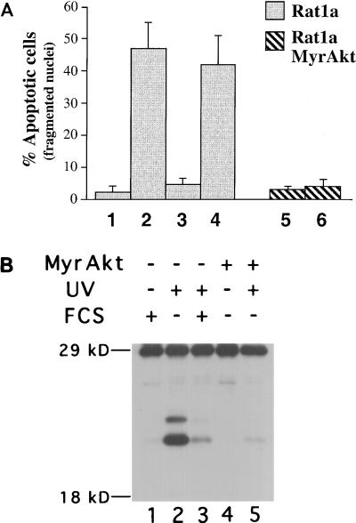
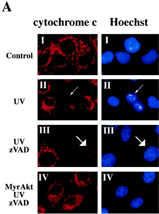
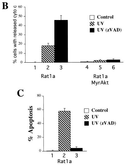
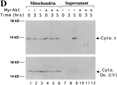
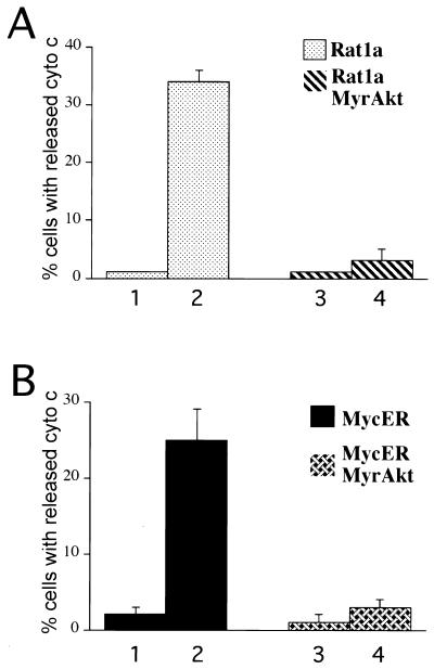
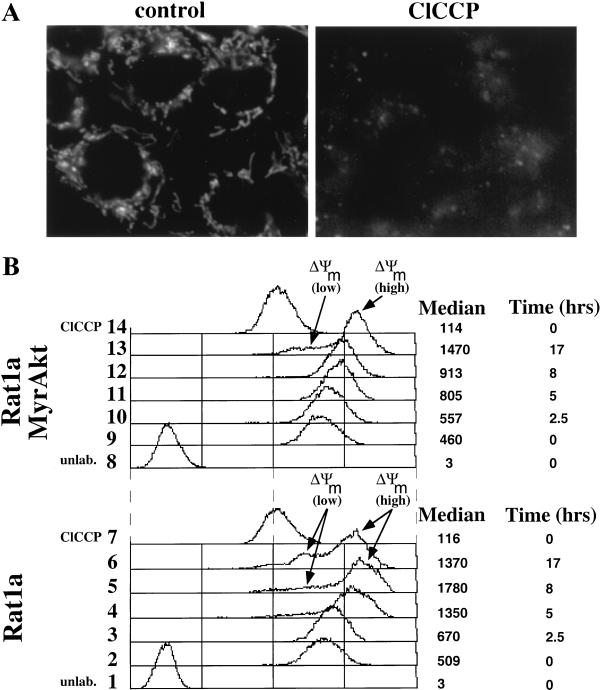
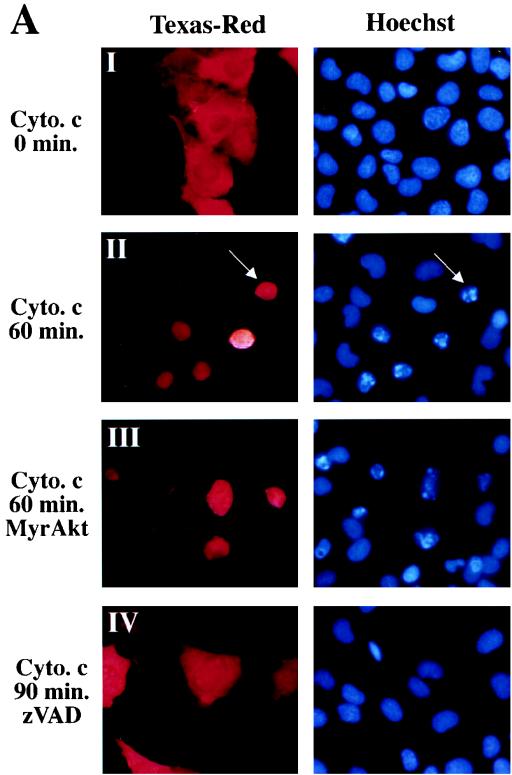
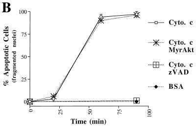
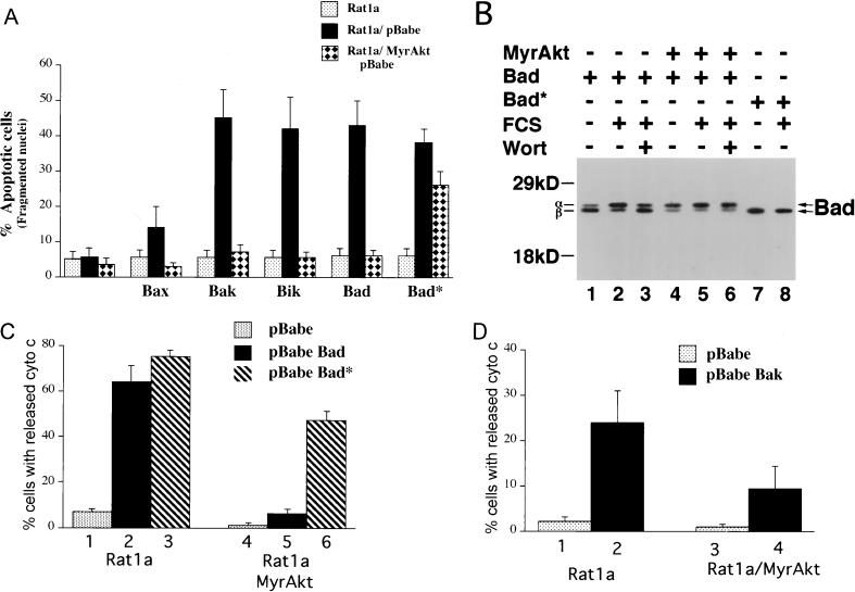
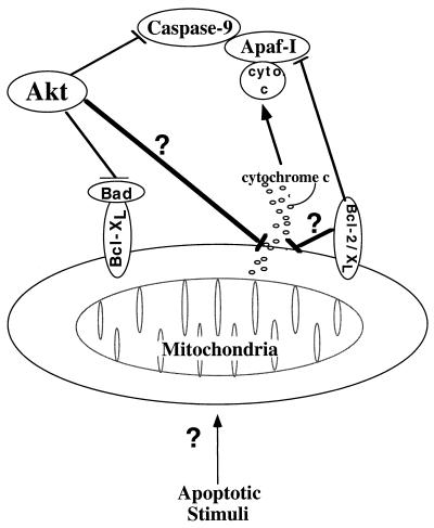
Growth factors signaling through the phosphoinositide 3-kinase/Akt pathway promote cell survival. The mechanism by which the serine/threonine kinase Akt prevents cell death remains unclear. We have previously shown that Akt inhibits the activity of DEVD-targeted caspases without changing the steady-state levels of Bcl-2 and Bcl-x(L). Here we show that Akt inhibits apoptosis and the processing of procaspases to their active forms by delaying mitochondrial changes in a caspase-independent manner. Akt activation is sufficient to inhibit the release of cytochrome c from mitochondria and the alterations in the inner mitochondrial membrane potential. However, Akt cannot inhibit apoptosis induced by microinjection of cytochrome c. We also demonstrated that Akt inhibits apoptosis and cytochrome c release induced by several proapoptotic Bcl-2 family members. Taken together, our results show that Akt promotes cell survival by intervening in the apoptosis cascade before cytochrome c release and caspase activation via a mechanism that is distinct from Bad phosphorylation.


Figures

FIG. 1
FIG. 1
Akt inhibits UV-mediated nuclei fragmentation, caspase activation, and apoptosis in Rat1a cells. (A) Percentage of apoptotic cells scored by DAPI staining of Rat1a (columns 1 to 4) and Rat1a/MyrAkt (columns 5 and 6) cells. Cells were either mock treated or treated with 200 nM wortmannin for 30 min (columns 4 and 6). Cells were then irradiated with UVC light (260 nm) at a dose of 50 Joules/m2 (lanes 2 to 6). Medium containing either 0% FCS (columns 1, 2, and 5 to 7) or 10% FCS (columns 3 and 4) was then added. After an additional 4.5 h, cells were DAPI stained and scored for nuclear condensation. Averages (± standard error) of at least 300 cells from three independent experiments are shown. (B) Akt inhibits the activation of at least two caspases during UV-mediated apoptosis. Rat1a (lanes 1 to 3) and Rat1a/MyrAkt (lanes 4 and 5) cells were deprived of serum for 2 h and irradiated with 50 J of UVC light. The medium was then changed to DMEM with 0% FCS (lanes 2, 4, and 5) or 10% FCS (lanes 1 and 3). After an additional 5 h, extracts were made and labeled with the biotin-conjugated YVAD-AMK (Calbiochem, La Jolla, Calif.). Extracts were subjected to sodium dodecyl sulfate-polyacrylamide gel electrophoresis (SDS-PAGE) on a 12% gel, and the Western blot was incubated with horseradish peroxidase-conjugated streptavidin (1 μg/ml).
FIG. 2
FIG. 2
Akt prevents the release of cytochrome c from mitochondria in a caspase-independent manner. (A) Cytochrome c immunostaining and Hoechst staining of Rat1a and Rat1a/MyrAkt cells. Fixation and staining of the cells was done 3.5 h after treatment as described in Materials and Methods. (I) Rat1a cells were deprived of serum for 20 min. (II) Rat1a cells were deprived of serum for 20 min and then irradiated with UV light. Arrows indicates diffused cytochrome c staining and nucleus with condensed chromatin in a cell undergoing apoptosis. (III) Rat1a cells were pretreated with 100 mM zVAD for 20 min, deprived of serum, and UV irradiated. Arrows indicate diffused cytochrome c staining and the corresponding nucleus of a cell protected from apoptosis. (IV) Rat1a/MyrAkt cells were treated as for panel III. (B) Quantitation of cells with diffused cytochrome (cyto) c staining. Averages (± standard error) of at least 100 cells from three independent experiments are shown. (C) Preincubation of Ra1a cells with zVAD is sufficient to prevent apoptosis as measured by DAPI staining. Cells were deprived of serum for 20 min and then either mock treated or treated with 100 μM zVAD for 20 min followed by UV irradiation (50 J/m2), as indicated. After an additional 5 h, cells were DAPI stained and scored for nuclear condensation. Averages (± standard errors) of at least 300 cells from three independent experiments are shown. (D) Rat1a (lanes 1 to 3 and 7 to 9) and Rat1a/MyrAkt (lanes 4 to 6 and 10 to 12) cells were deprived of serum for 20 min and then irradiated with 50 J of UV light as described for panel A. Cells were mechanically lysed and separated into mitochondrial and supernatant fractions 0, 3, and 5 h following irradiation as described in Materials and Methods. Fractions were subjected to SDS-PAGE (15% gel) followed by immunoblot analysis. Cytochrome c (Cyto. c) and COX IV [Cyto. Ox. subunit (IV)] are indicated by arrows. Positions of molecular weight markers are indicated on the left.
FIG. 2
FIG. 2
Akt prevents the release of cytochrome c from mitochondria in a caspase-independent manner. (A) Cytochrome c immunostaining and Hoechst staining of Rat1a and Rat1a/MyrAkt cells. Fixation and staining of the cells was done 3.5 h after treatment as described in Materials and Methods. (I) Rat1a cells were deprived of serum for 20 min. (II) Rat1a cells were deprived of serum for 20 min and then irradiated with UV light. Arrows indicates diffused cytochrome c staining and nucleus with condensed chromatin in a cell undergoing apoptosis. (III) Rat1a cells were pretreated with 100 mM zVAD for 20 min, deprived of serum, and UV irradiated. Arrows indicate diffused cytochrome c staining and the corresponding nucleus of a cell protected from apoptosis. (IV) Rat1a/MyrAkt cells were treated as for panel III. (B) Quantitation of cells with diffused cytochrome (cyto) c staining. Averages (± standard error) of at least 100 cells from three independent experiments are shown. (C) Preincubation of Ra1a cells with zVAD is sufficient to prevent apoptosis as measured by DAPI staining. Cells were deprived of serum for 20 min and then either mock treated or treated with 100 μM zVAD for 20 min followed by UV irradiation (50 J/m2), as indicated. After an additional 5 h, cells were DAPI stained and scored for nuclear condensation. Averages (± standard errors) of at least 300 cells from three independent experiments are shown. (D) Rat1a (lanes 1 to 3 and 7 to 9) and Rat1a/MyrAkt (lanes 4 to 6 and 10 to 12) cells were deprived of serum for 20 min and then irradiated with 50 J of UV light as described for panel A. Cells were mechanically lysed and separated into mitochondrial and supernatant fractions 0, 3, and 5 h following irradiation as described in Materials and Methods. Fractions were subjected to SDS-PAGE (15% gel) followed by immunoblot analysis. Cytochrome c (Cyto. c) and COX IV [Cyto. Ox. subunit (IV)] are indicated by arrows. Positions of molecular weight markers are indicated on the left.
FIG. 3
FIG. 3
Akt prevents the release of cytochrome c induced by multiple apoptotic stimuli. (A) Akt inhibits etoposide-mediated cytochrome c release. Rat1a (columns 1 and 2) and Rat1a/MyrAkt cells (columns 3 and 4) were either mock treated (columns 1 and 3) or treated with 10 μM etoposide (columns 2 and 4) for 3 h in the presence of 100 μM zVAD. Cells were then fixed and immunostained for cytochrome (cyto) c as described in Fig. 2. Averages (± standard errors) of at least 100 cells from four independent experiments are shown. (B) Akt inhibits Myc-mediated cytochrome c release. The percentage of cells demonstrating diffuse cytochrome c immunostaining was quantitated. Rat1a/MycER (columns 1 and 2) and Rat1a/MycER/MyrAkt cells (columns 3 and 4) were incubated overnight in DMEM with 2.5% FCS with (columns 2 and 4) or without (columns 1 and 3) 1 μM 4-HT. The next day, cells were placed in DMEM with 0.5% FCS and 100 μM zVAD for 3 h. Cells were then fixed and immunostained for cytochrome c as described for Fig. 2. Averages (± standard errors) of at least 100 cells from four independent experiments are shown.
FIG. 4
FIG. 4
Akt is sufficient to prevent alterations in mitochondrial dye uptake. (A) DiOC6 labeling of mitochondria requires an intact inner membrane potential. (B) FACS analysis of UV-irradiated Rat1a and Rat1a/MyrAkt cells incubated with DiOC6. Rat1a and Rat1a/MyrAkt cells were subjected to UV irradiation as described for Fig. 1, either mock treated or treated with 100 mM ClCCP for 5 min, and then loaded with DiOC6 as described in Materials and Methods. Proliferating Rat1a cells loaded with DiOC6 were defined as having a fluorescence of 500 arbitrary units.
FIG. 5
FIG. 5
Akt cannot block apoptosis induced by microinjection of cytochrome c. (A) Hoechst staining and Texas red fluorescence of microinjected cells. Rat1a (panels I, II, and IV) and Rat1a/MyrAkt (panel III) cells were mock treated or treated with 100 mM zVAD (panel IV) for 20 min and then microinjected with 0.2% Texas red and 1 mg of cytochrome (cyto.) c per ml. After the time indicated on the left, cells were fixed. (B) Time course of apoptosis in Rat1a and Rat1a/MyrAkt cells microinjected with cytochrome c. Cells exhibiting fragmented and condensed nuclei were scored as apoptotic. Rat1a and Rat1a/MyrAkt cells were injected with 1 mg of cytochrome c per ml and scored for nuclear condensation 0, 20, 60, and 90 min after microinjection. Alternatively, Rat1a cells were either mock treated or treated with 100 μM zVAD, then injected with BSA (1 mg/ml) or cytochrome c (1 mg/ml), fixed, and scored after 90 min. Averages (± standard errors) of at least 50 cells from three independent experiments are shown.
FIG. 5
FIG. 5
Akt cannot block apoptosis induced by microinjection of cytochrome c. (A) Hoechst staining and Texas red fluorescence of microinjected cells. Rat1a (panels I, II, and IV) and Rat1a/MyrAkt (panel III) cells were mock treated or treated with 100 mM zVAD (panel IV) for 20 min and then microinjected with 0.2% Texas red and 1 mg of cytochrome (cyto.) c per ml. After the time indicated on the left, cells were fixed. (B) Time course of apoptosis in Rat1a and Rat1a/MyrAkt cells microinjected with cytochrome c. Cells exhibiting fragmented and condensed nuclei were scored as apoptotic. Rat1a and Rat1a/MyrAkt cells were injected with 1 mg of cytochrome c per ml and scored for nuclear condensation 0, 20, 60, and 90 min after microinjection. Alternatively, Rat1a cells were either mock treated or treated with 100 μM zVAD, then injected with BSA (1 mg/ml) or cytochrome c (1 mg/ml), fixed, and scored after 90 min. Averages (± standard errors) of at least 50 cells from three independent experiments are shown.
FIG. 6
FIG. 6
Akt inhibits apoptosis and cytochrome c release induced by overexpression of the proapoptotic Bcl-2 family members. (A) Akt inhibits apoptosis accelerated by overexpression of Bax, Bak, Bik, and Bad. Rat 1a and Rat1a/MyrAkt cells were infected with eGFP retroviruses expressing Bax, Bak, Bik, Bad, and the Bad* (see Materials and Methods). Apoptosis was scored by DAPI staining 16 h following serum deprivation, a time point at which Rat 1a cells do not exhibit significant apoptosis. Cells were seeded at a density of 50,000 cells/3-cm-diameter dish. The next day cells were placed in DMEM containing 0% FCS for 16 h. Averages (± standard errors) of at least 300 cells from three independent experiments. (B) Akt induces a mobility shift of ectopically expressed Bad in Rat1a cells. Rat1a (lanes 1 to 3, 7, and 8) and Rat1a/MyrAkt (lanes 4 to 6) cells were infected with retrovirus expressing Bad or Bad*. Forty-eight hours following infection, cells were deprived of serum for 2 h. Cells were either mock treated (lanes 1, 2, 4, 5, 7, and 8) or treated with 200 nM wortmannin (Wort; lanes 3 and 6) for 30 min. Cells were then either mock treated (lanes 1, 4, and 7) or stimulated with 20% FCS for 30 min (lanes 2, 3, 5, 6, and 8). Cells were lysed, and extracts were subjected to SDS-PAGE (12% gel). Bad was detected with a hamster monoclonal anti-Bad antibody (PharMingen clone 13361S), and the different phosphorylation states are indicated with arrows. Positions of molecular weight standards are indicated on the left. (C) Akt prevents cytochrome c release accelerated by overexpression of Bad. Rat1a (columns 1 to 3) and Rat1a/MyrAkt (columns 4 to 6) cells demonstrating diffuse cytochrome c immunostaining were quantitated. Cells were treated as described for Fig. 2. Two hours (a time point at which there is no significant cytochrome c release in Rat 1a cells) after UV irradiation cells were fixed and immunostained for cytochrome c localization. Averages (± standard errors) of at least 100 cells from three independent experiments are shown. (D) Akt prevents cytochrome c release accelerated by overexpression of Bak. Cells were treated and scored for cytochrome c release as described for panel C.
FIG. 7
FIG. 7
Schematic illustration showing Akt antiapoptotic activities. Akt promotes cell survival by intervening in the apoptosis cascade upstream of cytochrome c (cyto c) release. Akt may maintain the integrity of the mitochondria by a general unknown mechanism or by a specific mechanism of Bad phosphorylation. By analogy to Bcl-2 and Bcl-xL, which inhibit apoptosis post-cytochrome c release through binding of Apaf-I, Akt can also inhibit apoptosis by phosphorylation and inactivation of caspase-9.

Similar articles

Cited by

References

    1. Adams J M, Cory S. The Bcl-2 protein family: arbiters of cell survival. Science. 1998;281:1322–1326. - PubMed
    1. Ahmed N N, Grimes H L, Bellacosa A, Chan T O, Tsichlis P N. Transduction of interleukin-2 antiapoptotic and proliferative signal via Akt protein kinase. Proc Natl Acad Sci USA. 1997;94:3627–3632. - PMC - PubMed
    1. Bergmann A, Agapite J, McCall K, Steller H. The Drosophila gene hid is a direct molecular target of Ras-dependent survival signaling. Cell. 1998;95:331–341. - PubMed
    1. Bossy-Wetzel E, Newmeyer D D, Green D R. Mitochondrial cytochrome c release in apoptosis occurs upstream of DEVD-specific caspase activation and independently of mitochondrial transmembrane depolarization. EMBO J. 1998;17:37–49. - PMC - PubMed
    1. Brunet A, Bonni A, Zigmond M J, Lin M Z, Juo P, Hu L S, Anderson M J, Arden K C, Blenis J, Greenberg M E. Akt promotes cell survival by phosphorylating and inhibiting a Forkhead transcription factor. Cell. 1999;96:857–868. - PubMed

Publication types

MeSH terms