. 1999 Aug;19(8):5811-22.
doi: 10.1128/MCB.19.8.5811.

Multiple roles of ligand in transforming the dioxin receptor to an active basic helix-loop-helix/PAS transcription factor complex with the nuclear protein Arnt

Affiliations

Multiple roles of ligand in transforming the dioxin receptor to an active basic helix-loop-helix/PAS transcription factor complex with the nuclear protein Arnt

M J Lees et al. Mol Cell Biol. 1999 Aug.

Abstract

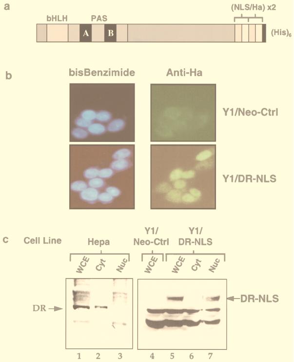
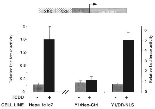
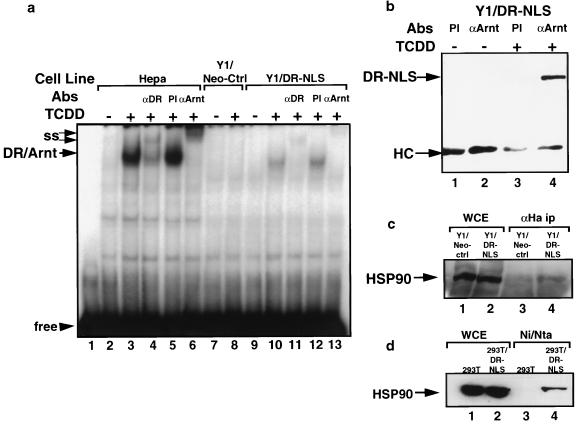
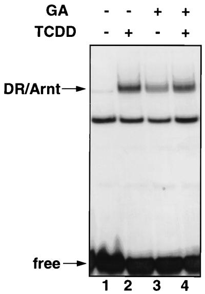
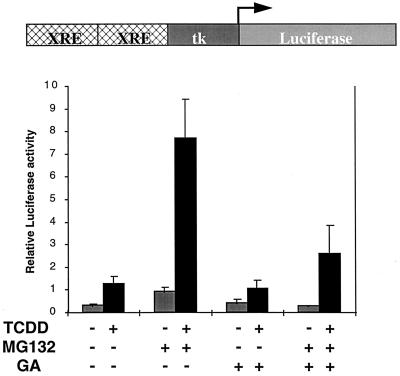
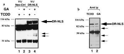
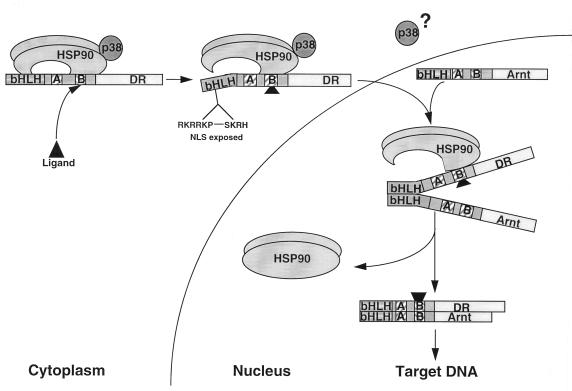
The dioxin receptor is a ligand-activated transcription factor belonging to an emerging class of basic helix-loop-helix/PAS proteins which show interaction with the molecular chaperone hsp90 in their latent states and require heterodimerization with a general cofactor, Arnt, to form active DNA binding complexes. Upon binding of polycyclic aromatic hydrocarbons typified by dioxin, the dioxin receptor translocates from the cytoplasm to the nucleus to allow interaction with Arnt. Here we have bypassed the nuclear translocation step by creating a cell line which expresses a constitutively nuclear dioxin receptor, which we find remains in a latent form, demonstrating that ligand has functional roles beyond initiating nuclear import of the receptor. Treatment of the nuclear receptor with dioxin induces dimerization with Arnt to form an active transcription factor complex, while in stark contrast, treatment with the hsp90 ligand geldanamycin results in rapid degradation of the receptor. Inhibition of degradation by a proteasome inhibitor allowed geldanamycin to transform the nuclear dioxin receptor to a heterodimer with Arnt (DR-Arnt). Our results indicate that unchaperoned dioxin receptor is extremely labile and is consistent with a concerted nuclear mechanism for receptor activation whereby hsp90 is released from the ligand-bound dioxin receptor concomitant with Arnt dimerization. Strikingly, artificial transformation of the receptor by geldanamycin provided a DR-Arnt complex capable of binding DNA but incapable of stimulating transcription. Limited proteolysis of DR-Arnt heterodimers indicated different conformations for dioxin versus geldanamycin-transformed receptors. Our studies of intracellular dioxin receptor transformation indicate that ligand plays multiple mechanistic roles during receptor activation, being important for nuclear translocation, transformation to an Arnt heterodimer, and maintenance of a structural integrity key for transcriptional activation.


Figures

FIG. 1
FIG. 1
Generation of a stable cell line expressing a nuclear DR. (A) Schematic representation of the C-terminally modified DR containing duplicate sequences of the nucleoplasmin NLS and the HA epitope followed by a hexahistidine tag. (B) The Y1/DR-NLS stable cell line expresses a constitutively nuclear DR. Y1/Neo-Ctrl and Y1/DR-NLS cells were seeded onto coverslips and fixed with methanol. Cells were incubated with the rat MAb 3F10 directed against the HA epitope, followed by incubation with a fluorescein isothiocyanate-conjugated goat anti-rat secondary antibody. Nuclei were visualized by bisbenzimide (blue) staining. (C) Immunoblot analysis of the DR in cell extracts. Protein extracts (100 μg) from whole cells (WCE), cytosol (Cyt), and nuclei (Nuc) of nontreated Hepa1c1c7, Y1/Neo-Ctrl, and Y1/DR-NLS cells were separated by SDS-PAGE (7.5% gel), transferred to nitrocellulose membranes, and immunoblotted with MAb RPT1 (specific for the native DR; lanes 1 to 3) or 12CA5 (specific for the HA tag; lanes 4 to 7). Positions of the native and NLS-HA-modified forms of the DR are indicated.
FIG. 2
FIG. 2
The nuclear DR requires ligand to activate transcription in Y1/DR-NLS cells. Hepa1c1c7, Y1/Neo-Ctrl, and Y1/DR-NLS cells were transiently transfected with an XRE-luciferase reporter gene and the renilla luciferase internal control vector pRL-TK. Cells were treated with dioxin (TCDD, 1 nM; dark bars) or vehicle alone (0.1% DMSO; light bars) for 24 h (Hepa1c1c7) or 30 h (Y1/Neo-Ctrl and Y1/DR-NLS). Luciferase activity was normalized against the internal control and is an average ± standard error of six transfection experiments. The left hand y axis pertains to the Hepa1c1c7 transfections, while the right-hand y axis relates to transfections in the modified Y1 cell lines.
FIG. 3
FIG. 3
The nuclear DR requires ligand to heterodimerize with Arnt and bind DNA. (a) The DR-NLS protein from Y1 cells is in a non-DNA binding form in the absence of ligand. Nuclear extracts (15 μg) from Hepa1c1c7 cells (lanes 2 to 6), Y1/Neo-Ctrl cells (lanes 7 and 8), or Y1/DR-NLS cells (lanes 9 to 13) treated with 1 nM TCDD or vehicle alone (0.1% DMSO) for 4 h were incubated with a 32P-labeled XRE probe and separated by nondenaturing PAGE (5.5% gel). Positions of the DR-Arnt band and free XRE probe are indicated. ss, supershifted bands generated by incubation with antibodies (Abs) directed against either the DR (αDR) or Arnt (αARNT). PI, preimmune serum. (b) The nuclear DR requires ligand to heterodimerize with Arnt. Y1/DR-NLS cells were treated with 1 nM TCDD or vehicle alone (0.1% DMSO) for 2 h. Nuclear extracts (100 μg) were immunoprecipitated with antiserum raised against Arnt (αArnt) or preimmune serum (PI), separated by SDS-PAGE (7.5% gel), and immunoblotted with anti-HA MAb 12CA5. Positions of the DR-NLS protein and immunoglobulin heavy chain (HC) are indicated. (c and d) The nuclear DR remains bound to hsp90. (c) Whole-cell extracts (WCE) from Y1/Neo-Ctrl or Y1/DR-NLS cells were immunoprecipitated (ip) with rat anti-HA MAb 3F10, separated by SDS-PAGE, and immunoblotted with an antibody specific for hsp90. (d) Whole-cell extracts from 293T or 293T/DR-NLS cells were purified by nickel affinity chromatography and separated by SDS-PAGE before being immunoblotted with an hsp90-specific MAb. The position of hsp90 is indicated with an arrow.
FIG. 4
FIG. 4
The hsp90 binding agent geldanamycin (GA) can induce formation of DR-Arnt heterodimers. (a) GA treatment of Y1/DR-NLS cells stimulates DR degradation which can be inhibited by the proteasome inhibitor MG132. Whole-cell extracts from Y1/DR-NLS cells treated with vehicle alone (0.1% DMSO; lane 1), GA (1 μg/ml; lane 2), or GA (1 μg/ml) plus MG132 (7.5 μM) (lane 3) for 2 h were analyzed for the presence of the DR-NLS protein by immunoblotting with anti-HA MAb 12CA5. The position of the DR-NLS protein is indicated, the two lower bands representing background proteins detected by 12CA5. (b) GA can induce a DR-Arnt heterodimer in the Y1/DR-NLS cell line. Y1/DR-NLS cells were treated with the indicated combinations of GA (1 μg/ml), TCDD (1 nM), and MG132 (7.5 μM) for 2 h. Nuclear extracts of treated cells were immunoprecipitated with a polyclonal antibody (Ab) raised against Arnt (A) or preimmune serum (PI), separated by SDS-PAGE (7.5% gel), and immunoblotted with the anti-HA MAb 12CA5. Locations of the DR-NLS protein and immunoglobulin heavy chain (HC) are indicated. (c) GA destabilizes DR-NLS–hsp90 complexes. Whole-cell extracts from 293T/DR-NLS cells treated for 30 min with DMSO (0.1%) or GA (1 μg/ml) in the presence of MG132 (7.5 μM) were purified by using Ni-NTA resin prior to immunoblotting with an hsp90 MAb. Ten percent of the eluted protein was run on a separate gel and immunoblotted with the anti-DR MAb RPT1 (lanes 5 and 6). Lanes 1 and 2 contain aliquots of the extracts prior to purification. The positions of hsp90 and DR-NLS are indicated.
FIG. 5
FIG. 5
Geldanamycin-induced DR-Arnt heterodimers are capable of binding DNA. Cytosolic extracts (15 μg) from Hepa1c1c7 cells were treated with DMSO vehicle (lane 1), TCDD (10 nM, lane 2), geldanamycin (GA; 10 μg/ml; lane 3), or a combination of TCDD (10 nM) and GA (10 μg/ml) (lane 4) for 2 h at room temperature, followed by incubation with a 32P-labeled XRE probe prior to separation by nondenaturing PAGE (5.5% gel). Positions of the DR-Arnt band and free probe are indicated.
FIG. 6
FIG. 6
The DR-Arnt complex induced by geldanamycin does not activate transcription. Y1/DR-NLS cells were cotransfected with the XRE-luciferase reporter gene and the renilla luciferase internal control vector pRL-TK. Cells were then treated with the indicated combinations of DMSO vehicle alone, TCDD (1 nM; dark bars), geldanamycin (GA; 1 μg/ml; light bars), and MG132 (7.5 μM) for 16 h. Luciferase activity was normalized against the internal control and is an average ± standard error of four transfection experiments.
FIG. 7
FIG. 7
The DR-Arnt complex induced by geldanamycin differs in conformation from heterodimers induced by dioxin. (A) Whole-cell extracts (100 μg) from Y1/Neo-Ctrl or Y1/DR-NLS cells treated for 2 h with TCDD (1 nM) or geldanamycin (GA; 1 μg/ml) in the presence of 7.5 μM MG132 were incubated with 150 ng of trypsin (20 min, 37°C), separated by SDS-PAGE (10% gel), and immunoblotted with anti-HA MAb 12CA5. (B) Whole-cell extracts from Y1/DR-NLS cells treated as for panel A were immunoprecipitated (ip) with anti-Arnt antibodies and digested with 25 ng of trypsin (15 min, 25°C) while bound to protein A-Sepharose. Proteolytic fragments were separated by SDS-PAGE (12.5% gel) and detected by immunoblotting with anti-HA MAb 12CA5. Geldanamycin-specific bands are indicated with small arrows.
FIG. 8
FIG. 8
Model for ligand-induced transformation of the cytosolic DR to the nuclear DR-Arnt heterodimer. Ligand binding stimulates release of hsp90 from the N terminus, exposing the NLS to promote nuclear import of the PAS B-bound hsp90-DR complex. Once in the nucleus, interaction of the free bHLH domain with Arnt initiates concomitant release of hsp90 and formation of the mature DR-Arnt heterodimer. See text for details.

Similar articles

Cited by

References

    1. Abbott B D, Probst M R. Developmental expression of two members of a new class of transcription factors. II. Expression of aryl hydrocarbon receptor nuclear translocator in the C57BL/6N mouse embryo. Dev Dyn. 1995;204:144–155. - PubMed
    1. Antonsson C, Whitelaw M L, McGuire J, Gustafsson J A, Poellinger L. Distinct roles of the molecular chaperone hsp90 in modulating dioxin receptor function via the basic helix-loop-helix and PAS domains. Mol Cell Biol. 1995;15:756–765. - PMC - PubMed
    1. Berghard A, Gradin K, Pongratz I, Whitelaw M, Poellinger L. Cross-coupling of signal transduction pathways: the dioxin receptor mediates induction of cytochrome P-450IA1 expression via a protein kinase C-dependent mechanism. Mol Cell Biol. 1993;13:677–689. - PMC - PubMed
    1. Brzozowski A M, Pike A C, Dauter Z, Hubbard R E, Bonn T, Engstrom O, Ohman L, Greene G L, Gustafsson J A, Carlquist M. Molecular basis of agonism and antagonism in the oestrogen receptor. Nature. 1997;389:753–758. - PubMed
    1. Carver L A, Bradfield C A. Ligand-dependent interaction of the aryl hydrocarbon receptor with a novel immunophilin homolog in vivo. J Biol Chem. 1997;272:11452–11456. - PubMed

Publication types

MeSH terms