. 1999 Jul;104(2):155-62.
doi: 10.1172/JCI6926.

Safety and antitumor activity of recombinant soluble Apo2 ligand

Affiliations

Safety and antitumor activity of recombinant soluble Apo2 ligand

A Ashkenazi et al. J Clin Invest. 1999 Jul.

Abstract

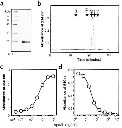
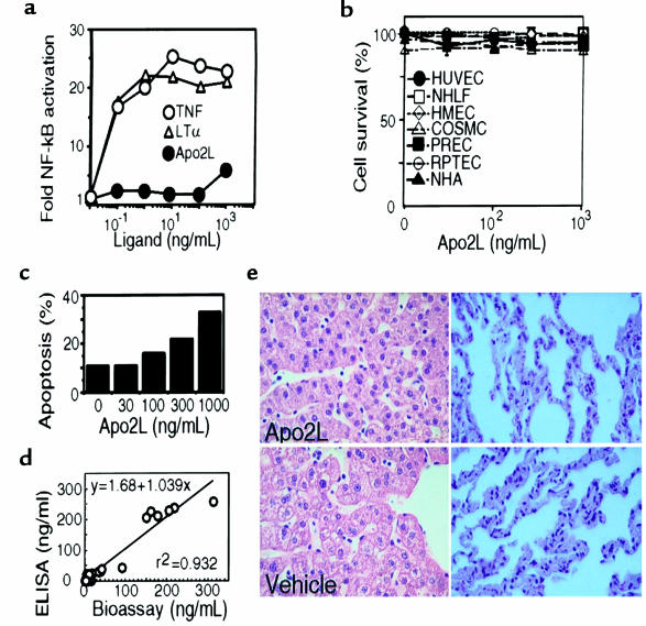
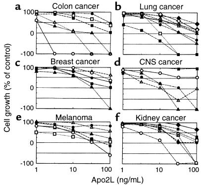
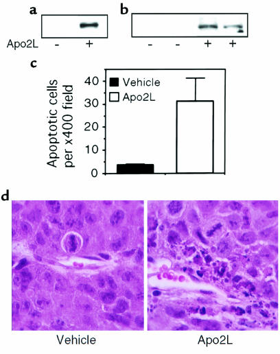
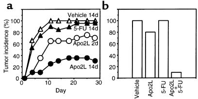
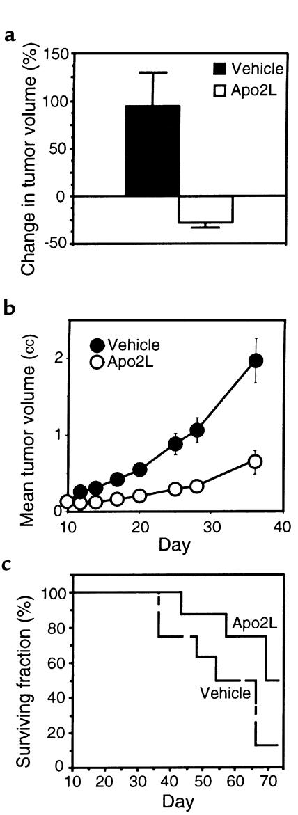
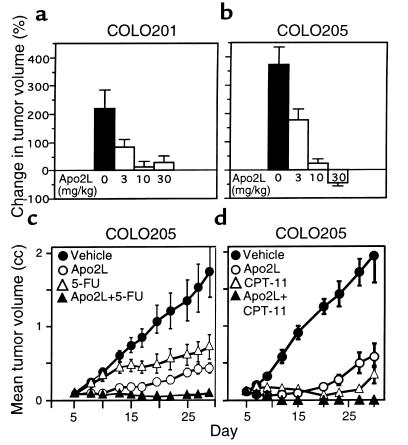
TNF and Fas ligand induce apoptosis in tumor cells; however, their severe toxicity toward normal tissues hampers their application to cancer therapy. Apo2 ligand (Apo2L, or TRAIL) is a related molecule that triggers tumor cell apoptosis. Apo2L mRNA is expressed in many tissues, suggesting that the ligand may be nontoxic to normal cells. To investigate Apo2L's therapeutic potential, we generated in bacteria a potently active soluble version of the native human protein. Several normal cell types were resistant in vitro to apoptosis induction by Apo2L. Repeated intravenous injections of Apo2L in nonhuman primates did not cause detectable toxicity to tissues and organs examined. Apo2L exerted cytostatic or cytotoxic effects in vitro on 32 of 39 cell lines from colon, lung, breast, kidney, brain, and skin cancer. Treatment of athymic mice with Apo2L shortly after tumor xenograft injection markedly reduced tumor incidence. Apo2L treatment of mice bearing solid tumors induced tumor cell apoptosis, suppressed tumor progression, and improved survival. Apo2L cooperated synergistically with the chemotherapeutic drugs 5-fluorouracil or CPT-11, causing substantial tumor regression or complete tumor ablation. Thus, Apo2L may have potent anticancer activity without significant toxicity toward normal tissues.


Figures

Figure 1
Figure 1
Oligomeric state and bioactivity of bacterially produced soluble human Apo2L. (a) Denaturing SDS-PAGE analysis of purified soluble Apo2L (2 μg) without (–) or with (+) reduction by 25 mM of DTT. Molecular weight markers are shown in the left lane. The gel was stained with silver. Identical results were obtained when reduced and nonreduced Apo2L was run on separate gels (not shown). (b) Nondenaturing chromatographic analysis of purified soluble Apo2L. Purified Apo2L (2 μg) was run on a Superose 12 size-exclusion HPLC column in a buffer containing 400 mM ammonium sulfate and 13 mM sodium phosphate (pH 6.5) and was detected by absorbance at 214 nm. The elution time of molecular weight markers is indicated by arrows. The peak appearing at 29–30 minutes is from buffer elution at the included volume of the column. (c) Measurement of Apo2L concentration. Representative standard curve for Apo2L concentration as determined by ELISA with 2 monoclonal anti-human Apo2L antibodies that recognize epitopes in Apo2L’s extracellular region. (d) Measurement of Apo2L bioactivity. Representative standard curve for Apo2L cytotoxicity toward SK-MES-1.
Figure 2
Figure 2
Safety of Apo2L. (a) Human umbilical cord endothelial cells (HUVEC) were incubated with purified soluble TNF, LT-α, or Apo2L for 1 hour, and NF-κB activity in nuclear extracts was determined by electrophoretic mobility-shift assay (38). (b) HUVEC, normal human lung fibroblasts (NHLF), human mammary epithelial cells (HMEC), colon smooth muscle cells (COSMC), prostate epithelial cells (PREC), renal proximal tubule epithelial cells (RPTEC), or normal human astrocytes (NHA) were incubated for 24 hours with Apo2L (1 μg/mL), and cell death was measured by FACS® analysis of propidium iodide staining. (c) Baboon CB1 cells were treated with vehicle or Apo2L in the presence of 10 μg/mL cycloheximide for 16 hours, and apoptosis was determined as in b. (d) Serum samples from cynomolgus monkeys treated with various doses of Apo2L were analyzed for Apo2L concentration by ELISA, and for Apo2L activity by bioassay, as in Figure 1, c and d. (e) Histological sections of liver and lung tissue from cynomolgus monkeys treated with vehicle or Apo2L (10 mg/kg/d) for 7 days.
Figure 3
Figure 3
Sensitivity of cancer cell lines to Apo2L. Cells were incubated with serial dilutions of Apo2L for 48 hours, and cell growth relative to buffer controls was determined. (a) Colon cancer lines: COLO205 (open circles), HCT15 (open triangles), HCT116 (filled triangles), HCC-2998 (filled squares), SW620 (open squares), HT29 (filled circles). (b) Lung cancer cell lines: NCI-H460 (filled squares), HOP-92 (open squares), HOP-62 (filled triangles), NCI-H226 (filled circles), NCI-H322M (open triangles), EKVX (plus signs), NCI-H522 (open circles), A549 (open diamonds), NCI-H23 (filled diamonds). (c) Breast cancer cell lines: MDA-MB-231 (filled triangles), MDA-N (filled squares), MCF7 (filled circles), BT-549 (open circles), NCI/ADR-RES (open triangles). (d) Central nervous system (CNS) cancer cell lines: SF-539 (filled triangles), SF-295 (open triangles), U251 (filled circles), SF-268 (open circles), SNB-75 (filled squares). (e) Melanoma cell lines: MALME-3M (open circles), M14 (filled squares), SK-MEL-28 (open squares), SK-MEL-2 (filled triangles), UACC-257 (open triangles), SK-MEL-5 (filled circles). (f) Renal cancer cell lines: RXF-393 (open circles), ACHN (filled triangles), TK-10 (open triangles), A498 (filled circles), CAKI-1 (filled circles), SN12C (open squares), UO-31 (filled squares), 786-0 (filled diamonds).
Figure 4
Figure 4
Induction of tumor cell apoptosis by Apo2L in vivo. Nude mice with subcutaneous HCT116 tumors were treated with vehicle (–) or Apo2L (+) (15 mg/kg) by intratumoral (a) or intraperitoneal (bd) injection. After 3 hours (a) or 6 hours (b), tumor cell extracts were assayed for caspase-3 activity by immunoblot analysis of the 85-kDa cleavage product of PARP. (c) Histological sections of tumors from vehicle- or Apo2L-treated animals were stained with hematoxylin and eosin, and apoptotic figures were counted in 10 random ×400 fields per tumor (mean ± SD). (d) Hematoxylin and eosin–stained tumor sections from vehicle- or Apo2L-treated animals.
Figure 5
Figure 5
Effect of Apo2L on tumor formation. (a) Nude mice (10 per group) were injected subcutaneously with HCT116 cells; 1 day later, they were injected intraperitoneally with vehicle or Apo2L (15 mg/kg/d) for 2 days or 14 days, or with 5-FU (25 mg/kg/d) for 14 days. The appearance of tumors was assessed every few days for a period of 4 weeks. (b) Nude mice (10 per group) were injected with HCT116 cells. On days 1 and 2 after inoculation, the mice received intraperitoneal injections of vehicle, Apo2L (0.5 mg/kg/d), 5-FU (5 mg/kg/d), or Apo2L plus 5-FU. The appearance of tumors of measurable size was assessed on day 15.
Figure 6
Figure 6
Effect of Apo2L on growth of established HCT116 tumors and on survival. (a) Nude mice carrying HCT116 tumors (5 per group) received vehicle or Apo2L (5 mg/kg/d) over 3 days through intraperitoneal infusion minipumps, and tumor volume was measured. (b) Nude mice carrying HCT116 tumors (10 per group) received intraperitoneal injections of vehicle or Apo2L (10 mg/kg/d) on days 10–14 and 16–20 after tumor cell inoculation, and tumor volume was measured (mean ± SEM). (c) Independent groups of mice were treated as in b with vehicle (dashed line) or Apo2L (solid line), and were followed for survival.
Figure 7
Figure 7
Effect of Apo2L on growth of established COLO205 tumors. (a and b) Nude mice carrying COLO201 (a) or COLO205 (b) tumors (10 per group) were injected intraperitoneally with vehicle or Apo2L (3, 10, or 30 mg/kg/d) over 4 days, and the tumor volume was measured. (c) Nude mice carrying COLO205 tumors (10 per group) received intraperitoneal injections of vehicle (closed circles) or Apo2L (30 mg/kg/d; open circles), 5-FU (25 mg/kg/d; open triangles), or Apo2L plus 5-FU (closed triangles) on days 5-9 and 12-16, and tumor volume was determined. (d) Nude mice carrying COLO205 tumors (9 per group) received intraperitoneal injections of vehicle (closed circles) or Apo2L (30 mg/kg/d; open circles) on days 5-9 and 12-16, or CPT-11 (80 mg/kg/d; open triangles) on days 5, 9, and 13, or Apo2L plus CPT-11 (closed triangles), and tumor volume was determined. The data are mean ± SEM.

References

    1. Jacobson MD, Weil M, Raff MC. Programmed cell death in animal development. Cell. 1997;88:347–354. - PubMed
    1. Nagata S. Apoptosis by death factor. Cell. 1997;88:355–365. - PubMed
    1. Ashkenazi A, Dixit VM. Death receptors: signaling and modulation. Science. 1998;281:1305–1308. - PubMed
    1. Levine AJ. p53, the cellular gatekeeper for growth and division. Cell. 1997;88:323–331. - PubMed
    1. Tartaglia L, Goeddel D. Two TNF receptors. Immunol Today. 1992;13:151–153. - PubMed

MeSH terms