. 1999 Jul;104(2):173-80.
doi: 10.1172/JCI6909.

Rapid generation of broad T-cell immunity in humans after a single injection of mature dendritic cells

Affiliations

Rapid generation of broad T-cell immunity in humans after a single injection of mature dendritic cells

M V Dhodapkar et al. J Clin Invest. 1999 Jul.

Abstract

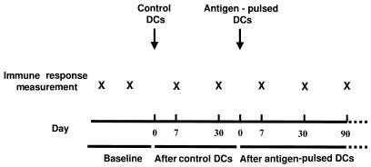
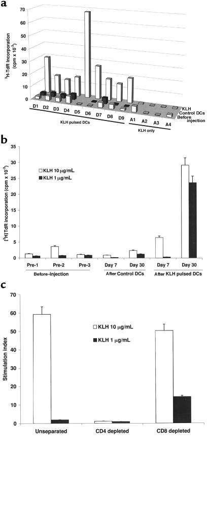
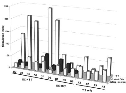
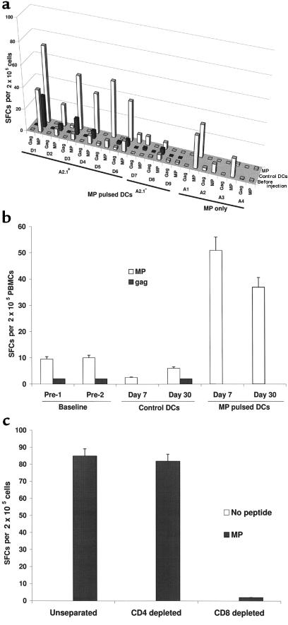
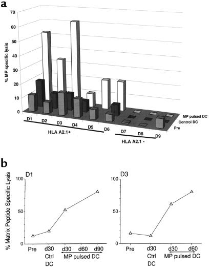
Dendritic cells (DCs) are potent antigen-presenting cells that initiate protective T-cell immunity in mice. To study the immunogenicity of DCs in humans, we injected 9 healthy subjects subcutaneously with a control injection of autologous monocyte-derived, mature DCs, followed 4-6 weeks later by DCs pulsed with keyhole limpet hemocyanin (KLH), HLA-A*0201-positive restricted influenza matrix peptide (MP), and tetanus toxoid (TT). Four more subjects received these antigens without DCs. Injection of unpulsed DCs, or antigens alone, failed to immunize. Priming of CD4(+) T cells to KLH was observed in all 9 subjects injected with KLH-pulsed DCs, and boosting of TT-specific T-cell immunity was seen in 5 of 6 subjects injected with TT-pulsed DCs. Injection of antigen-pulsed DCs led to a severalfold increase in freshly isolated MP-specific, IFN-gamma-secreting CD8(+) T cells in all 6 HLA-A*0201-positive subjects, as early as 7 days after injection. When T cells were boosted in culture, there was an increase in MHC tetramer-binding cells and cytotoxic T cells after DC vaccination. These data provide the first controlled evidence of the immunogenicity of DCs in humans, and demonstrate that a single injection of mature DCs rapidly expands T-cell immunity.


Figures

Figure 1
Figure 1
Study schema for DC injection. Four additional controls received antigen only.
Figure 2
Figure 2
(a) Priming to KLH after KLH-pulsed DCs. KLH-specific proliferation at baseline and at 30 days after control DC injection and KLH-pulsed DC injection are shown (except in D5, who demonstrated priming only at day 60). For A1–A4, data shown are for baseline and 30 days after antigen injection. For each measurement shown, 105 PBMCs were incubated with 10 μg/mL KLH for 5 days. Results are expressed as [3H]TdR incorporation in counts per minute. SEM for all measurements was <20%. (b) Kinetics of KLH priming as determined by antigen-specific proliferation in a representative subject (D1). For each assay, 3 × 10⁴ or 1 × 10⁵ PBMCs per well were incubated with 1 or 10 μg/mL KLH for 5 days. Results, as shown for 105 PBMCs per well, are expressed as [3H]TdR incorporation in counts per minute. (c) CD4 nature of the KLH proliferative response. KLH-specific proliferation in unseparated PBMCs was compared with that after CD4+ and CD8+ T-cell depletion by panning.
Figure 3
Figure 3
Boosting of TT-specific immunity after TT-pulsed DCs. TT-specific proliferation was measured at baseline and at 30 days after control DC injection and TT-pulsed DC injection. In D1, D3, and D8, TT was omitted from the antigen-pulsed DCs because of strong baseline DTH responses. For A1–A4, data shown are for baseline and 30 days after antigen-only injection. For each measurement shown, 105 PBMCs were incubated with 3 μg/mL TT for 5 days. Results are expressed as stimulation index. SEM for all measurements was <20%.
Figure 4
Figure 4
(a) Boosting of HLA-A*0201 restricted influenza MP-reactive T cells after MP-pulsed DCs. MP-reactive T cells were quantified using an ELISPOT assay. HLA-A*0201 restricted gag peptide served as control. Results are shown as the number of SFCs per 2 × 10⁵ PBMCs, at baseline and 30 days after the injection of control DCs, MP-pulsed DCs, or MP alone without DCs. SEM for all measurements was <20 %. (b) Kinetics of boosting of MP-reactive T cells in a representative subject (D6). MP-reactive T cells were quantified using an ELISPOT assay. HLA-A*0201 restricted gag peptide served as control. For each assay, graded doses of PBMCs (25 × 103 to 2 × 10⁵ cells per well) were incubated with 10 μM influenza MP. Results are shown as the number of SFCs per 2 × 10⁵ PBMCs per well, at various time points. (c) CD8+ nature of the MP-reactive T cells in the ELISPOT assay. The presence of MP-reactive T cells in unseparated PBMCs was compared with that after CD4 and CD8 depletion by panning. Data are from subject D1.
Figure 5
Figure 5
(a) Boosting of MP-specific CTLs after MP-pulsed DCs. T cells were cocultured with DCs pulsed with 1 μM MP (unpulsed DCs as controls) for 7 days (DC/T ratio = 30:1), and lytic activity was tested against appropriate targets as described in Methods (E/T ratio = 20:1). Data shown are from before injection and 30 days after control DC injection and antigen-pulsed DC injection, and represent percent MP-specific lysis after subtracting the background lysis with unpulsed DCs. SEM for all measurements was <20%. (b) Enhancement of MP-specific memory CTLs after MP-pulsed DC injection. T cells were cocultured with DCs pulsed with MP (unpulsed DCs as controls) for 7 days (DC/T ratio = 1:30), and lytic activity was tested against MP-pulsed or unpulsed T2 targets (E/T ratio = 20:1). Data shown represent percent MP-specific lysis after subtracting the background lysis with unpulsed DCs, in 2 representative subjects (D1 and D3), with longer follow-up. SEM for all measurements was <10%.
Figure 6
Figure 6
MP-specific memory CD8+ T cells are expanded after MP-pulsed DC injection. PBMCs taken before or after immunization with MP-pulsed DCs were thawed together and cultured with autologous unpulsed [DC(–)] or MP-pulsed [DC(MP)] DCs. After a 7-day culture, the number of MP-specific CD8+ T cells was quantified by A*0201-MP tetramer binding. (Top) D5, before and 7 days after immunization; (middle) D6, before and 7 days after immunization; (bottom) D1, before and 90 days after immunization. Percentage of tetramer-binding CD8+ T cells is noted in the upper-right quadrant.

References

    1. Raychaudhuri S, Rock KL. Fully mobilizing host defense: building better vaccines. Nat Biotechnol. 1998;16:1025–1031. - PubMed
    1. Banchereau J, Steinman RM. Dendritic cells and the control of immunity. Nature. 1998;392:245–252. - PubMed
    1. Cella M, Sallusto F, Lanzavecchia A. Origin, maturation and antigen presenting function of dendritic cells. Curr Opin Immunol. 1997;9:10–16. - PubMed
    1. Celluzzi CM, Mayordomo JI, Storkus WJ, Lotze MT, Falo LD., Jr Peptide-pulsed dendritic cells induce antigen-specific, CTL-mediated protective tumor immunity. J Exp Med. 1996;183:283–287. - PMC - PubMed
    1. Mayordomo JI, et al. Bone marrow–derived dendritic cells serve as potent adjuvants for peptide-based antitumor vaccines. Stem Cells. 1997;15:94–103. - PubMed

Publication types