

A role for coenzyme M (2-mercaptoethanesulfonic acid) in a bacterial pathway of aliphatic epoxide carboxylation
- PMID: 10411892
- PMCID: PMC17533
- DOI: 10.1073/pnas.96.15.8432
A role for coenzyme M (2-mercaptoethanesulfonic acid) in a bacterial pathway of aliphatic epoxide carboxylation
Abstract
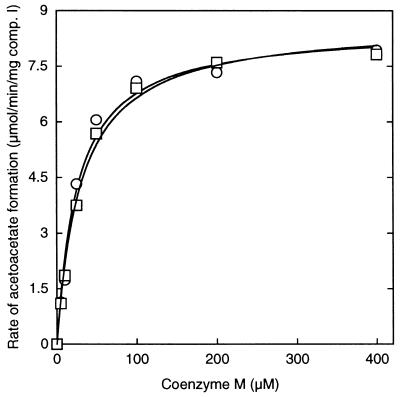
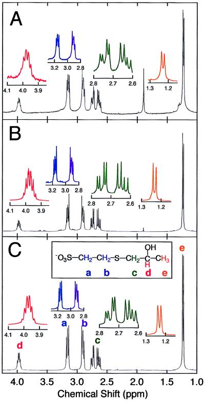
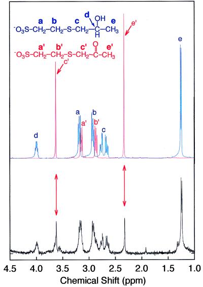
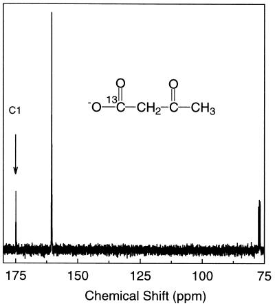
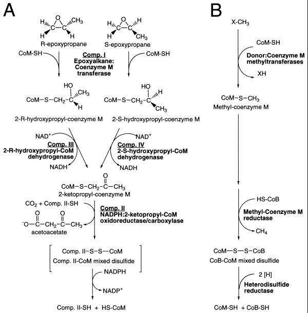
The bacterial metabolism of short-chain aliphatic alkenes occurs via oxidation to epoxyalkanes followed by carboxylation to beta-ketoacids. Epoxyalkane carboxylation requires four enzymes (components I-IV), NADPH, NAD(+), and a previously unidentified nucleophilic thiol. In the present work, coenzyme M (2-mercaptoethanesulfonic acid), a compound previously found only in the methanogenic Archaea where it serves as a methyl group carrier and activator, has been identified as the thiol and central cofactor of aliphatic epoxide carboxylation in the Gram-negative bacterium Xanthobacter strain Py2. Component I catalyzed the addition of coenzyme M to epoxypropane to form a beta-hydroxythioether, 2-(2-hydroxypropylthio)ethanesulfonate. Components III and IV catalyzed the NAD(+)-dependent stereoselective dehydrogenation of R- and S-enantiomers of 2-(2-hydroxypropylthio)ethanesulfonate to form 2-(2-ketopropylthio)ethanesulfonate. Component II catalyzed the NADPH-dependent cleavage and carboxylation of the beta-ketothioether to form acetoacetate and coenzyme M. These findings evince a newfound versatility for coenzyme M as a carrier and activator of alkyl groups longer in chain-length than methane, a function for coenzyme M in a catabolic pathway of hydrocarbon oxidation, and the presence of coenzyme M in the bacterial domain of the phylogenetic tree. These results serve to unify bacterial and Archaeal metabolism further and showcase diverse biological functions for an elegantly simple organic molecule.
Figures
Similar articles
-
Aliphatic epoxide carboxylation.Annu Rev Biochem. 2003;72:55-76. doi: 10.1146/annurev.biochem.72.121801.161820. Epub 2003 Jan 8. Annu Rev Biochem. 2003. PMID: 12524213 Review.
-
Two short-chain dehydrogenases confer stereoselectivity for enantiomers of epoxypropane in the multiprotein epoxide carboxylating systems of Xanthobacter strain Py2 and Nocardia corallina B276.Biochemistry. 1999 Jan 5;38(1):247-56. doi: 10.1021/bi982114h. Biochemistry. 1999. PMID: 9890905
-
Characterization of five catalytic activities associated with the NADPH:2-ketopropyl-coenzyme M [2-(2-ketopropylthio)ethanesulfonate] oxidoreductase/carboxylase of the Xanthobacter strain Py2 epoxide carboxylase system.Biochemistry. 2000 Feb 15;39(6):1294-304. doi: 10.1021/bi992282p. Biochemistry. 2000. PMID: 10684609
-
Evidence that a linear megaplasmid encodes enzymes of aliphatic alkene and epoxide metabolism and coenzyme M (2-mercaptoethanesulfonate) biosynthesis in Xanthobacter strain Py2.J Bacteriol. 2001 Apr;183(7):2172-7. doi: 10.1128/JB.183.7.2172-2177.2001. J Bacteriol. 2001. PMID: 11244054 Free PMC article.
-
Getting a handle on the role of coenzyme M in alkene metabolism.Microbiol Mol Biol Rev. 2008 Sep;72(3):445-56. doi: 10.1128/MMBR.00005-08. Microbiol Mol Biol Rev. 2008. PMID: 18772284 Free PMC article. Review.
Cited by
-
Poplar phyllosphere harbors disparate isoprene-degrading bacteria.Proc Natl Acad Sci U S A. 2018 Dec 18;115(51):13081-13086. doi: 10.1073/pnas.1812668115. Epub 2018 Nov 29. Proc Natl Acad Sci U S A. 2018. PMID: 30498029 Free PMC article.
-
Involvement of coenzyme M during aerobic biodegradation of vinyl chloride and ethene by Pseudomonas putida strain AJ and Ochrobactrum sp. strain TD.Appl Environ Microbiol. 2006 May;72(5):3756-8. doi: 10.1128/AEM.72.5.3756-3758.2006. Appl Environ Microbiol. 2006. PMID: 16672529 Free PMC article.
-
Estimation of methanogen biomass by quantitation of coenzyme M.Appl Environ Microbiol. 1999 Dec;65(12):5541-5. doi: 10.1128/AEM.65.12.5541-5545.1999. Appl Environ Microbiol. 1999. PMID: 10584015 Free PMC article.
-
Catalysis of methyl group transfers involving tetrahydrofolate and B(12).Vitam Horm. 2008;79:293-324. doi: 10.1016/S0083-6729(08)00410-X. Vitam Horm. 2008. PMID: 18804699 Free PMC article. Review.
-
Cytochrome P450 initiates degradation of cis-dichloroethene by Polaromonas sp. strain JS666.Appl Environ Microbiol. 2013 Apr;79(7):2263-72. doi: 10.1128/AEM.03445-12. Epub 2013 Jan 25. Appl Environ Microbiol. 2013. PMID: 23354711 Free PMC article.
References
-
- Sawada S, Totsuka T. Atmos Environ. 1986;20:821–832.
-
- Hartmans S, de Bont J A M, Harder W. FEMS Microbiol Rev. 1989;63:235–264. - PubMed
Publication types
MeSH terms
Substances
Grants and funding
LinkOut - more resources
Full Text Sources
Molecular Biology Databases
