. 1999 Jul 20;96(15):8443-8.
doi: 10.1073/pnas.96.15.8443.

Cell cycle regulation of Dfp1, an activator of the Hsk1 protein kinase

Affiliations

Cell cycle regulation of Dfp1, an activator of the Hsk1 protein kinase

G W Brown et al. Proc Natl Acad Sci U S A. .

Abstract

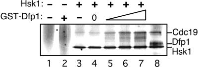
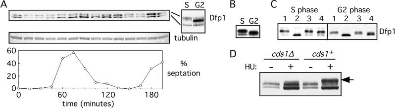
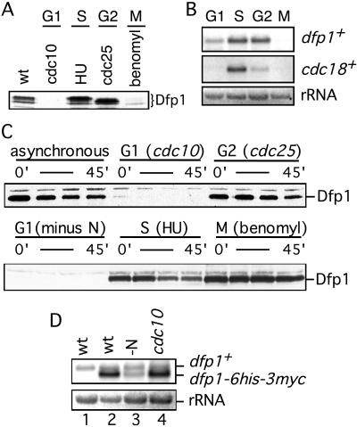
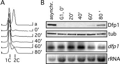
In fission yeast, the Hsk1 protein kinase is essential for the initiation of DNA replication. We have shown previously that Hsk1 forms a heterodimeric complex with the regulatory subunit, Dfp1. In this report we describe the further characterization of Dfp1. Reconstitution experiments with purified proteins indicate that Dfp1 is necessary and sufficient to activate Hsk1 phosphorylation of exogenous substrates, such as the Schizosaccharomyces pombe minichromosome maintenance protein Cdc19. The dfp1(+) gene is essential for viability of S. pombe, and depletion of the Dfp1 protein significantly delays the onset of S phase. Dfp1 is a phosphoprotein in vivo and becomes hyperphosphorylated when cells are blocked in S phase by treatment with the DNA synthesis inhibitor hydroxyurea. Hyperphosphorylation in S phase depends on the checkpoint kinase Cds1. The abundance of Dfp1 varies during progression through the cell cycle. The protein is absent when cells are arrested in G(1) phase. When cells are released into the cell cycle, Dfp1 appears suddenly at the G(1)/S transition, coincident with the initiation of DNA replication. The absence of Dfp1 before S phase is due largely, but not exclusively, to posttranscriptional regulation. We propose that cell cycle-regulated activation of Dfp1 expression at the G(1)/S transition results in activation of the Hsk1 protein kinase, which, in turn, leads to the initiation of DNA replication.


Figures

Figure 1
Figure 1
Activation of Hsk1 kinase by recombinant Dfp1. Increasing amounts (0, 10, 25, or 50 ng) of GST-Dfp1 were added to purified monomeric Hsk1 (≈5 ng, lanes 4–7). Proteins were incubated under kinase assay conditions. All reactions contained 100 ng of GST-Cdc19 substrate. Control reactions contained no added protein (lane 1), GST-Dfp1 (50 ng) alone (lane 2), GST (100 ng) with Hsk1 monomer (lane 3), or heterodimeric Hsk1⋅Dfp1 (lane 8).
Figure 2
Figure 2
Deletion of dfp1+ delays entry into S phase. (A) Tetrad analysis of the diploid dfp1+/dfp1ura4+ disruption strain GBY405. (B) Representative microcolonies from one tetrad from GBY405. Microcolonies (Upper Right and Lower Left) were uracil auxotrophs and, therefore, dfp1+. Germinated spores (Upper Left and Lower Right) did not give rise to colonies. (C) DNA contents from flow cytometry of germinating spores of GBY405. Spores were germinated in medium containing uracil (+ura) to permit germination of all spores or in medium lacking uracil (−ura) to allow germination of spores bearing the dfp1 disruption only. The positions of 1C and 2C DNA contents are indicated. (D) Depletion of Dfp1. Expression of dfp1 in the strain GBY429 was repressed by the addition of thiamine to 5 μg/ml at t = 0 hr. DNA content was measured by flow cytometry every hour after repression.
Figure 3
Figure 3
Expression and phosphorylation of Dfp1 during cell cycle progression. (A) A culture of the strain GBY398 was blocked in G2 and then released synchronously into the cell cycle. The culture was sampled every 15 min, and extracts were prepared. Dfp1 was detected by immunoblot analysis. The blot was reprobed for tubulin as a loading control. Synchrony was assessed by measuring the percentage of cells with a septum (% septation). The 60- (S) and 135-min (G2) samples are also shown side by side for comparison of the different Dfp1 isoforms. (B) Extracts were prepared from cells blocked in S phase (GBY397) or in G2 phase (GBY398) by using denaturing conditions. Proteins were immunoblotted, probing for Dfp1. (C) Native extracts were prepared from cells blocked in S or G2 phase. Hsk1 and associated Dfp1 were immunoprecipitated, and the immunoprecipitates were incubated with λ phosphatase (lanes 2), without λ phosphatase (lanes 3), or with λ phosphatase plus the inhibitor vanadate (lanes 4). The untreated controls are in lanes 1. Reaction products were subjected to immunoblot analysis, probing for Dfp1. Note that the fastest-migrating Dfp1 isoform seen in A and B is extracted inefficiently under native conditions. (D) The phosphorylation state of Dfp1 in S phase cells was analyzed in cds1+ or cds1Δ strains. Samples were taken from cultures arrested in S phase with hydroxyurea (+HU) or from asynchronous cultures (−HU). The position of the S phase Dfp1 phosphoisomer is indicated by the arrow.
Figure 4
Figure 4
Dfp1 expression is down-regulated during G1 phase. (A) Dfp1 levels in cells blocked in G1 (GBY401), S (GBY397), G2 (GBY398), or M (GBY397) phases were compared with levels in asynchronous culture (wt, GBY397) by using immunoblot analysis. The Dfp1 species are indicated. Equal loading of all lanes was confirmed by reprobing immunoblots with anti-tubulin antibody (not shown). (B) Total RNA was prepared from cells blocked as in A and fractionated on formaldehyde-agarose gels. A Northern blot was probed for dfp1+ mRNA and cdc18+ mRNA. The ethidium bromide-stained rRNA is shown as a control for equal loading. (C) Strains (GBY410, 411, 412) expressing dfp1–6his-3myc from the repressible nmt1 promoter were blocked at the indicated cell cycle positions. Expression of dfp1–6his-3myc was repressed by the addition of thiaminee, and Dfp1 protein levels at 0, 15, 30, and 45 min after repression was analyzed by immunoblotting. Equal loading of all lanes was confirmed by reprobing immunoblots with anti-tubulin antibody (not shown). (D) RNA was prepared from a logarithmically growing culture of wild-type cells (wt, lane 1), from GBY410 in logarithmically growing culture (wt, lane 2), from GBY410 cultured in the absence of nitrogen for 4 hr (-N, lane 3), or from GBY411 cultured at 36°C for 4 hr (cdc10, lane 4). A Northern blot of this RNA was probed with the dfp1+ ORF. The positions of the dfp1+ and dfp1–6his-3myc mRNAs are indicated. The ethidium bromide-stained rRNA is shown as a control for equal loading.
Figure 5
Figure 5
Expression of Dfp1 at the G1/S transition. Cells blocked in G1 (GBY401) were released into the cell cycle and sampled every 20 min. (A) DNA content was measured by flow cytometry. The positions of 1C and 2C DNA contents are indicated. (B) Dfp1 levels were analyzed by immunoblot analysis, probing with the anti-HA antibody 16B12 (Dfp1). The blot was reprobed with anti-tubulin antibodies as a loading control (tub). RNA levels were analyzed by Northern hybridization analysis, probing with the dfp1 ORF (dfp1). The ethidium bromide-stained rRNA is shown as a control for loading (rRNA).

Similar articles

Cited by

References

    1. Culotti J, Hartwell L H. Exp Cell Res. 1971;67:389–401. - PubMed
    1. Hollingsworth R E, Sclafani R A. Proc Natl Acad Sci USA. 1990;87:6272–6276. - PMC - PubMed
    1. Yoon H-J, Campbell J L. Proc Natl Acad Sci USA. 1991;88:3574–3578. - PMC - PubMed
    1. Masai H, Miyake T, Arai K. EMBO J. 1995;14:3094–3104. - PMC - PubMed
    1. Sato N, Arai K-I, Masai H. EMBO J. 1997;16:4340–4351. - PMC - PubMed

Publication types

LinkOut - more resources