

doi: 10.1073/pnas.96.15.8511.
Suppression of apoptosis signal-regulating kinase 1-induced cell death by 14-3-3 proteins
Affiliations
- PMID: 10411906
- PMCID: PMC17547
- DOI: 10.1073/pnas.96.15.8511
Item in Clipboard
Suppression of apoptosis signal-regulating kinase 1-induced cell death by 14-3-3 proteins
Proc Natl Acad Sci U S A.
.
Abstract
Apoptosis signal-regulating kinase 1 (ASK1) is a pivotal component of a signaling pathway induced by many death stimuli, including tumor necrosis factor alpha, Fas, and the anticancer drugs cisplatin and paclitaxel. Here we report that ASK1 proapoptotic activity is antagonized by association with 14-3-3 proteins. We found that ASK1 specifically bound 14-3-3 proteins via a site involving Ser-967 of ASK1. Interestingly, overexpression of 14-3-3 in HeLa cells blocked ASK1-induced apoptosis whereas disruption of the ASK1/14-3-3 interaction dramatically accelerated ASK1-induced cell death. Targeting of ASK1 by a 14-3-3-mediated survival pathway may provide a novel mechanism for the suppression of apoptosis.
Figures

Human ASK1 specifically binds 14-3-3 in vivo and in vitro. HeLa cells were transiently transfected with plasmids coding for HA-tagged ASK1wt, ASK1S967A, ASK1K709R, or the control CAB1 (Clan of ARF-Binder 1; A. L. Boman and R. A. Kahn, unpublished data). CAB1 is an ADP ribosylation factor-associated protein that does not bind 14-3-3. Forty-eight hours after transfection, cell lysates were prepared. (A) ASK1 immunocomplexes contain 14-3-3 proteins. ASK1 complexes were immunoprecipitated by using an anti-HA antibody and assayed for the presence of 14-3-3 by its ability to activate the ADP ribosyltransferase activity of ExoS. The 14-3-3-dependent ExoS activity was expressed as pmol of ADP ribose incorporated into the substrate per min per pmol of ExoS. Data shown are representative of three experiments. Error bars represent standard error (n = 3). (B) 14-3-3 immunocomplexes contain ASK1. Endogenous 14-3-3 proteins were isolated from the same HeLa cell lysates as in A by using anti-14-3-3 serum. The 14-3-3 IPs were blotted for HA-ASK1 by using an anti-HA antibody (Upper). Lower shows similar expression levels of the HA-tagged proteins in total cell lysates. (C) Interaction of ASK1 with 14-3-3ζ is disrupted by binding-site mutations of 14-3-3ζ. Immobilized His-tagged 14-3-3ζ or control β-gal proteins (5 μg each) were incubated with 35S-labeled ASK1. After washing, bound proteins were resolved by using SDS/PAGE, and ASK1 was revealed by autoradiography (Upper). Similar amounts of immoblized proteins were used as revealed by Coomassie blue staining (Lower). (D) Peptide ligands of 14-3-3 inhibit ASK1–14-3-3 interactions. Peptides were preincubated with immobilized His–14-3-3ζ (0.2 μg) before adding 35S-labeled ASK1. After washing, 14-3-3ζ-bound ASK1 was quantified by PhosphorImager. The percentage of ASK1 bound to 14-3-3 relative to peptide-free samples is plotted against increasing concentrations of the test peptides. Error bars represent standard error (n = 3).
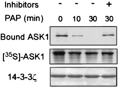
Interaction of ASK1 with 14-3-3 is phosphorylation-dependent. 35S-labeled ASK1 was treated with potato acid phosphatase (PAP) for the indicated times with or without phosphatase inhibitors, and then incubated with 14-3-3ζ-coated beads. After washing, 14-3-3ζ-bound ASK1 was subjected to SDS/PAGE and autoradiography (Top). Middle shows phosphatase-treated ASK1. The amount of 14-3-3ζ in the beads was revealed by Coomassie blue staining (Bottom).
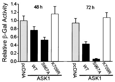
The mutation S967A accelerates ASK1-induced cell death. HeLa cells were cotransfected with expression vectors for HA-tagged ASK1wt, ASK1S967A, or ASK1K709R and a vector for β-gal. At the indicated times, attached cells were harvested and assayed for β-gal activity. This activity reflects the viability of transfected cells only, because dead cells will detach from the culture dish. The control sample was transfected with an amount of pcDNA3 equal to the amount of ASK1 DNA. Results shown are mean ± standard error (n = 3) and are representative of three independent experiments.

14-3-3 binding inhibits ASK1-induced apoptosis. HeLa cells were cotransfected with an eGFP expression vector and test plasmids as indicated. Thirty-six hours posttransfection, cells were subjected to analysis. (A) ASK1-transfected cells show nuclear morphology characteristic of apoptosis, as determined by DAPI staining and fluorescence microscopy. Representative cells with apoptotic nuclei are indicated by arrows. (B) ASK1-induced apoptosis is modulated by 14-3-3 binding. Top shows the quantitation of apoptotic cell death by using nuclear morphology. The ratio of apoptotic transfected cells to total counted transfected cells is indicated above each bar. Middle shows expression levels of HA–ASK1 and Flag-tagged 14-3-3ζ proteins as determined by immunoblot. Bottom depicts the DNA integrity of transfected cells. Total DNA from the transfected samples was isolated, and an equal amount of DNA from each was separated on a 1.5% agarose gel.
References
-
- Jacobson M D, Weil M, Raff M C. Cell. 1997;88:347–354. - PubMed
-
- Rudin C M, Thompson C B. Annu Rev Med. 1997;48:267–281. - PubMed
-
- Ellis R E, Yuan J Y, Horvitz H R. Annu Rev Cell Biol. 1991;7:663–698. - PubMed
-
- Cryns V, Yuan J. Genes Dev. 1998;12:1551–1570. - PubMed
-
- Ashkenazi A, Dixit V M. Science. 1998;281:1305–1308. - PubMed
Publication types
MeSH terms
Substances
Grants and funding
LinkOut - more resources
Full Text Sources
Other Literature Sources
Molecular Biology Databases
Research Materials
Miscellaneous
