

Cytotoxic T lymphocyte antigen-4 (CTLA-4) regulates primary and secondary peptide-specific CD4(+) T cell responses
- PMID: 10411922
- PMCID: PMC17563
- DOI: 10.1073/pnas.96.15.8603
Cytotoxic T lymphocyte antigen-4 (CTLA-4) regulates primary and secondary peptide-specific CD4(+) T cell responses
Abstract
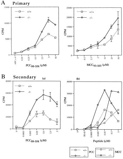
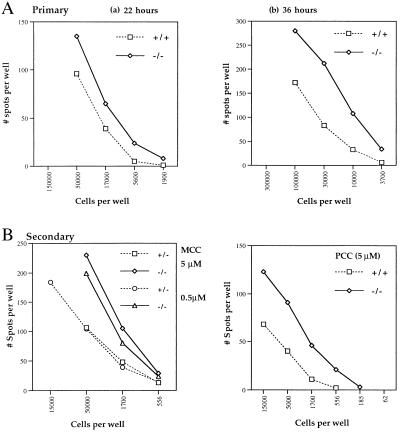
CTLA-4-deficient mice develop a fatal lymphoproliferative disorder, characterized by polyclonal expansion of peripheral lymphocytes. To examine the effect of restricting the CD4(+) TCR repertoire on the phenotype of CTLA-4-deficient mice and to assess the influence of CTLA-4 on peptide-specific CD4(+) T cell responses in vitro, an MHC class II-restricted T cell receptor (AND TCR) transgene was introduced into the CTLA-4(-/-) animals. The expression of the AND TCR transgene by CD4(+) T cells delays but does not prevent the lymphoproliferation in the CTLA-4(-/-) mice. The CD4(+) T cells become preferentially activated and expand. Interestingly, young AND TCR(+) CTLA-4(-/-) mice carrying a null mutation in the rag-1 gene remain healthy and the T cells maintain a naive phenotype until later in life. We demonstrate that CTLA-4 regulates the peptide-specific proliferative response generated by naive and previously activated AND TCR(+) RAG(-/-) T cells in vitro. The absence of CTLA-4 also augments the responder frequency of cytokine-secreting AND TCR(+) RAG(-/-) T cells. These results demonstrate that CTLA-4 is a key regulator of peptide-specific CD4(+) T cell responses and support the model that CTLA-4 plays a differential role in maintaining T cell homeostasis of CD4(+) vs. CD8(+) T cells.
Figures
Similar articles
-
Secondary but not primary T cell responses are enhanced in CTLA-4-deficient CD8+ T cells.Eur J Immunol. 1998 Oct;28(10):3137-43. doi: 10.1002/(SICI)1521-4141(199810)28:10<3137::AID-IMMU3137>3.0.CO;2-X. Eur J Immunol. 1998. PMID: 9808182
-
Lymphoproliferation in CTLA-4-deficient mice is mediated by costimulation-dependent activation of CD4+ T cells.Immunity. 1997 Dec;7(6):885-95. doi: 10.1016/s1074-7613(00)80406-9. Immunity. 1997. PMID: 9430233
-
Blockade of T cell activation using a surface-linked single-chain antibody to CTLA-4 (CD152).J Immunol. 2000 May 1;164(9):4433-42. doi: 10.4049/jimmunol.164.9.4433. J Immunol. 2000. PMID: 10779742
-
The role of CTLA-4 in the regulation of T cell immune responses.Immunol Cell Biol. 1999 Feb;77(1):1-10. doi: 10.1046/j.1440-1711.1999.00795.x. Immunol Cell Biol. 1999. PMID: 10101680 Review.
-
Function and regulation of memory CD4 T cells.Immunol Res. 1999;19(2-3):127-41. doi: 10.1007/BF02786482. Immunol Res. 1999. PMID: 10493168 Review.
Cited by
-
Induction of cytotoxic T lymphocyte antigen 4 (CTLA-4) restricts clonal expansion of helper T cells.J Exp Med. 2001 Oct 1;194(7):893-902. doi: 10.1084/jem.194.7.893. J Exp Med. 2001. PMID: 11581312 Free PMC article.
-
Intracellular concentrations of Ca(2+) modulate the strength of signal and alter the outcomes of cytotoxic T-lymphocyte antigen-4 (CD152)-CD80/CD86 interactions in CD4(+) T lymphocytes.Immunology. 2009 Mar;126(3):363-77. doi: 10.1111/j.1365-2567.2008.02902.x. Epub 2008 Aug 14. Immunology. 2009. PMID: 18710402 Free PMC article.
-
Role of Immune Cells and Immunotherapy in Multiple Myeloma.Life (Basel). 2024 Apr 1;14(4):461. doi: 10.3390/life14040461. Life (Basel). 2024. PMID: 38672732 Free PMC article. Review.
-
At the bench: preclinical rationale for CTLA-4 and PD-1 blockade as cancer immunotherapy.J Leukoc Biol. 2013 Jul;94(1):25-39. doi: 10.1189/jlb.1212621. Epub 2013 Apr 26. J Leukoc Biol. 2013. PMID: 23625198 Free PMC article. Review.
-
Immune Checkpoints, a Novel Class of Therapeutic Targets for Autoimmune Diseases.Front Immunol. 2021 Apr 21;12:645699. doi: 10.3389/fimmu.2021.645699. eCollection 2021. Front Immunol. 2021. PMID: 33968036 Free PMC article. Review.
References
-
- Lenschow D J, Walunas T L, Bluestone J A. Annu Rev Immunol. 1996;14:233–258. - PubMed
-
- Wülfing C, Davis M M. Science. 1998;282:2266–2269. - PubMed
-
- Viola A, Schroeder S, Sakakibara Y, Lanzavecchia A. Science. 1999;283:680–682. - PubMed
-
- Chambers C A, Allison J P. Curr Opin Cell Biol. 1999;11:203–210. - PubMed
-
- Marengére L E M, Waterhouse P, Duncan G S, Mittrucker H-W, Feng G-S, Mak T W. Science. 1996;272:1170–1173. - PubMed
Publication types
MeSH terms
Substances
Grants and funding
LinkOut - more resources
Full Text Sources
Other Literature Sources
Molecular Biology Databases
Research Materials
