

Induction of hyporesponsiveness to intact foreign protein via retroviral-mediated gene expression: the IgG scaffold is important for induction and maintenance of immune hyporesponsiveness
- PMID: 10411923
- PMCID: PMC17564
- DOI: 10.1073/pnas.96.15.8609
Induction of hyporesponsiveness to intact foreign protein via retroviral-mediated gene expression: the IgG scaffold is important for induction and maintenance of immune hyporesponsiveness
Abstract
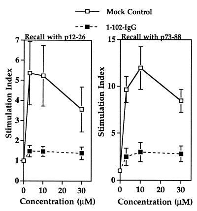
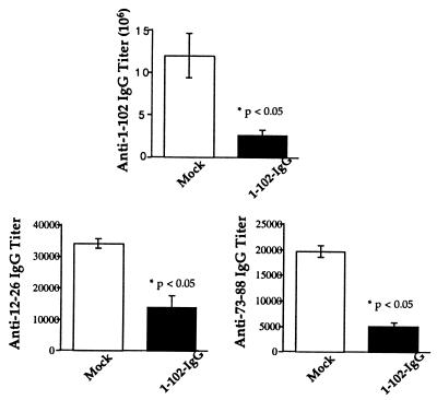
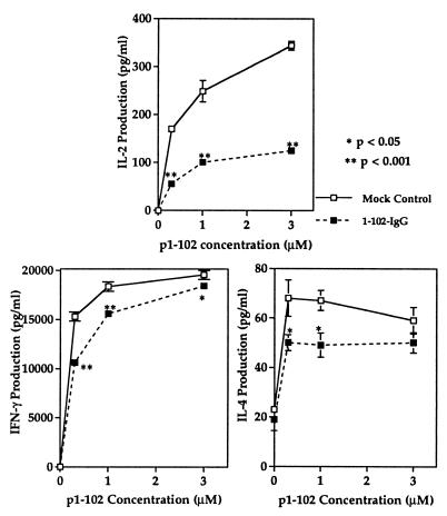
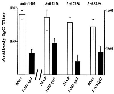
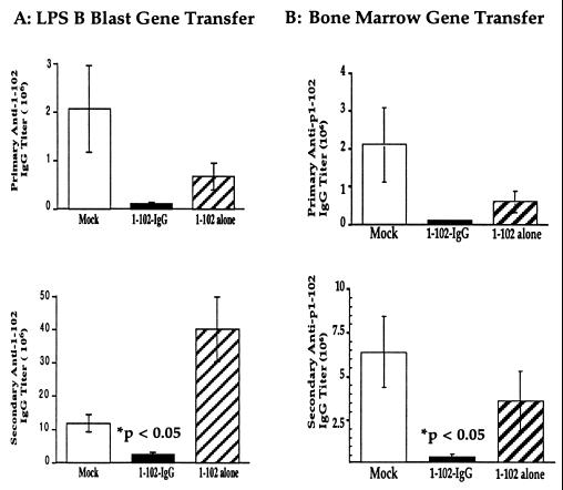
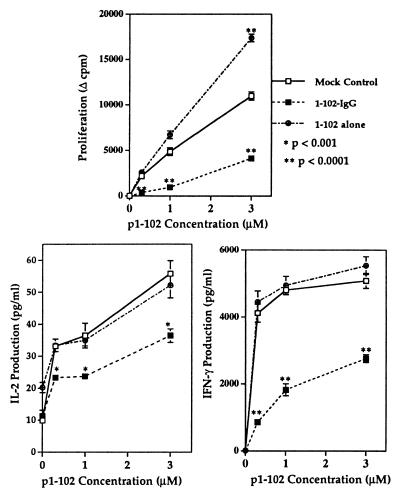
IgG molecules can be highly tolerogenic carriers for associated antigens. Previously, we reported that recipients of bone marrow or lipopolysaccharide-stimulated B-cell blasts, both of which were retrovirally gene-transferred with an immunodominant peptide in-frame with the variable region of a murine IgG heavy chain, were rendered profoundly unresponsive to that epitope. To further investigate whether tolerance to larger molecules can be achieved via this approach and whether the IgG scaffold is important for induction and maintenance of immunological tolerance, we engineered two retroviral constructs encoding the cI lambda repressor (MBAE-1-102 and MBAE-1-102-IgG) for gene transfer. Our results show that recipients of bone marrow or peripheral B cells, transduced with the MBAE-1-102-IgG recombinant, are hyporesponsive to p1-102. In addition, the self-IgG scaffold enhanced the induction and maintenance of such an immune hyporesponsiveness. Thus, our studies demonstrate that in vivo-expressed IgG heavy chain fusion protein can be processed and presented on the appropriate MHC class II, resulting in hyporesponsiveness to that antigen and offering an additional therapeutic approach to autoimmune diseases.
Figures
Similar articles
-
Tolerance induction via a B-cell delivered gene therapy-based protocol: optimization and role of the Ig scaffold.Cell Immunol. 2005 May;235(1):12-20. doi: 10.1016/j.cellimm.2005.06.007. Epub 2005 Aug 10. Cell Immunol. 2005. PMID: 16098495
-
Genetically transferred central and peripheral immune tolerance via retroviral-mediated expression of immunogenic epitopes in hematopoietic progenitors or peripheral B lymphocytes.Mol Med. 1997 Mar;3(3):212-24. Mol Med. 1997. PMID: 9100227 Free PMC article.
-
Mechanisms of tolerance induction by a gene-transferred peptide-IgG fusion protein expressed in B lineage cells.J Immunol. 2000 Nov 15;165(10):5631-6. doi: 10.4049/jimmunol.165.10.5631. J Immunol. 2000. PMID: 11067919
-
Tolerance induction by gene transfer to lymphocytes.Curr Gene Ther. 2007 Oct;7(5):369-80. doi: 10.2174/156652307782151443. Curr Gene Ther. 2007. PMID: 17979683 Review.
-
Gene therapy for immunologic tolerance: using bone marrow-derived cells to treat autoimmunity and hemophilia.Curr Stem Cell Res Ther. 2011 Mar;6(1):38-43. doi: 10.2174/157488811794480753. Curr Stem Cell Res Ther. 2011. PMID: 20955157 Review.
Cited by
-
Progress toward inducing immunologic tolerance to factor VIII.Blood. 2013 May 30;121(22):4449-56. doi: 10.1182/blood-2013-01-478669. Epub 2013 Mar 15. Blood. 2013. PMID: 23502223 Free PMC article. Review.
-
B-Cell Gene Therapy for Tolerance Induction: Host but Not Donor B-Cell Derived IL-10 is Necessary for Tolerance.Front Microbiol. 2011 Jul 15;2:154. doi: 10.3389/fmicb.2011.00154. eCollection 2011. Front Microbiol. 2011. PMID: 21811487 Free PMC article.
-
Retroviral gene therapy with an immunoglobulin-antigen fusion construct protects from experimental autoimmune uveitis.J Clin Invest. 2000 Jul;106(2):245-52. doi: 10.1172/JCI9168. J Clin Invest. 2000. PMID: 10903340 Free PMC article.
-
B cells "transduced" with TAT-fusion proteins can induce tolerance and protect mice from diabetes and EAE.Clin Immunol. 2011 Sep;140(3):260-7. doi: 10.1016/j.clim.2011.04.009. Epub 2011 Apr 20. Clin Immunol. 2011. PMID: 21546313 Free PMC article.
-
Inhibitors - cellular aspects and novel approaches for tolerance.Haemophilia. 2014 May;20 Suppl 4(0 4):80-6. doi: 10.1111/hae.12407. Haemophilia. 2014. PMID: 24762281 Free PMC article. Review.
References
-
- Schönrich G, Momburg F, Malissen M, Schmitt-Verhulst A-M, Malissen B, Hämmerling G J, Arnold B. Int Immunol. 1992;4:581–590. - PubMed
-
- Tisch R, Yang X-D, Singer S M, Liblau R S, Fugger L, McDevitt H O. Nature (London) 1993;366:72–75. - PubMed
-
- Higgins P J, Weiner H. J Immunol. 1988;140:440–445. - PubMed
-
- Critchfield J M, Racke M K, Zuniger-Pflucker J C, Cannella B, Raine C S, Goverman J, Lenardo M J. Science. 1994;263:1139–1143. - PubMed
Publication types
MeSH terms
Substances
Grants and funding
LinkOut - more resources
Full Text Sources
Other Literature Sources
Research Materials
