. 1999 Jul 20;96(15):8627-32.
doi: 10.1073/pnas.96.15.8627.

Dipeptidyl peptidase I is required for the processing and activation of granzymes A and B in vivo

Affiliations

Dipeptidyl peptidase I is required for the processing and activation of granzymes A and B in vivo

C T Pham et al. Proc Natl Acad Sci U S A. .

Abstract

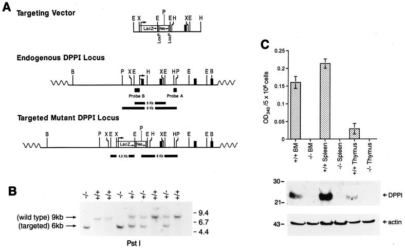
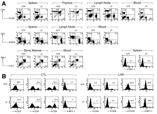
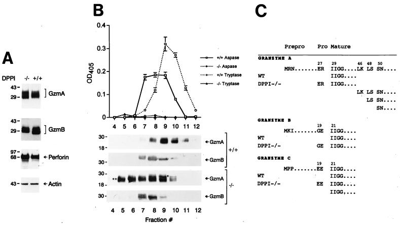
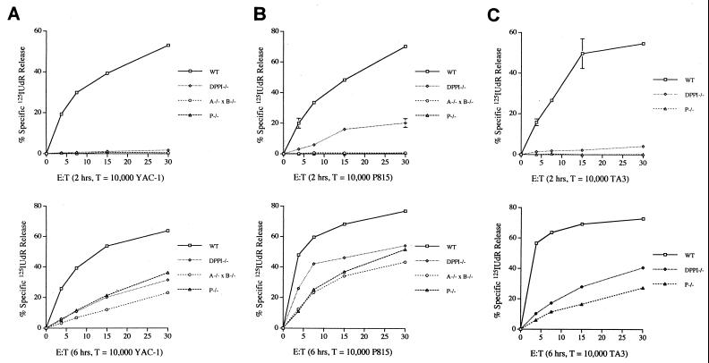
Dipeptidyl peptidase I (DPPI) is a lysosomal cysteine protease that has been implicated in the processing of granzymes, which are neutral serine proteases exclusively expressed in the granules of activated cytotoxic lymphocytes. In this report, we show that cytotoxic lymphocytes derived from DPPI-/- mice contain normal amounts of granzymes A and B, but these molecules retain their prodipeptide domains and are inactive. Cytotoxic assays with DPPI-/- effector cells reveal severe defects in the induction of target cell apoptosis (as measured by [(125)I]UdR release) at both early and late time points; this defect is comparable to that detected in perforin-/- or granzyme A-/- x B-/- cytotoxic lymphocytes. DPPI therefore plays an essential role in the in vivo processing and activation of granzymes A and B, which are required for cytotoxic lymphocyte granule-mediated apoptosis.


Figures

Figure 1
Figure 1
Targeting of the DPPI gene. (A) Creation of the targeting vector. The DPPI locus is shown in the Middle and the targeting construct is depicted at Top. The locations and transcriptional orientations of the β-galactosidase (LacZ) and PGK-Neo cassettes are indicated. The targeting construct was designed to replace exon 1 and part of intron 1 with the selectable marker cassette. The locations of the external (probe A) and internal (probe B) probes used to detect homologous recombination are shown. (B) Southern blot analysis of tail DNA from wild-type, DPPI+/−, and DPPI−/− animals. DNA was digested with PstI and hybridized with probe A. The wild-type allele is found within a 9-kb fragment; the targeted allele is reduced to 6 kb because of an internal PstI site in the PGK-Neo cassette. (C) DPPI activity in wild-type and DPPI−/− tissues. Freshly isolated bone marrow cells, splenocytes, and thymocytes were lysed and assayed for DPPI activity as determined by the hydrolysis of Gly-Phe-β-naphthylamide. Activity was defined as the OD at 405 nm for 5 × 10⁶ cells. Results represent the mean of duplicate determinations. Equal amounts of total lysates were also fractionated on 10% SDS/PAGE gels and immunoblotted with a specific rabbit anti-mouse DPPI antibody. A β-actin antibody was used to control for protein content and loading.
Figure 2
Figure 2
Normal immune system development and cytotoxic lymphocyte activation in DPPI mutant mice. (A) Flow cytometric analyses of lymphoid and myeloid cells in wild-type and DPPI−/− mice. Cells harvested from spleen, thymus, mesenteric lymph nodes, and peripheral blood were analyzed for the expression of the indicated surface markers. The percentages of cells expressing CD3, CD4, CD8, B220, and NK1.1 were similar for all mice. Note that there was a moderate increase in the Mac-1+/Gr-1+ in the example shown here; 3 of 11 tested mice had a similar finding. (B) Normal proliferation and activation of DPPI−/− splenocytes in primary MLR and LAK activation. Splenocytes from wild-type and DPPI−/− animals were activated in a 5-day mixed lymphocyte reaction culture or in the presence of high-dose IL-2 to generate LAK cells. The expansion of the CD8+ CTL and NK1.1+ LAK cell populations were essentially equivalent in DPPI+/+ and −/− splenocytes.
Figure 3
Figure 3
Lack of granzyme processing and activity in LAK cells derived from DPPI−/− mice. (A) Granzyme A and B levels are normal in DPPI−/− LAK cells. Equal amounts of protein derived from wild type DPPI−/− LAK cells were analyzed by Western blotting by using polyclonal antibodies directed against mouse granzymes A and B and mAbs against mouse perforin and β-actin. The specificities of these antibodies have been determined previously (see Materials And Methods). (B) Granzyme A and B activities in LAK cell lysates. Wild-type and DPPI−/− LAK cell lysates were fractionated on a cationic exchange column by using a linear 0.25–2 M NaCl gradient, and each fraction was analyzed for aspase and tryptase activity. These same fractions were separated by SDS/PAGE, transferred to nitrocellulose, and incubated with anti-granzyme A and B antibodies to confirm the abundance and sizes of the proteins. (C) Granzymes are abnormally processed in DPPI−/− LAK cells. Column fractions from B were fractionated by using nonreducing SDS/10% PAGE and transferred to poly(vinylidene difluoride) membranes; specific bands were excised, eluted, and subjected to microsequencing to determine the N termini. These results were confirmed in two independent experiments.
Figure 4
Figure 4
Defective [125I]UdR release exhibited by DPPI−/− CTL and LAK cells. Standard cytotoxicity assays of wild-type (□), DPPI−/− (◊), granzyme A−/− × B−/− (○), and perforin−/− (▵) LAK cells tested against YAC-1 target cells at increasing effector/target (E:T) ratios (Left); CTL were tested against TA3 and P815 target cells (Center and Right). Release of [125I]UdR is measured after 2 (Upper) and 6 (Lower) hr. The results are represented as mean ± SEM of duplicate samples. This experiment was repeated three times with similar results. Note that DPPI−/− LAK and CTL have an early defect in [125I]UdR release that is due to the loss of active granzyme B, and a persistent defect at 6 hr that is caused by the loss of active granzyme A.

Comment in

Similar articles

Cited by

References

    1. Henkart P A. Immunity. 1994;1:343–346. - PubMed
    1. Masson D, Tschopp J. J Biol Chem. 1985;260:9069–9072. - PubMed
    1. Podack E R, Young J D, Cohn Z A. Proc Natl Acad Sci USA. 1985;82:8629–8633. - PMC - PubMed
    1. Liu C-C, Walsh C M, Young J D. Immunol Today. 1995;16:194–201. - PubMed
    1. Shi L, Mai S, Israels S, Browne K, Trapani J A, Greenberg A H. J Exp Med. 1997;185:855–866. - PMC - PubMed

Publication types

MeSH terms

LinkOut - more resources