. 1999 Jul 20;96(15):8645-50.
doi: 10.1073/pnas.96.15.8645.

Defective CTLA-4 cycling pathway in Chediak-Higashi syndrome: a possible mechanism for deregulation of T lymphocyte activation

Affiliations

Defective CTLA-4 cycling pathway in Chediak-Higashi syndrome: a possible mechanism for deregulation of T lymphocyte activation

F J Barrat et al. Proc Natl Acad Sci U S A. .

Abstract

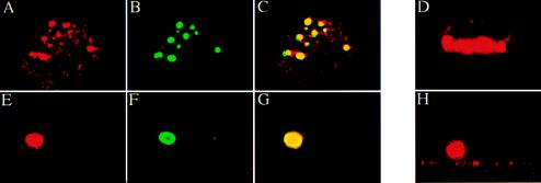
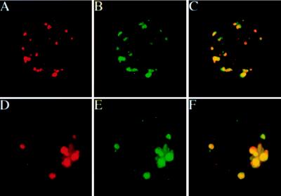
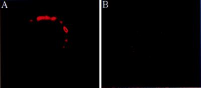
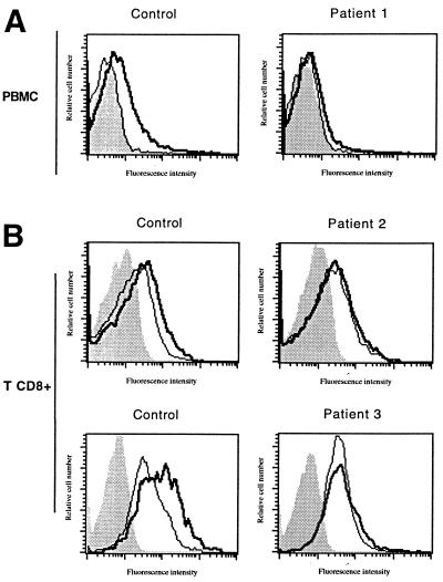
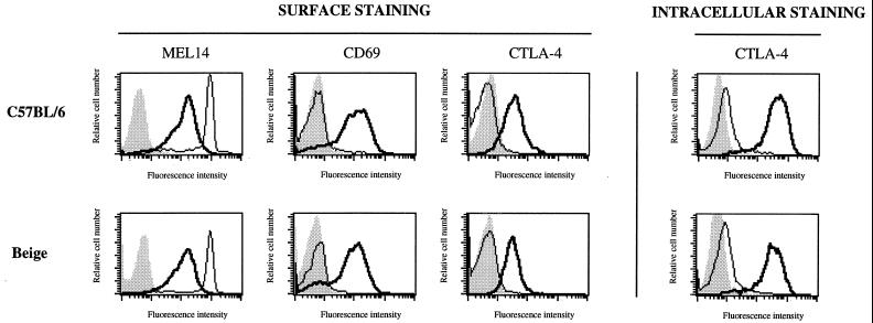
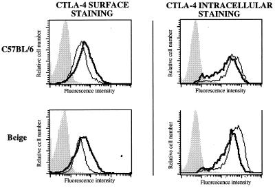
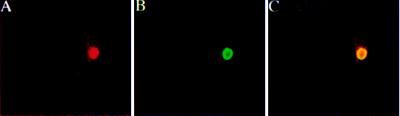
Cytotoxic T lymphocyte-associated antigen 4 (CTLA-4, also known as CD152) has been shown to play a major role in the regulation of T cell activation. Its membrane expression is highly regulated by endocytosis and trafficking through the secretory lysosome pathway. Chediak-Higashi syndrome (CHS) is an inherited disorder caused by mutations in the lysosomal trafficking regulator gene, LYST. It results in defective membrane targeting of the proteins present in secretory lysosomes, and it is associated with a variety of features, including a lymphoproliferative syndrome with hemophagocytosis. The murine equivalent of CHS, beige mice, present similar characteristics but do not develop the lymphoproliferative syndrome. We show herein that CTLA-4 is present in enlarged, abnormal vesicles in CHS T cells and is not properly expressed at the cell surface after T cell activation, whereas its surface expression is not impaired. It is therefore proposed that the defective surface expression of CTLA-4 by CHS T cells is involved in the generation of lymphoproliferative disease. This observation may provide insight into the role of CTLA-4 in humans.


Figures

Figure 1
Figure 1
Detection of CTLA-4 in perforin-containing vesicles and polarization toward sites of T cell activation. PMBC from control (A–D) and CHS patient 1 (E–H) were activated by incubation on glass coverslips coated with anti-CD3 and anti-CD28 mAbs for 48 h. They were fixed, permeabilized, and stained for CTLA-4 (A, D, E, H) (detected by Texas red-coupled goat anti-mouse IgG2a) and FITC-coupled anti-perforin (B, F) mAbs. Immunofluorescence was examined by confocal microscopy. The overlapping distribution is shown in yellow (C, G). Vertical (X, Z) optical sections are shown (D, H). Similar results were observed with patient 2 and patient 3.
Figure 2
Figure 2
Intracellular expression pattern of CTLA-4 in activated T cells of C57BL/6 and beige mice. T cells isolated from the lymph nodes of C57BL/6 (A–C) and beige (D–F) mice were activated for 48 h with an anti-CD3ɛ mAb (10 μg/ml), fixed, permeabilized, and stained for CTLA-4 (A, D) and FITC-coupled anti-Lamp-1 (B, E) mAbs. The overlapping distribution between CTLA-4 and Lamp-1 is shown in yellow (C, F).
Figure 3
Figure 3
CTLA-4 surface expression on activated T cells from CHS patient 1 and control T cells. T cells from control (A) and CHS patient 1 (B) were activated by incubation on glass coverslips coated with anti-CD3 and anti-CD28 mAb for 48 h. They were stained for CTLA-4 and then fixed to detect the surface expression of CTLA-4. Confocal analysis showed that CTLA-4 molecules were polarized at the cell surface in the control, whereas they were undetectable in cells from CHS patient 1. After activation, more than 90% of patient and control cells were CD69+.
Figure 4
Figure 4
CTLA-4 surface expression on CHS and control cells after allogeneic activation. (A) PBMCs isolated from patient 1 with lymphoproliferative syndrome and control PBMCs were stained for CTLA-4 (thick lines) after 72 h of allogeneic cell stimulation and pervanadate treatment. More than 90% of the cells from patient 1 and the control were CD69+. (B) PBMCs isolated from patients 2 and 3 and control PBMCs were co-stained for CD8 and CTLA-4 (thick lines). In both patients and control, more than 75% of the CD8+ T cells expressed CD69 and HLA-DR after activation. Control staining was carried out with an isotype-matched irrelevant mAb (thin lines). Shaded areas represent unstained cells.
Figure 5
Figure 5
CTLA-4 surface and intracellular expression by T cells from C57BL/6 and beige mice. (Left) T cells isolated from the lymph nodes of C57BL/6 and beige mice were activated for 48 h with an anti-CD3ɛ (10 μg/ml) and stained for the CD62L antigen of the L-selectin (Mel14), the activation marker CD69, and the CTLA-4 antigen before (thin lines) and after activation (thick lines). (Right) Cells were also stained for intracellular CTLA-4 before (thin lines) and after (thick lines) activation. Shaded areas represent unstained cells.
Figure 6
Figure 6
CTLA-4 surface and intracellular expression of pervanadate-treated T cells from C57BL/6 and beige mice. T cells isolated from the lymph nodes of C57BL/6 and beige mice were stained for surface and intracellular CTLA-4 after being activated for 96 h with γ-irradiated (2000 rads) spleen cells prepared from DBA/2 mice. Cells were incubated with (thick lines) or without (thin lines) pervanadate.
Figure 7
Figure 7
Intracellular CTLA-4 expression in CHS T cells before in vitro cell activation. Nonactivated T cells from CHS patient 2 were fixed, permeabilized, and stained as in Fig. 1 for CTLA-4 (A) and perforin (B) and examined by confocal microscopy. There was no CTLA-4 staining in control cells (not shown). Staining in cells from patient 2 was associated with perforin-containing vesicles (C). The same results were obtained when T cells from patient 1 and patient 3 were used (not shown).

Similar articles

Cited by

References

    1. Thompson C B, Allison J P. Immunity. 1997;7:445–450. - PubMed
    1. Bluestone J A. J Immunol. 1997;158:1989–1993. - PubMed
    1. Harding F A, McArthur J G, Gross J A, Raulet D H, Allison J P. Nature (London) 1992;356:607–609. - PubMed
    1. Shahinian A, Pfeffer K, Lee K P, Kundig T M, Kishihara K, Wakeham A, Kawai K, Ohashi P S, Thompson C B, Mak T W. Science. 1993;261:609–612. - PubMed
    1. Green J M, Noel P J, Sperling A I, Walunas T L, Gray G S, Bluestone J A, Thompson C B. Immunity. 1994;1:501–508. - PubMed

Publication types

MeSH terms

LinkOut - more resources