. 1999 Jul 20;96(15):8711-5.
doi: 10.1073/pnas.96.15.8711.

Cancer dormancy and cell signaling: induction of p21(waf1) initiated by membrane IgM engagement increases survival of B lymphoma cells

Affiliations

Cancer dormancy and cell signaling: induction of p21(waf1) initiated by membrane IgM engagement increases survival of B lymphoma cells

R Marches et al. Proc Natl Acad Sci U S A. .

Abstract

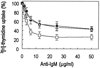
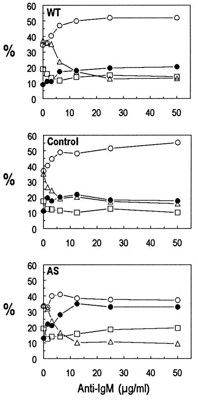
The p21(WAF1) (p21) cyclin-dependent kinase inhibitor plays a major role in regulating cell cycle arrest. It was recently reported that the p53-independent elevation of p21 protein levels is essential in mediating the G(1) arrest resulting from signal transduction events initiated by the crosslinking of membrane IgM on Daudi Burkitt lymphoma cells. Although the role of p21 in cell cycle regulation is well documented, there is little information concerning its role in antibody-mediated apoptosis. In the present study, we examined the involvement of p21 in the regulation of apoptosis by suppressing its induction in anti-IgM-treated Daudi cells through a p21 antisense expression construct approach. Reduction in induced p21 protein levels resulted in diminished G(1) arrest and increased apoptosis. The increased susceptibility to anti-IgM-mediated apoptosis was associated with increased caspase-3-like activity and poly-(ADP)ribose polymerase cleavage. These data suggest that p21 may directly interfere with the caspase cascade, thus playing a dual role in regulating both cell cycle progression and apoptosis.


Figures

Figure 1
Figure 1
The level of p21 protein in antisense p21-transfected Daudi cells after treatment with anti-IgM. Equal amounts (50 μg of total protein) of cell lysates from WT, control, and AS cells treated or untreated for 24 hours with 20 μg/ml anti-IgM were electrophoresed on a 12% SDS/PAGE gel and were transferred to poly(vinylidene difluoride) membranes. The p21 protein in each sample was detected with 1 μg/ml anti-p21 mAb followed by enhanced chemiluminescence detection. Values represent the relative induction of p21 as normalized to p21 protein levels in untreated WT cells as detected by densitometric scanning of the bands. A representative experiment of three is shown.
Figure 2
Figure 2
Dose-dependent inhibition of DNA synthesis in Daudi cells by anti-IgM. AS (-□-), WT (-●-), or control transfected (-▵-) Daudi cells (5 × 10⁴) were incubated at 37°C for 24 hours with the indicated concentrations of anti-IgM. DNA synthesis was assessed by measuring the level of 3H-thymidine incorporation. Values represent the average of four independent experiments.
Figure 3
Figure 3
Effect of anti-IgM on cell cycle status and viability in antisense p21-transfected Daudi cells. Daudi cells (1 × 10⁶/ml) were incubated for 24 hours with the indicated concentrations of anti-IgM, after which the cells were stained with 7-AAD and Hoechst. The percentages of apoptotic (dead) (-●-) and viable cells in G1 (-○-), S-phase (-▵-), and G2-M (-□-) were determined by flow cytometric analysis of the DNA content and membrane permeability. A representative experiment of three is shown.
Figure 4
Figure 4
Expression of mIgM on transfected Daudi cells. Cell surface expression of IgM was analyzed by flow cytometry of cells that were stained directly with FITC-conjugated IgG1 anti-human μ mAb (HB57). A total of 5 × 10⁵ cells were stained for each sample with a preliminary-determined optimal concentration of mAb. The thin line indicates the background staining with a FITC-conjugated IgG1 anti-HER2 control mAb. A representative experiment of three is shown.
Figure 5
Figure 5
Proteolytic cleavage of PARP and caspase activity in Daudi cells after treatment for 24 h with 20 μg/ml anti-IgM. Fifty micrograms of protein of total cell lysate per lane were separated by 10% SDS/PAGE. Immunodetection of PARP was carried out by using a 1:2,000 dilution of a Rb anti-PARP serum (Upper). Caspase-3-like protease activity was detected with ApoAlert CPP32 protease Assay kit (Lower). A representative experiment of three is shown.

References

    1. Page D, DeFranco A. Mol Cell Biol. 1990;10:3003–3012. - PMC - PubMed
    1. Uhr J W, Tucker T, May R D, Siu H, Vitetta E S. Cancer Res. 1991;51:5045S–5053S. - PubMed
    1. Yefenof E, Picker L J, Scheuermann R H, Tucker T F, Vitetta E S, Uhr J W. Proc Natl Acad Sci USA. 1993;90:1829–1833. - PMC - PubMed
    1. Racila E, Scheuermann R H, Picker L J, Yefenof E, Tucker T, Chang W, Marches R, Street N E, Vitetta E S, Uhr J W. J Exp Med. 1995;181:1539–1550. - PMC - PubMed
    1. Marches R, Racila E, Tucker T F, Picker L, Mongini P, Hsueh R, Vitetta E S, Scheuermann R H, Uhr J W. Ther Immunol. 1996;2:125–136. - PubMed

Publication types

MeSH terms