Review
. 1999 Aug;83(8):980-6.
doi: 10.1136/bjo.83.8.980.

Ganglion cell death in glaucoma: what do we really know?

Affiliations
Review

Ganglion cell death in glaucoma: what do we really know?

N N Osborne et al. Br J Ophthalmol. 1999 Aug.
No abstract available


Figures

Figure 1
Figure 1
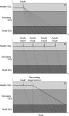
Three hypothetical views of how retinal ganglion cell death may occur in glaucoma. In (A), all ganglion cells are initiated to die at more or less the same time but individual cells die at variable rates. In (B), a series of acute insults occurs at different periods and this leads to the death of subsets of ganglion cells all dying at similar rates. In (C), an acute insult leads to the death of a subset of ganglion cells: the other ganglion cells then die because of secondary degeneration rather than because of a defined insult, at variable rates.
Figure 2
Figure 2
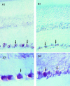
Immunohistochemical staining showing the presence of brain derived neurotrophic growth factor (BDNF) (A) and the TrkB receptor (B) in the retina. Clearly the immunofluorescence is associated with the ganglion cells in each case.
Figure 3
Figure 3
A schematic representation of how ganglion cells may die by overexcitation as would occur in hypoxia/ischaemia. In the normal healthy retina (A-C) the degree of excitation of a ganglion cell will be a balance between the level of excitatory and inhibitory neurotransmitters (solid and open circles, respectively) and the complement of excitatory (for example, ionotropic glutamate) and inhibitory (for example, GABAA) receptors (grey and black buckets, respectively). The degree of stimulation will depend on the complement of receptors. However, the cells are not overstimulated whether they have the equivalent number of excitatory and inhibitory (A), more excitatory (B), or more inhibitory (C) receptors, because the level of neurotransmitters are at a low and controlled level. In hypoxia/ischaemia (D-F), extracellular neurotransmitters are elevated and the potential for overstimulation of receptors can occur. Cells that contain equivalent numbers of excitatory and inhibitory receptors can become slightly overexcited (that is, suffer slightly more excitatory than inhibitory input than normal) and may therefore become sick or unhealthy (D). In contrast, cells that have very much more excitatory than inhibitory receptors (E) will be overstimulated to a greater extent and this could lead to immediate death. Theoretically, neurons that have more inhibitory than excitatory receptors will not be overexcited in hypoxia/ischaemia (F) and are therefore less likely to suffer from the insult.

References

    1. FASEB J. 1995 Oct;9(13):1277-87 - PubMed
    1. Arch Ophthalmol. 1981 Apr;99(4):635-49 - PubMed
    1. Curr Opin Ophthalmol. 1997 Apr;8(2):38-41 - PubMed
    1. Int Rev Cytol. 1980;68:251-306 - PubMed
    1. Surv Ophthalmol. 1998 Jan-Feb;42(4):367-72 - PubMed

Publication types