. 1999 Aug 1;19(15):6475-87.
doi: 10.1523/JNEUROSCI.19-15-06475.1999.

Ultrastructural localization of the alpha4-subunit of the neuronal acetylcholine nicotinic receptor in the rat substantia nigra

Affiliations

Ultrastructural localization of the alpha4-subunit of the neuronal acetylcholine nicotinic receptor in the rat substantia nigra

M M Arroyo-Jim nez et al. J Neurosci. .

Abstract

The distribution of the alpha4-subunit of the neuronal nicotinic acetylcholine receptor (nAChR) in the rat brain was examined at light and electron microscopy levels using immunohistochemical staining. In the present study we demonstrate the specificity, in both tissue homogenates and brain sections, of a polyclonal antibody raised against the rat nAChR alpha4-subunit. The characterization of this antibody involved: (1) Western blot analysis of rat brain homogenates and membrane extracts from cells previously transfected with diverse combinations of neuronal nAChR subunits, and (2) immunohistochemistry using transfected cells and rat brain tissue. At the light microscope level, the alpha4-subunit-like-immunoreactivity (LI) was widely distributed in the rat brain and matched the distribution of the alpha4-subunit transcripts observed previously by in situ hybridization. Strong immunohistochemical labeling was detected in the mesencephalic dopaminergic nuclei. The nAChRs in this region are thought to be responsible for the modulation of dopaminergic transmission. The neurotransmitter identity of alpha4-immunolabeled neurons in the substantia nigra pars compacta (SNpc) and the ventral tegmental area was thus assessed by investigating the possible colocalization of the nAChR alpha4-subunit with tyrosine hydroxylase using confocal microscopy. The double labeling experiments unambiguously indicated that the alpha4-subunit-LI is present in dopaminergic neurons. At the electron microscope level, the neurons in the SNpc exhibited alpha4-subunit-LI in association with a minority of postsynaptic densities, suggesting that the alpha4-subunit may be a component of functional nAChRs mediating synaptic transmission between midbrain cholinergic neurons and mesencephalic dopaminergic neurons.


Figures

Fig. 1.
Fig. 1.
Analysis of the α4-subunit antibody specificity by immunoblot of total homogenates of rat brain and lung. Equivalent amounts of protein from each fraction (50 μg) were subjected to SDS-PAGE, transferred to nitrocellulose, and probed with the polyclonal antibody against the α4-subunit of the nAChR. A, After incubation with the anti-α4-subunit antibody, one specific band was positive in brain homogenates and was not present in lung homogenates.B, Incubation with the α4-subunit antibody pre-adsorbed with the corresponding synthetic peptide used for immunization has led to a dramatic diminution in the labeling intensity of this band in brain homogenates.
Fig. 2.
Fig. 2.
Immunoblot analysis of the α4-subunit antibody specificity in membranes extracted from HEK-293 cells transfected with different cDNAs of nAChRs. We detected one band of the expected molecular weight range only in the cells that were transfected with either α4β4 rat cDNAs (left) or α4β2 human cDNAs (right). This band was not present in either α3β4 rat cDNAs, in α7–5HT3-transfected, or in nontransfected cell extracts (NT).
Fig. 3.
Fig. 3.
Staining of transfected HEK-293 cells with the α4-subunit antibody. Immunofluorescence positive signal was present in HEK-293 cells transfected with either (A) α4β2 human cDNAs or (B) α4β4 rat cDNAs. Scale bar, 10 μm.
Fig. 4.
Fig. 4.
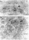
Distribution of nAChR α4-subunit-LI neurons in the adult rat substantia nigra. A, Coronal sections containing rat SNpc and SNpr were stained with the α4-subunit antibody detected using immunoperoxidase method. Scale bar, 200 μm.B, The α4-subunit-LI was present in neuronal perikarya and dendrites, but absent from cell nuclei as shown in this detail of SNpc-immunopositive cells. Scale bar, 50 μm.
Fig. 5.
Fig. 5.
Distribution of α4-subunit-LI in cerebral cortex, globus pallidus (GP), and striatum (ST) after immunoperoxidase labeling.A, Coronal sections of rat somatosensory cortex show a laminar distribution of α4-subunit-LI, mainly in neurons of the infragranular layers. Cell bodies and apical dendrites of pyramidal neurons show a prominent labeling. Scale bar, 100 μm.B, Cell bodies of globus pallidus were clearly stained with α4-subunit antibody but few neurons were positive in the striatum. Scale bar, 100 μm.
Fig. 6.
Fig. 6.
Confocal microscopy shows colocalization of α4-subunit-LI and TH in dopaminergic neurons in the SNpc (AC) and VTA (DF). A,D, Cy2-conjugated streptavidin decorates neurons immunolabeled with the antibody raised against the α4-subunit.B, E, Cy5-conjugated anti-mouse IgG-stained neurons containing TH. Superimposition of the images (C, F) show that most of the neurons were double-labeled. Scale bar, 20 μm.
Fig. 7.
Fig. 7.
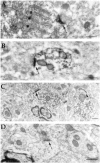
Ultrastructural localization of the α4-subunit of the nAChR in the rat SNpc, using immunoperoxidase labeling.A, Dense deposits of DAB are observed in spots of the somatodendritic compartments (asterisks).B, Immunoreactivity for the α4-subunit is distributed heterogeneously. In the perikarya, immunolabeling is frequently associated with endoplasmic reticulum (asterisk), but not with the Golgi apparatus (Go). Scale bar, 0.4 μm.
Fig. 8.
Fig. 8.
Postsynaptic localization of the α4-subunit-LI in the rat SNpc (AC). Dense deposits of immunoreaction product are associated with postsynaptic densities (arrows) and not with perisynaptic plasma membrane.A, Densely immunolabeled axosomatic synaptic contact (arrow) is close to a lightly labeled endoplasmic reticulum (asterisks). B, Immunolabeled synaptic profile (arrow) on a dendritic shaft. Dendroplasm is also lightly immunolabeled. C, In the same field as in B and C unlabeled synaptic contacts (arrowhead) were present beside labeled synaptic profiles (arrow). D, No labeling was apparent in controls (arrow) in which the first antibody was either omitted or preadsorbed with the peptide used as antigen. Scale bar, 0.4 μm.
Fig. 9.
Fig. 9.
Postsynaptic localization of the α4-subunit-LI in the rat substantia nigra (AG). Consistent with our observations using immunoperoxidase, the immunogold labeling is observed at some synaptic profiles (arrowheads). Most of the postsynaptic densities were decorated with two to four gold particles. To estimate the nonspecific labeling, the primary antibody was either omitted or preadsorbed with the antigen peptide before incubations. In that case, most of the synaptic profiles (arrows) were unlabeled (H). Scale bar, 0.25 μm.
Fig. 10.
Fig. 10.
Histogram distribution of synaptic profiles decorated with one or more gold particles per profile (before applying the morphological and gold particle distribution criteria), in both experimental (dark gray) and control (light gray) conditions (A). In experimental conditions (B) this distribution (open diamonds) rapidly diverges from a Poisson distribution (black diamonds) for two and more gold particles per synaptic profile.
Fig. 11.
Fig. 11.
Histogram showing the radial distribution of gold particles over synaptic profiles sampled in the SNpc. The distances between the center of each gold particles and the postsynaptic plasma membrane were grouped into bins 10 nm wide. The values along thex-axis indicate bin centers, with bin “0” centered on the postsynaptic plasmic membrane. Minus signsindicate direction of the presynaptic domain. These data were pooled from 44 synaptic profiles.

References

    1. Beninato M, Spencer RF. A cholinergic projection to the rat substantia nigra from the pedunculopontine tegmental nucleus. Brain Res. 1987;412:169–174. - PubMed
    1. Beninato M, Spencer RF. The cholinergic innervation of the rat substantia nigra: a light and electron microscopic immunohistochemical study. Exp Brain Res. 1988;72:178–184. - PubMed
    1. Bertrand D, Changeux JP. Nicotinic receptor: an allosteric protein specialized for intercellular communication. Semin Neurosci. 1995;7:75–90.
    1. Blaha C, Winn P. Modulation of dopamine efflux in the striatum following cholinergic stimulation of the substantia nigra in intact and pedunculopontine tegmental nucleus-lesioned rats. J Neurosci. 1993;13:1035–1044. - PMC - PubMed
    1. Blaha C, Allen L, Das S, Inglis W, Latimer M, Vincent S, Winn P. Modulation of dopamine efflux in the nucleus accumbens after cholinergic stimulation of the ventral tegmental area in intact, pedunculopontine tegmental nucleus-lesioned, and laterodorsal tegmental nucleus-lesioned rats. J Neurosci. 1996;16:714–722. - PMC - PubMed

Publication types

LinkOut - more resources