. 1999 Aug;67(8):4191-200.
doi: 10.1128/IAI.67.8.4191-4200.1999.

Plasma membrane expression of heat shock protein 60 in vivo in response to infection

Affiliations

Plasma membrane expression of heat shock protein 60 in vivo in response to infection

C Belles et al. Infect Immun. 1999 Aug.

Abstract

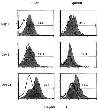
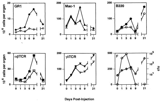
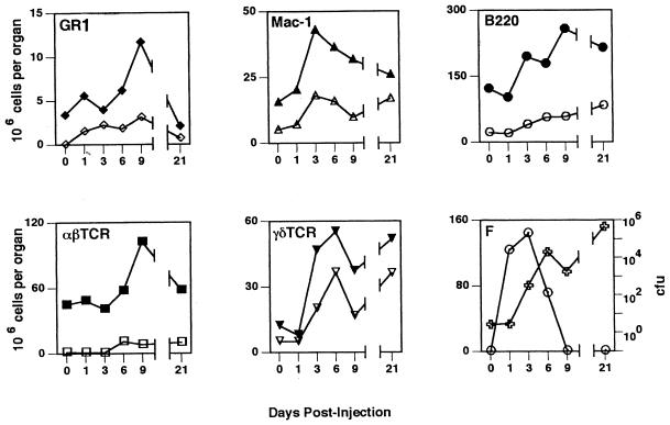
Heat shock protein 60 (hsp60) is constitutively expressed in the mitochondria of eukaryotic cells. However, it has been identified in other subcellular compartments in several disease states and in transformed cells, and it is an immunogenic molecule in various infectious and autoimmune diseases. To better understand the factors that influence expression of hsp60 in normal cells in vivo, we analyzed its cellular and subcellular distribution in mice infected with the intracellular bacterium Listeria monocytogenes. Western blotting of subcellular fractionated spleen cells showed that although endogenous hsp60 was restricted to the mitochondria in noninfected animals, it was associated with the plasma membrane as a result of infection. The low levels of plasma membrane-associated hsp60 seen in the livers in noninfected animals subsequently increased during infection. Plasma membrane hsp60 expression did not correlate with bacterial growth, being most evident during or after bacterial clearance and persisting at 3 weeks postinfection. Using flow cytometry, we determined that Mac-1(+), T-cell receptor gammadelta(+), and B220(+) cells represented the major Hsp60(+) populations in spleens of infected mice. By contrast, B220(+) cells were the predominant hsp60(+) population in livers of infected mice. Of the immune cells analyzed, the kinetic profile of the gammadelta T-cell response most closely matched that of hsp60 expression in both the spleen and liver. Collectively, these findings show that during infection hsp60 can be localized to the plasma membrane of viable cells, particularly antigen-presenting cells, providing a means by which hsp60-reactive lymphocytes seen in various infectious disease and autoimmune disorders may be generated and maintained.


Figures

FIG. 1
FIG. 1
Detection of hsp60 expression in spleens of Listeria-infected mice. Spleens from BALB/c mice 3 (top) and 6 (bottom) days after infection with L. monocytogenes (0.38 LD50) were sectioned, fixed in acetone, and stained with antibodies specific for macrophages (Mac-1-biotin plus avidin-AP; red cells) and hsp60 (LK2-FITC; light green cells). The bottom photomicrograph shows that the majority of Mac-1+ cells are also hsp60+ and appear yellow-orange. The staining patterns shown are typical of those obtained in more than six independent experiments.
FIG. 2
FIG. 2
Separation of mitochondria and plasma membrane of spleen and liver cells. Cells were fractionated, and individual fractions were tested for activity of marker enzymes for mitochondria (SR; ■) and plasma membrane APD; ○). Fractions containing highest levels of each enzyme (arrows labeled M for mitochondria and PM for plasma membrane) were combined and used as the final mitochondrial and plasma membrane fractions in Western blot experiments. Profiles are typical of those obtained in all experiments.
FIG. 3
FIG. 3
Subcellular distribution of hsp60 in vivo during infection. Spleen and liver cells from mice at various times (days [d]) after infection with L. monocytogenes (0.34 LD50) were fractionated, and 15 μg of protein from the nuclear (N), mitochondrial (M), cytoplasm (C), and plasma membrane (P) fractions were analyzed for hsp60 expression by Western blotting. Molecular weight markers (std) and total cell lysates of L. monocytogenes (20 μg; L) and EL4 (15 μg; E) were included on each gel. The results shown are representative of those obtained in three independent experiments. The horizontal lines on each blot represent positions of molecular weight standards (97.4, 66, and 46 kDa).
FIG. 4
FIG. 4
Subcellular distribution of the PDC in spleens and livers of Listeria-infected mice. Western blots originally probed with anti-hsp60 antibody (Fig. 3) were stripped and reprobed with an antiserum from a patient with primary biliary cirrhosis containing high-titer antibodies to PDC. Bound antibody was visualized by chemiluminescence as described in the legend to Fig. 3. The subunits of PDC present in the samples are indicated on the left. The horizontal lines on each blot represent positions of molecular weight standards (97.4, 66, 46, and 30 kDa). d1, d3, and d6, 1, 3, and 6 days postinfection.
FIG. 5
FIG. 5
Increased hsp60 expression on macrophages in livers and spleens of infected mice. Spleen and liver cells obtained from mice at various times after infection were analyzed by flow cytometry. hsp60 expression by monocytes/macrophages was determined by gating on Mac-1+ cells and analyzing for hsp60 expression. The filled and open plots show profiles of staining obtained with anti-hsp60 (filled profiles) and an isotype-matched antibody, respectively; numbers represent percentages of Mac-1+ cells which are hsp60+.
FIG. 6
FIG. 6
Kinetic profiles of cellular response, hsp60 expression, and bacteria in the liver. Before and at various times after infection with L. monocytogenes (0.30 LD50), the numbers of granulocytes (GR1), monocytes/macrophages (Mac-1), B cells (B220), αβ T cells (αβTcR) and γδ T cells (γδTcR) present in the liver were determined from total cell counts, and the proportion of cells reactive with lineage-specific antibodies as determined by flow cytometry. Closed symbols, total cell numbers; open symbols, numbers of hsp60+ cells of each cell subtype. Panel F shows the bacterial CFU (○) and total number of surface hsp60+ cells (✙). All values are means from four animals. Standard deviations were <20% in all cases and for the sake of clarity are not shown.
FIG. 7
FIG. 7
Kinetic profiles of the cellular response, hsp60 expression, and bacteria in the spleen. Spleen cells were analyzed and the different populations were enumerated as described in the legend to Fig. 6. Closed symbols, total cell numbers; open symbols, numbers of hsp60+ cells of each cell subtype. Panel F shows bacterial CFU (○) and total number of surface hsp60+ cells (✙). All values are means from four animals. Standard deviations were <20% in all cases and for the sake of clarity are not shown.

References

    1. Balch W E, Rothman J E. Characterization of protein transport between successive compartments of the Golgi apparatus: asymmetric properties of donor and acceptor activities in a cell-free system. Arch Biochem Biophys. 1985;240:413–425. - PubMed
    1. Barrios C, Lussow A R, van Embden J, van der Zee R, Rappuoli R, Constantino P, Louis J A, Lambert P-H, Del Giudice G. Mycobacterial heat-shock proteins as carrier molecules II: the use of the 70-kDa mycobacterial heat-shock protein as carrier for conjugated vaccines can circumvent the need for adjuvants and Bacillus Calmette Guerin priming. Eur J Immunol. 1992;22:1365–1372. - PubMed
    1. Belles, C., and S. R. Carding. Unpublished observations.
    1. Belles C, Kuhl A L, Donoghue A J, Sano Y, O’Brien R L, Born W, Bottomly K, Carding S R. Bias in the γδ cell response to Listeria monocytogenes: Vδ6.3+ cells are a major component of the γδ T cell response to Listeria monocytogenes. J Immunol. 1996;156:4280–4289. - PubMed
    1. Boog C J P, de Graeff-Meeder E R, Lucassen M A, van der Zee R, Voorhorst-Ogink M M, van Kooten J S, Geuze H J, van Eden W. Two monoclonal antibodies generated against human HSP60 show reactivity with synovial membranes of patients with juvenile chronic arthritis. J Exp Med. 1992;175:1805–1810. - PMC - PubMed

Publication types

Substances