

Interleukin-1 and tumor necrosis factor activities partially account for calvarial bone resorption induced by local injection of lipopolysaccharide
- PMID: 10417196
- PMCID: PMC96729
- DOI: 10.1128/IAI.67.8.4231-4236.1999
Interleukin-1 and tumor necrosis factor activities partially account for calvarial bone resorption induced by local injection of lipopolysaccharide
Abstract
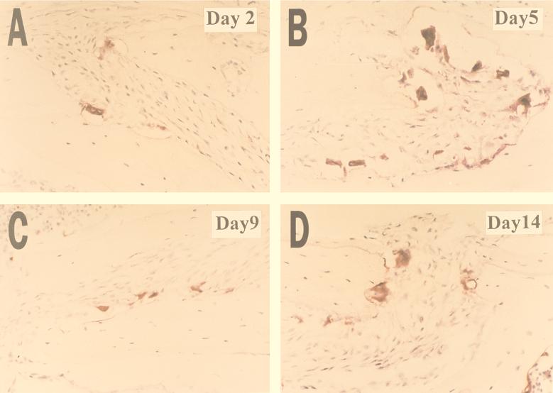
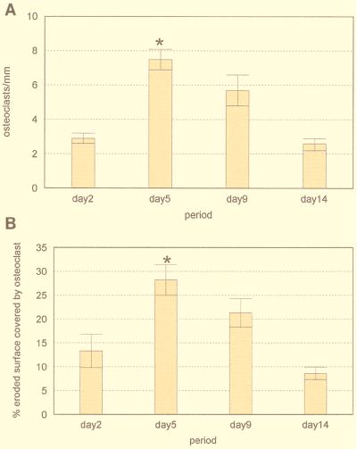
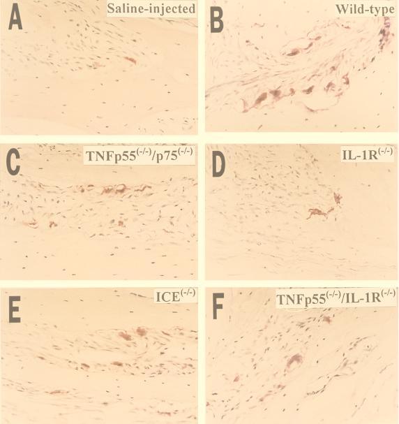
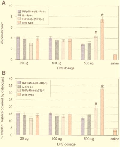
The present study was undertaken to test the hypothesis that tumor necrosis factor (TNF) and/or interleukin-1 (IL-1) activity mediates lipopolysaccharide (LPS)-induced bone resorption in vivo. To test this hypothesis, Escherichia coli LPS or Porphyromonas gingivalis LPS was injected into the subcutaneous tissues overlying mouse calvariae. Histological sections, prepared from the center of the lesion, were stained for tartrate-resistant acid phosphatase, and histomorphometric analysis was performed to quantify the osteoclast number and the area of bone resorption. In time course experiments using normal mice, a peak of bone resorption occurred 5 days after endotoxin stimulation. In dose-response experiments, IL-1 receptor type 1 deletion (IL-1R(-/-)), TNF double-receptor p55/p75 deletion (TNF p55(-/-)/p75(-/-)), combined TNF p55 and IL-1 receptor type 1 deletion (TNF p55(-/-)/IL-1R(-/-)), and IL-1beta-converting enzyme-deficient (ICE(-/-)) mice and the respective wild-type mice were injected with 500, 100, or 20 micrograms of P. gingivalis LPS and sacrificed 5 days after LPS injection. At the highest dose (500 micrograms), significant decreases in osteoclast number occurred in mutant mice compared to wild-type mice: (i) a 64% reduction for the TNF p55(-/-)/IL-1R(-/-) mice, (ii) a 57% reduction for the IL-1R(-/-) mice, (iii) a 41% reduction for the TNF p55(-/-)/p75(-/-) mice, and (iv) a 38% reduction for the ICE(-/-) mice. At the two lower doses, bone resorption was apparent but no significant differences between mutant and wild-type animals were observed. The present data indicate that at higher doses, LPS-induced bone resorption is substantially mediated by IL-1 and TNF receptor signaling. Furthermore, IL-1 receptor signaling appears to be slightly more important than TNF receptor signaling. At lower LPS doses, other pathways leading to osteoclast activity that are independent of TNF and IL-1 are involved.
Figures
References
-
- Agarwal S, Piesco N P, Johns L P, Riccelli A E. Differential expression of IL-1 beta, TNF- alpha, IL-6 and IL-8 in human monocytes in response to lipopolysaccharides from different microbes. J Dent Res. 1995;74:1057–1065. - PubMed
-
- Amar S, Van Dyke T E, Eugster H P, Schultze N, Koebel P, Bluethmann H. Tumor necrosis factor (TNF)-induced cutaneous necrosis is mediated by TNF receptor 1. J Inflamm. 1996;47:180–189. - PubMed
-
- Assuma R, Oates T, Cochran D, Amar S, Graves D T. IL-1 and TNF antagonists inhibit the inflammatory response and bone loss in experimental periodontitis. J Immunol. 1998;16:403–409. - PubMed
-
- Aznar C, Fitting C, Cavaillon J M. LPS-induced production of cytokines by bone marrow-derived macrophages: destruction between intracellular IL-1 production and IL-1 release. Cytokine. 1990;2:259–265. - PubMed
Publication types
MeSH terms
Substances
Grants and funding
LinkOut - more resources
Full Text Sources
Other Literature Sources
Research Materials
