. 1999 Aug;181(15):4561-7.
doi: 10.1128/JB.181.15.4561-4567.1999.

The structure of multiple polypeptide domains determines the signal recognition particle targeting requirement of Escherichia coli inner membrane proteins

Affiliations

The structure of multiple polypeptide domains determines the signal recognition particle targeting requirement of Escherichia coli inner membrane proteins

J A Newitt et al. J Bacteriol. 1999 Aug.

Abstract

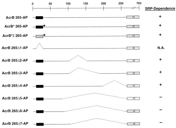
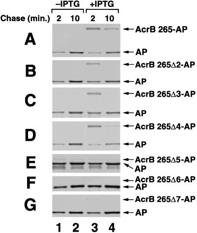
The signal recognition particle (SRP) targeting pathway is required for the efficient insertion of many polytopic inner membrane proteins (IMPs) into the Escherichia coli inner membrane, but in the absence of SRP protein export proceeds normally. To define the properties of IMPs that impose SRP dependence, we analyzed the targeting requirements of bitopic IMPs that are structurally intermediate between exported proteins and polytopic IMPs. We found that disruption of the SRP pathway inhibited the insertion of only a subset of bitopic IMPs. Studies on a model bitopic AcrB-alkaline phosphatase fusion protein (AcrB 265-AP) showed that the SRP requirement for efficient insertion correlated with the presence of a large periplasmic domain (P1). As previously reported, perturbation of the SRP pathway also affected the insertion of a polytopic AcrB-AP fusion. Even exhaustive SRP depletion, however, failed to block the insertion of any AcrB derivative by more than 50%. Taken together, these data suggest that many proteins that are normally targeted by SRP can utilize alternative targeting pathways and that the structure of both hydrophilic and membrane-spanning domains determines the degree to which the biogenesis of a protein is SRP dependent.


Figures

FIG. 1
FIG. 1
Insertion of a subset of bitopic IMPs is inhibited by overexpression of ftsY(G385A). MC4100 cells transformed with pTRC-ftsY(G385A) and a second plasmid encoding an AP fusion were grown to mid-log phase. The cultures were then divided in half; one portion was left untreated (lanes 1 and 2), and IPTG was added to the other portion (lanes 3 and 4) to induce ftsY(G385A) overexpression. The insertion of AcrB 265-AP (A), Tsr 164-AP (B), YfgA 139-AP (C), and MdoH 173-AP (D) was analyzed 40 min after IPTG addition by pulse-chase labeling and immunoprecipitation of AP-containing polypeptides from protease-treated spheroplasts as described in Materials and Methods. The length of the chase is indicated. The diagrams illustrate the structure of each IMP. In all cases, the N terminus is located in the cytoplasm, and the C terminus is in the periplasm.
FIG. 2
FIG. 2
Summary of the AcrB 265-AP derivatives used in this study. The AcrB 265-AP protein (top line) contains a TM domain (amino acid residues 9 to 29 [solid rectangle]) and an AP moiety fused at residue 265 (shaded rectangle). The construction of all derivatives is described in Materials and Methods. AcrB* 265-AP (second line) contains a leucine-to-arginine point mutation (★) at residue 30, and AcrB*Σ 265-AP (third line) contains both the L30R mutation and the first TM domain of Tsr (hatched rectangle) in place of the native AcrB TM domain. The location of deletions in seven additional derivatives (Δ1–Δ7) is indicated by dashed lines. The effect of the disruption of the SRP pathway on the insertion of each derivative is indicated. +, Defect observed; −, defect not observed; N.A., not testable.
FIG. 3
FIG. 3
Insertion of an AcrB 265-AP derivative containing a Tsr TM domain is inhibited by overexpression of ftsY(G385A). The insertion of AcrB 265-AP (A), AcrB* 265-AP (B), AcrB*Σ 265-AP (C), and AcrB 265Δ1-AP (D) in MC4100 was analyzed as described in the legend to Fig. 1. IPTG was added to the cells in lanes 3 and 4 to induce ftsY(G385A) overexpression. The length of the chase is indicated. The position of the point mutation in AcrB* 265-AP and AcrB*Σ 265-AP is indicated (★), and each distinct TM domain is represented by different shading.
FIG. 4
FIG. 4
Large deletions in the AcrB 265-AP periplasmic domain abolish SRP dependence. The insertion of AcrB 265-AP (A), AcrB 265Δ2-AP (B), AcrB 265Δ3-AP (C), AcrB 265Δ4-AP (D), AcrB 265Δ5-AP (E), AcrB 265Δ6-AP (F), and AcrB 265Δ7-AP (G) in MC4100 was analyzed as described in the legend to Fig. 1. The deletions in each plasmid are summarized in Fig. 2. IPTG was added to the cells in lanes 3 and 4 to induce ftsY(G385A) overexpression. The length of the chase is indicated.
FIG. 5
FIG. 5
Exhaustive depletion of SRP does not completely block the insertion of a polytopic IMP-AP fusion. (A) Growth of WAM113 transformed with a plasmid expressing AcrB 576-AP in the presence of arabinose (▴) or glucose (○). (B) Cells grown in the presence of arabinose or glucose in the experiment depicted in panel A were removed from cultures at the indicated time point, and the Ffh levels were measured by Western blot. The amount of Ffh in cells grown in glucose is expressed as a percentage of the amount of Ffh in cells grown in arabinose at the indicated time point. (C) The insertion of AcrB 576-AP was examined in the experiment depicted in panel A after 7 h of Ffh depletion (arrow) by pulse-chase labeling and immunoprecipitation of AP-containing polypeptides from protease-treated spheroplasts as described in Materials and Methods. Lanes: 1 to 3, cells grown in arabinose; 4 to 6, cells grown in glucose. The length of the chase is indicated.

References

    1. Bernstein H D, Poritz M A, Strub K, Hoben P J, Brenner S, Walter P. Model for signal sequence recognition from amino-acid sequence of 54K subunit of signal recognition particle. Nature. 1989;340:482–486. - PubMed
    1. Brown S, Fournier M J. The 4.5S RNA gene of Escherichia coli is essential for cell growth. J Mol Biol. 1984;178:533–550. - PubMed
    1. Bukau B, Horwich A L. The Hsp70 and Hsp60 chaperone machines. Cell. 1998;92:351–366. - PubMed
    1. Casadaban M J. Transposition and fusion of the lac genes to selected promoters in Escherichia coli using bacteriophage lambda and Mu. J Mol Biol. 1976;104:541–555. - PubMed
    1. Claros M G, von Heijne G. TopPred II: an improved software for membrane protein structure predictions. Comput Appl Biosci. 1994;10:685–686. - PubMed

MeSH terms

LinkOut - more resources