. 1999 Aug;181(15):4576-83.
doi: 10.1128/JB.181.15.4576-4583.1999.

Cloning, sequencing, and characterization of the cgmB gene of Sinorhizobium meliloti involved in cyclic beta-glucan biosynthesis

Affiliations

Cloning, sequencing, and characterization of the cgmB gene of Sinorhizobium meliloti involved in cyclic beta-glucan biosynthesis

P Wang et al. J Bacteriol. 1999 Aug.

Abstract

Periplasmic cyclic beta-glucans of Rhizobium species provide important functions during plant infection and hypo-osmotic adaptation. In Sinorhizobium meliloti (also known as Rhizobium meliloti), these molecules are highly modified with phosphoglycerol and succinyl substituents. We have previously identified an S. meliloti Tn5 insertion mutant, S9, which is specifically impaired in its ability to transfer phosphoglycerol substituents to the cyclic beta-glucan backbone (M. W. Breedveld, J. A. Hadley, and K. J. Miller, J. Bacteriol. 177:6346-6351, 1995). In the present study, we have cloned, sequenced, and characterized this mutation at the molecular level. By using the Tn5 flanking sequences (amplified by inverse PCR) as a probe, an S. meliloti genomic library was screened, and two overlapping cosmid clones which functionally complement S9 were isolated. A 3.1-kb HindIII-EcoRI fragment found in both cosmids was shown to fully complement mutant S9. Furthermore, when a plasmid containing this 3.1-kb fragment was used to transform Rhizobium leguminosarum bv. trifolii TA-1JH, a strain which normally synthesizes only neutral cyclic beta-glucans, anionic glucans containing phosphoglycerol substituents were produced, consistent with the functional expression of an S. meliloti phosphoglycerol transferase gene. Sequence analysis revealed the presence of two major, overlapping open reading frames within the 3.1-kb fragment. Primer extension analysis revealed that one of these open reading frames, ORF1, was transcribed and its transcription was osmotically regulated. This novel locus of S. meliloti is designated the cgm (cyclic glucan modification) locus, and the product encoded by ORF1 is referred to as CgmB.


Figures

FIG. 1
FIG. 1
Southern hybridization analysis of S. meliloti genomic DNA. (A) Blots of S. meliloti 1021 genomic DNA (lanes 1, 2, and 3) and S. meliloti mutant S9 genomic DNA (lanes 4, 5, and 6) were hybridized with a 1-kb probe corresponding to an internal fragment of Tn5. Genomic DNA was digested with BamHI and EcoRI (lanes 1 and 4); EcoRI (lanes 2 and 5); and BamHI (lanes 3 and 6). (B) Blots of EcoRI-digested genomic DNA from S. meliloti mutant S9 (lane 1) and S. meliloti 1021 (lane 2) were hybridized with a 3.3-kb probe corresponding to DNA sequences flanking the Tn5 insert within mutant S9; the 3.3-kb product of inverse PCR (see text) served as a positive control (lane 3).
FIG. 2
FIG. 2
Thin-layer chromatogram of cyclic β-(1,2)-glucans isolated from S. meliloti mutant S9 complemented with cosmids 12A7 and 13H9. Periplasmic glucans were extracted from cells by using 70% ethanol, and half of each extract was treated with alkali before thin-layer chromatographic analysis as previously described (8). Alkali treatment results in the selective removal of succinyl substituents, whereas phosphoglycerol substituents are resistant to alkali treatment. Thus, cyclic β-(1,2)-glucans containing phosphoglycerol substituents are revealed by thin-layer chromatography after alkali treatment as anionic cyclic β-(1,2)-glucans. From left to right: S. meliloti S9 containing cosmid 13H9 (lanes 1 and 2); S. meliloti S9 containing cosmid 12A7 (lanes 3 and 4); S. meliloti 1021 (lanes 5 and 6); and S. meliloti S9 (lanes 7 and 8). +, extracts subjected to alkali treatment prior to thin-layer chromatography; −, extracts not subjected to alkali treatment. Anionic cyclic β-(1,2)-glucans containing either or both succinyl and phosphoglycerol substituents have the same relative mobility on this thin-layer chromatography system.
FIG. 3
FIG. 3
Restriction maps of genomic DNA inserts within cosmids 12A7 and 13H9 and plasmid subclones. (A) EcoRI restriction maps of genomic DNA inserts within cosmids 12A7 and 13H9. The genomic DNA fragments are present in the same orientation in both cosmids, and the right ends of the inserts shown are near the cos site of the cosmid vector pLAFRI. (B) Enlarged view of an approximately 12-kb region within cosmids 12A7 and 13H9 (corresponding to the shaded area in A) that contains the Tn5 insert. Recognition sites for EcoRI, BamHI, SalI, and ApaI are shown. Plasmid pEW1 is a derivative of pTR100 containing a 2.9-kb BamHI fragment. Plasmid pEW2 is a derivative of pTR100 containing a 3.1-kb HindIII fragment. The 3.1-kb HindIII fragment was isolated from plasmid pFL6 and contains the 50-bp EcoRI-HindIII polylinker region from pUC19 as well as the 3.1-kb HindIII-EcoRI fragment derived from the 12-kb region within cosmids 12A7 and 13H9 (see Table 1). Note that not all HindIII recognition sites have been determined for this 12-kb region. Thus, the HindIII restriction map is not shown. The dark, horizontal bar indicates the 3,524 bp sequenced in the present study. (C) Further enlargement of sequenced region (3,524 bp). The horizontal arrows correspond to the location of the two open reading frames identified in the present study and also indicate the direction of transcription. A, ApaI; B, BamHI; E, EcoRI; H, HindIII; S, SalI. Vertical arrows indicate the location of the Tn5 insertion within mutant S9.
FIG. 4
FIG. 4
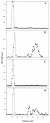
DEAE-cellulose anion-exchange column chromatography profiles of the cell-associated cyclic β-(1,2)-glucans from S. meliloti mutant S9 and R. leguminosarum biovar trifolii TA-1JH. Cell-associated glucans were extracted from cells as described in the text and purified by gel filtration column chromatography on Sephadex G-50 followed by desalting on a Sephadex G-15 column as previously described (8). Prior to fractionation on DEAE-cellulose, extracts were treated with alkali to selectively remove succinyl substituents (as described in the legend to Fig. 2). These samples were again desalted on Sephadex G-15 prior to application to DEAE-cellulose. Because phosphoglycerol substituents are resistant to alkali treatment, cyclic β-(1,2)-glucans containing these substituents remain anionic after treatment and elute from the column only upon application of a salt gradient (beginning at approximately 40 ml). Neutral cyclic β-(1,2)-glucans elute at the void volume between 10 and 20 ml. (A) S. meliloti mutant S9. No anionic cyclic β-(1,2)-glucans were detected after alkali treatment. (B) S. meliloti mutant S9 carrying plasmid pEW2. Several peaks corresponding to anionic, glycerophosphorylated cyclic β-(1,2)-glucans were observed. The phosphorus content was determined to correspond to 1.7 mol per mol of total cyclic β-(1,2)-glucan, assuming an average size of 20 glucose residues per glucan molecule. (C) R. leguminosarum bv. trifolii TA-1JH. No anionic cyclic β-(1,2)-glucans were detected after alkali treatment. (D) R. leguminosarum bv. trifolii TA-1JH carrying plasmid pEW2. Several peaks corresponding to anionic, glycerophosphorylated cyclic β-(1,2)-glucans were observed. The phosphorus content was determined to correspond to 1.3 mol per mol of total cyclic β-(1,2)-glucan, assuming an average size of 20 glucose residues per glucan molecule. OD, optical density.
FIG. 5
FIG. 5
Nucleotide sequence of the region upstream of ORF1. Nucleotides 1 to 720 of the 3,524-nucleotide sequenced region are shown. Inverted repeat sequences are indicated by shading and dashed arrows and are numbered. Direct repeat sequences are shown by overlining. The transcription start site at nucleotide 578 is indicated by a boldface asterisk. The deduced amino acid sequence of the N terminus of the protein encoded by ORF1 is indicated with single-letter amino acid symbols aligned above the second nucleotide of each codon. A potential ribosome binding site is underlined and boldfaced.
FIG. 6
FIG. 6
Localization of the ORF1 transcription start site by primer extension analysis. Primer extension reactions were performed with RNA isolated from S. meliloti 1021 grown under low- (lanes 1 to 3) and high- (lanes 4 to 6) osmolarity conditions. Reaction mixtures contained 25 (lanes 1 and 4), 50 (lanes 2 and 5), or 100 μg (lanes 3 and 6) of RNA. The oligonucleotide primer used was 18-mer with the sequence 5′-CGGTTTGTGAACGGACTC-3′, corresponding to nucleotides 693 to 676 in Fig. 5. Dideoxy sequencing ladders were generated with the same primer used for the primer extension reaction. The nucleotide corresponding to the transcription start site is indicated by an asterisk. Cells were grown in GMS medium for low-osmolarity growth conditions and in GMS medium containing 0.4 M NaCl for high-osmolarity growth conditions.
FIG. 7
FIG. 7
Transcription of the cgmB gene in R. leguminosarum bv. trifolii TA-1JH. Primer extension reactions were performed by using 15-μg samples of RNA isolated from R. leguminosarum bv. trifolii TA-1JH containing pTR100 (lane 1), R. leguminosarum bv. trifolii TA-1JH containing pEW2 (lane 2), and S. meliloti 1021 (lane 3). The oligonucleotide primer used in primer extension reactions and for generation of dideoxy sequencing ladders is the same as that used in Fig. 6.

Similar articles

Cited by

References

    1. Altschul S F, Gish W, Miller W, Myers E W, Lipman D J. Basic local alignment search tool. J Mol Biol. 1990;215:403–410. - PubMed
    1. Altschul S F, Madden T L, Schäffer A A, Zhang J, Zhang Z, Miller W, Lipman D J. Gapped BLAST and PSI-BLAST: a new generation of protein database search programs. Nucleic Acids Res. 1997;25:3389–3402. - PMC - PubMed
    1. Anderson M L M, Young B D. Quantitative filter hybridization. In: Hames B D, Higgins S J, editors. Nucleic acid hybridization: a practical approach. Oxford, England: IRL Press; 1985. pp. 73–111.
    1. Becker A A, Kleickmann A A, Arnold W, Puhler A. Analysis of the Rhizobium meliloti exoH/exoK/exoL fragment: ExoK shows homology to excreted endo-beta-1,3-1,4-glucanases and ExoH resembles membrane proteins. Mol Gen Genet. 1993;238:145–154. - PubMed
    1. Bhagwat A A, Gross K C, Tully R E, Keister D L. β-glucan synthesis in Bradyrhizobium japonicum: characterization of a new locus (ndvC) influencing β-(1→6) linkages. J Bacteriol. 1996;178:4635–4642. - PMC - PubMed

Publication types

Associated data

LinkOut - more resources