. 1999 Aug;181(15):4617-27.
doi: 10.1128/JB.181.15.4617-4627.1999.

Effects of growth conditions on expression of mycobacterial murA and tyrS genes and contributions of their transcripts to precursor rRNA synthesis

Affiliations

Effects of growth conditions on expression of mycobacterial murA and tyrS genes and contributions of their transcripts to precursor rRNA synthesis

J A Gonzalez-y-Merchand et al. J Bacteriol. 1999 Aug.

Abstract

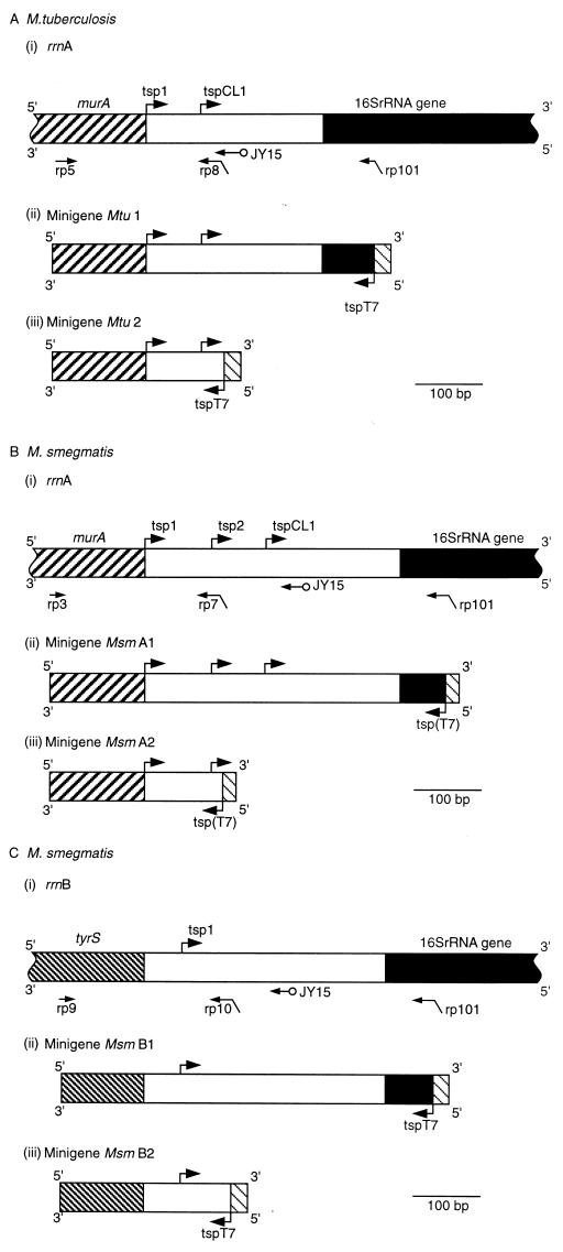
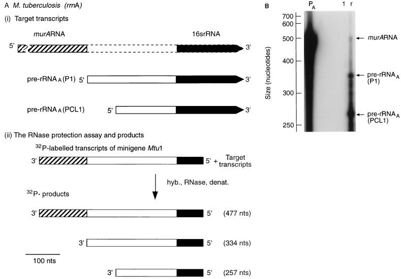
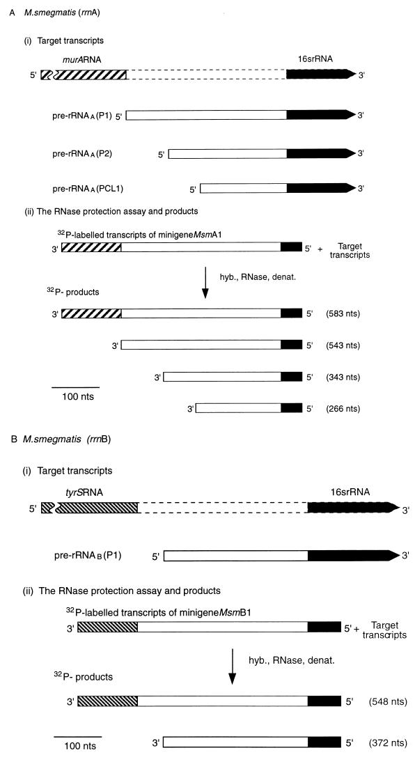
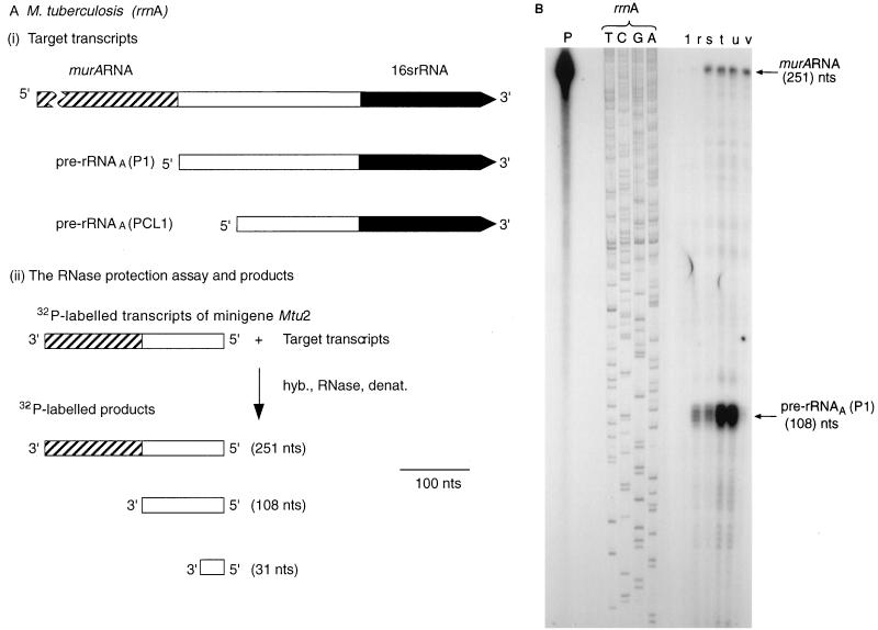
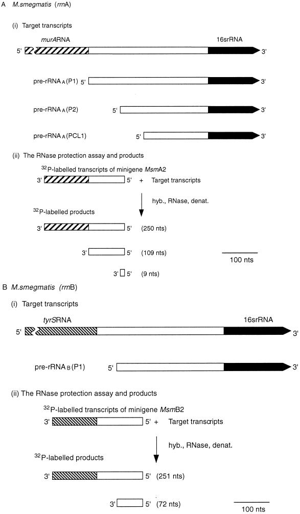
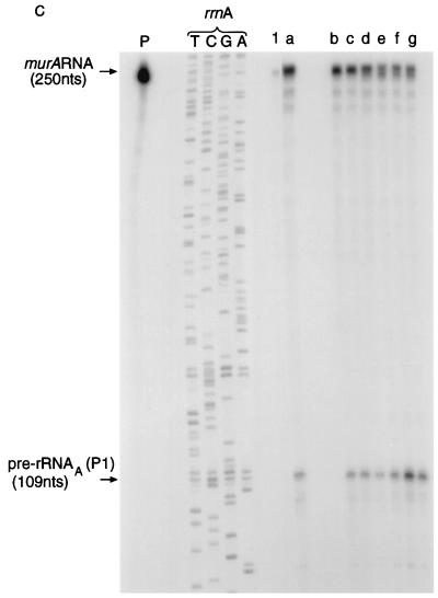
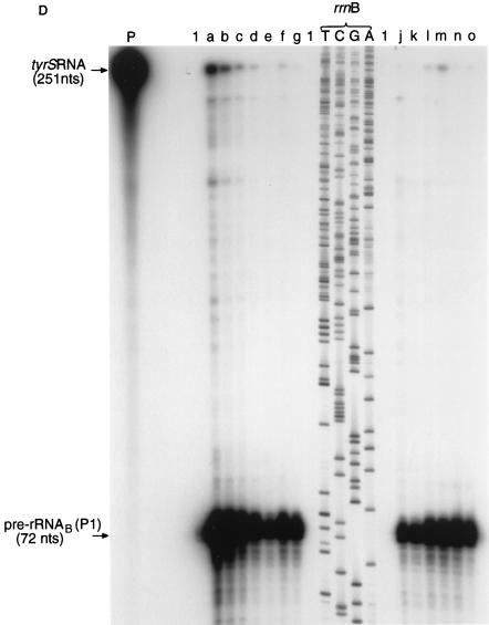
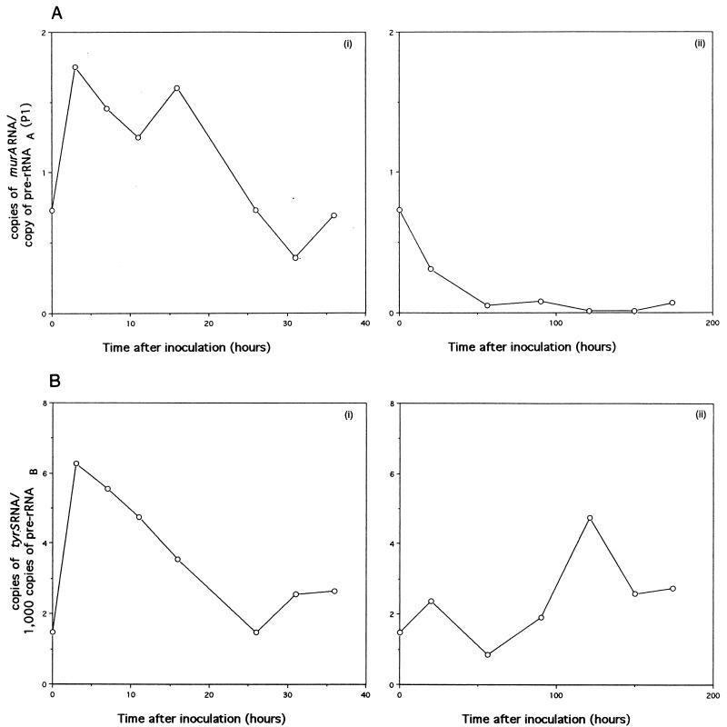
All mycobacteria studied to date have an rRNA operon, designated rrnA, located downstream from a single copy of the murA gene, which encodes an enzyme (EC 2.5.1.7) important for peptidoglycan synthesis. The rrnA operon has a promoter, P1(A), located within the coding region of murA, near the 3' end. Samples of RNA were isolated from Mycobacterium tuberculosis at different stages of the growth cycle and from Mycobacterium smegmatis grown under different conditions. RNase protection assays were used to investigate transcripts of both murA and rrnA. Transcription of murA was found to continue into the 16S rRNA gene, as if murA and rrnA form a hybrid (protein coding-rRNA coding) operon. During the growth of M. tuberculosis, the hybrid operon contributed approximately 2% to total pre-rRNA. Analysis of M. smegmatis RNA revealed that the level of murA RNA depended on the growth rate and that the patterns of expression during the growth cycle were different for murA and rrnA. M. smegmatis has a second rRNA operon, rrnB, located downstream from a single copy of the tyrS gene, encoding tyrosyl-tRNA synthetase. Transcription of tyrS was found to continue into the 16S rRNA gene rrnB. The hybrid tyrS-rrnB operon contributed 0.2 to 0.6% to rrnB transcripts. The pattern of tyrS expression during the growth cycle matched the pattern of rrnB expression, reflecting the essential role of TyrS and rRNA in protein biosynthesis.


Figures

FIG. 1
FIG. 1
Upstream regions of mycobacterial rrn operons and constructs used in RNase protection assays. A horizontal arrow indicates the location of the binding site for the primer specified; a diagonally bent arrow indicates the location of the binding site for the primer specified (horizontal section of the arrow) and the presence of the T7 promoter sequence (angled section); a horizontal arrow with a circle at the end indicates the binding site for primer JY15, used in DNA sequencing reactions. An arrow bent at a right angle denotes the location of a tsp (vertical section) and the direction of transcription (horizontal section). ▨, murA; ▧, tyrS; ■, 16S rRNA gene; ▧, T7 promoter; tspT7 or tsp(T7), tsp for the T7 promoter. Mtu, M. tuberculosis; Msm, M. smegmatis. (A) rrnA operon of M. tuberculosis. (i) Upstream region of the rrnA operon. The scheme shows the locations of murA, tsp’s (arrows bent at right angles labelled tsp1 and tspCL1) for rrnA, binding sites for the primers used to construct the minigenes, and the binding site for primer JY15. (ii) Minigene Mtu 1 constructed by amplification of a section of the upstream region with primers rp5 and rp101. (iii) Minigene Mtu 2 constructed by amplification of a section of the upstream region with primers rp5 and rp8. The same alignment and scale (shown by the bar) are used in panels i to iii. (B) rrnA operon of M. smegmatis. (i) Upstream region of rrnA. The scheme shows the locations of murA, tsp’s (arrows labelled tsp1, tsp2, and tspCL1) for rrnB binding sites for the primers used to construct the minigenes, and the binding site for primer JY15. (ii) Minigene Msm A1 constructed by amplification with primers rp3 and rp101. (iii) Minigene Msm A2 constructed by amplification with primers rp3 and rp7. The same alignment and scale (shown by the bar) are used in panels i to iii. (C) rrnB operon of M. smegmatis. (i) Upstream region of rrnB. The scheme shows the locations of tyrS, the tsp (arrow labelled tsp1) for the rrnB operon, the binding sites for the primers used to construct the minigenes, and the binding site for primer JY15. (ii) Minigene Msm B1 constructed with primers rp9 and rp101. (iii) Minigene Msm B2 constructed with primers rp9 and rp10. The same alignment and scale (shown by the bar) are used in panels i to iii.
FIG. 2
FIG. 2
Transcription of M. tuberculosis murA continues into the rRNA coding region of the downstream rrn operon. (A) Design of RNase protection experiments. The scale is shown by the bar. nts, nucleotides. hyb., RNase, denat., respectively, denote the main steps in the RNase protection assay, namely, hybridization, RNase treatment, and denaturation. (i) Target transcripts. The broken lines denote the initial uncertainty in the length of the murA transcript. (ii) RNase protection assay and products. The 32P-labelled probe was obtained by transcribing minigene Mtu 1 (Fig. 1A). The radioactive products (see panel B) were the result of protection of the probe by the target transcripts shown in panel i. (B) Autoradiograph of separated products. The assay described in panel A was carried out, the products were separated, and the sizes of the products were established by use of molecular weight markers as described in Materials and Methods. Lanes: PA, 32P-labelled transcripts of minigene Mtu 1; 1, control sample containing tRNA (10 μg); r, sample (10 μg) of RNA isolated from M. tuberculosis after 5 days of culturing.
FIG. 3
FIG. 3
Transcription of M. smegmatis murA and tyrS continues into the rRNA coding regions of the downstream rrn operons. (A and B) Schemes for RNase protection experiments. The scale is shown by the bars. nts, nucleotides. hyb., RNase, denat., respectively, denote the main steps in the RNase protection assay, namely, hybridization, RNase treatment, and denaturation. (A) Expression of murA and rrnA. (i) Target transcripts. The broken lines denote the initial uncertainty in the length of the murA transcript. Pre-rRNAA(P2) is a transcript of rrnA initiated at the P2 promoter and starting from tsp2 (Fig. 1B). (ii) RNase protection assay and products. The 32P-labelled probe was obtained by transcription of minigene Msm A1 (Fig. 1B). The radioactive products (see panel C) were the result of protection of the probe by the target transcripts shown in panel i. (B) Expression of tyrS and rrnB. (i) Target transcripts. The broken lines denote the initial uncertainty in the length of the tyrS transcript. (ii) RNase protection assay and products. The 32P-labelled probe was obtained by transcribing minigene Msm B1 (Fig. 1C). The radioactive products (see panel C) resulted from protection of the probe by the target transcripts shown in panel i. (C) Autoradiograph of products after separation. The assays described in panels A and B were carried out, the products were separated, and the sizes of the products were established by use of molecular weight markers as described in Materials and Methods. Lanes: PA, 32P-labelled transcripts of minigene Msm A1 (Fig. 1B); PB, 32P-labelled transcripts of minigene Msm B1 (Fig. 1C); 1, control sample containing tRNA (30 μg); b, sample (30 μg) of RNA isolated from M. smegmatis after 7 h of culturing and hybridized with PA; b′, sample (30 μg) of RNA isolated from M. smegmatis after 7 h of culturing and hybridized with PB.
FIG. 4
FIG. 4
Expression of the murA gene during the growth of M. tuberculosis. (A) Design of RNase protection experiments. The scale is shown by the bar. nts, nucleotides. hyb., RNase, denat., respectively, denote the main steps in the RNase protection assay, namely, hybridization, RNase treatment, and denaturation. (i) Target transcripts. (ii) RNase protection assay and products. The 32P-labelled probe was obtained by transcribing minigene Mtu 2 (Fig. 1A). The radioactive products (see panel B) resulted from protection of the probe by the target transcripts shown in panel i. (B) Autoradiograph of products after separation. The assays described in panel A were carried out, and the products were separated and identified by autoradiography as described in Materials and Methods. The sizes of the products were established by use of the products of sequencing reactions as markers. T, C, G, and A denote reactions carried out with the rrnA operon of M. tuberculosis as a substrate and JY15 as a primer. Lanes: P, 32P-labelled transcripts of minigene Mtu 2; 1, control sample containing tRNA (10 μg). r, s, t, u, and v, respectively, samples (10 μg) of RNA isolated from M. tuberculosis after 5, 10, 13, 22, and 40 days of culturing.
FIG. 5
FIG. 5
Expression of murA, rrnA, tyrS, and rrnB during the growth of M. smegmatis. (A and B) Schemes for RNase protection experiments. The scale is shown by the bars. nts, nucleotides. hyb., RNase, denat., respectively, denote the main steps in the RNase protection assay, namely, hybridization, RNase treatment, and denaturation. (A) Expression of murA and rrnA. (i) Target transcripts. Pre-rRNAA(P2) is a transcript of rrnA initiated at the P2 promoter and starting from tsp2 (Fig. 1B). (ii) RNase protection assay and products. The 32P-labelled probe was obtained by transcribing minigene Msm A2 (Fig. 1B). The radioactive products (see panel C) resulted from protection of the probe sequences by the target transcripts shown in panel i. (B) Expression of tyrS and rrnB. (i) Target transcripts. (ii) RNase protection assay and products. The 32P-labelled probe was obtained by transcribing minigene Msm B2 (Fig. 1C). The radioactive products (see panel D) were the result of protection of probe sequences by the target transcripts shown in panel i. (C) Autoradiograph of murA RNA- and pre-rRNAA-protected products after separation. The assay described in panel A was carried out, and the products were separated as described in Materials and Methods. The sizes of the protected products were established by use of the products of sequencing reactions as markers. T, C, G, and A denote reactions carried out with the rrnA operon of M. smegmatis as a substrate and JY15 as a primer. Lanes: P, 32P-labelled transcripts of minigene Msm A2; 1, control sample containing tRNA (30 μg); a, b, c, d, e, f, and g, respectively, samples containing RNA (30 μg) isolated from M. smegmatis after 3, 7, 11, 16, 26, 31, and 36 h of culturing in Lemco broth. (D) Autoradiograph of tyrS RNA- and pre-rRNAB-protected products after separation. The assay described in panel B was carried out, and the products were separated as described in Materials and Methods. The sizes of the protected products were established by use of the products of sequencing reactions as markers. T, C, G, and A denote reactions carried out with the rrnB operon of M. smegmatis as a substrate and JY15 as a primer. Lanes: P, 32P-labelled transcripts of minigene Msm B2; 1, control sample containing tRNA (30 μg); a, b, c, d, e, f, and g, respectively, samples containing RNA (30 μg) isolated from M. smegmatis after 3, 7, 11, 16, 26, 31, and 36 h of growth in Lemco broth; j, k, l, m, n, and o, respectively, samples containing RNA (30 μg) isolated from M. smegmatis after 20, 36, 56, 90, 121, and 150 h of growth in Kohn-Harris glucose medium.
FIG. 5
FIG. 5
Expression of murA, rrnA, tyrS, and rrnB during the growth of M. smegmatis. (A and B) Schemes for RNase protection experiments. The scale is shown by the bars. nts, nucleotides. hyb., RNase, denat., respectively, denote the main steps in the RNase protection assay, namely, hybridization, RNase treatment, and denaturation. (A) Expression of murA and rrnA. (i) Target transcripts. Pre-rRNAA(P2) is a transcript of rrnA initiated at the P2 promoter and starting from tsp2 (Fig. 1B). (ii) RNase protection assay and products. The 32P-labelled probe was obtained by transcribing minigene Msm A2 (Fig. 1B). The radioactive products (see panel C) resulted from protection of the probe sequences by the target transcripts shown in panel i. (B) Expression of tyrS and rrnB. (i) Target transcripts. (ii) RNase protection assay and products. The 32P-labelled probe was obtained by transcribing minigene Msm B2 (Fig. 1C). The radioactive products (see panel D) were the result of protection of probe sequences by the target transcripts shown in panel i. (C) Autoradiograph of murA RNA- and pre-rRNAA-protected products after separation. The assay described in panel A was carried out, and the products were separated as described in Materials and Methods. The sizes of the protected products were established by use of the products of sequencing reactions as markers. T, C, G, and A denote reactions carried out with the rrnA operon of M. smegmatis as a substrate and JY15 as a primer. Lanes: P, 32P-labelled transcripts of minigene Msm A2; 1, control sample containing tRNA (30 μg); a, b, c, d, e, f, and g, respectively, samples containing RNA (30 μg) isolated from M. smegmatis after 3, 7, 11, 16, 26, 31, and 36 h of culturing in Lemco broth. (D) Autoradiograph of tyrS RNA- and pre-rRNAB-protected products after separation. The assay described in panel B was carried out, and the products were separated as described in Materials and Methods. The sizes of the protected products were established by use of the products of sequencing reactions as markers. T, C, G, and A denote reactions carried out with the rrnB operon of M. smegmatis as a substrate and JY15 as a primer. Lanes: P, 32P-labelled transcripts of minigene Msm B2; 1, control sample containing tRNA (30 μg); a, b, c, d, e, f, and g, respectively, samples containing RNA (30 μg) isolated from M. smegmatis after 3, 7, 11, 16, 26, 31, and 36 h of growth in Lemco broth; j, k, l, m, n, and o, respectively, samples containing RNA (30 μg) isolated from M. smegmatis after 20, 36, 56, 90, 121, and 150 h of growth in Kohn-Harris glucose medium.
FIG. 6
FIG. 6
Correlation of murA RNA with pre-rRNAA(P1) synthesis and of tyrS RNA with pre-rRNAB synthesis during the growth of M. smegmatis. The radioactivities of the products shown in Fig. 5C and D were measured by phosphorimaging. (A) Number of copies of murA RNA per copy of pre-rRNAA(P1). The ordinate is equivalent to the radioactivity of murA RNA divided by 2.3 times the radioactivity of pre-rRNAA(P1). The factor 2.3 is the ratio of the sizes (250 nucleotides and 109 nucleotides, respectively) of murA RNA- and pre-rRNAA(P1)-protected products; this factor allows pre-rRNAA(P1) synthesis to be compared with murA RNA synthesis. (i) growth in Lemco broth. (ii) Growth in Kohn-Harris glucose medium. (B) Number of copies of tyrS RNA per 1,000 copies of pre-rRNAB. The ordinate is equivalent to 1,000 times the radioactivity of tyrS RNA divided by 3.5 times the radioactivity of pre-rRNAB. The factor 3.5 is the ratio of the sizes (251 nucleotides and 72 nucleotides, respectively) of tyrS RNA- and pre-rRNAB-protected products; this factor allows pre-rRNAB synthesis to be compared with tyrS RNA synthesis. (i) growth in Lemco broth. (ii) Growth in Kohn-Harris glucose medium.

Similar articles

Cited by

References

    1. Boros I, Csordás-Tóth E, Kiss A, Török I, Udvardy K, Venetianer P. Identification of two new promoters probably involved in the transcription of a ribosomal RNA gene of Escherichia coli. Biochim Biophys Acta. 1983;739:173–180. - PubMed
    1. Bremer H, Dennis P P. Modulation of chemical composition and other parameters of the cell growth rate. In: Neidhardt F C, Ingraham J L, Low K B, Magasanik B, Schaechter M, Umbarger H E, editors. Escherichia coli and Salmonella typhimurium: cellular and molecular biology. Washington, D.C: American Society for Microbiology; 1987. pp. 1527–1542.
    1. Clarke P H, Meadow P M. Evidence for the occurrence of permeases for tricarboxylic acid intermediates in Pseudomonas aeruginosa. J Gen Microbiol. 1959;20:144–155. - PubMed
    1. Cole S T, Brosch R, Parkhill J, Garnier T, Churcher C, Harris D, et al. Deciphering the biology of Mycobacterium tuberculosis from the complete genome sequence. Nature. 1998;393:537–544. - PubMed
    1. Condon C, Squires C, Squires C L. Control of rRNA transcription in Escherichia coli. Microbiol Rev. 1995;59:623–645. - PMC - PubMed

Publication types

MeSH terms

LinkOut - more resources