In vivo pH and metabolite changes during a single contraction in rat uterine smooth muscle
- PMID: 10420014
- PMCID: PMC2269472
- DOI: 10.1111/j.1469-7793.1999.0783p.x
In vivo pH and metabolite changes during a single contraction in rat uterine smooth muscle
Abstract
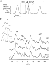
1. We have used 31P NMR spectroscopy to measure metabolites and pHi at three periods during a phasic contraction of the uterus, in vivo, to determine whether they change as a consequence of contraction. The regular uterine contractions were recorded via a balloon catheter in the uterine lumen. Each phasic contraction was divided into three parts: the period between contractions (rest), the development of force (up) and the relaxation of force (down). The NMR data were summed separately from each of these three periods over 20-40 successive contractions. 2. Significant changes in ATP, phosphocreatine (PCr) and inorganic phosphate (Pi) occurred during the contraction. [ATP] fell from 2.0 to 1.6 mM and [PCr] from 2.6 to 2.0 mM during the up period, while [Pi] increased from 2.2 to 2.8 mM. Recovery of ATP and PCr occurred during the relaxation part of the contraction, whereas Pi did not fully recover until the contraction was complete. 3. Significant acidification from pH 7.28 +/- 0.02 at rest to 7.16 +/- 0.02, occurred with contraction. This acidification is greater than that previously reported for in vitro uterine preparations. Measurements of uterine blood flow show that it decreased with contraction. Therefore, ischaemia, in addition to the metabolic consequences of contraction, may account for the larger acidification observed in vivo. 4. Lowering pHi in an in vitro uterine preparation by a similar level to that found in vivo produced a significant reduction of the phasic contractions. Thus we propose that these changes, especially the fall in pHi during force development, feed back negatively on the contraction to limit its strength, and may help prevent uterine ischaemia and fetal hypoxia during labour.
Figures


References
-
- Adams GR, Dillon PF. Glucose dependence of sequential norepinephrine contractions of vascular smooth muscle. Blood Vessels. 1989;26:77–83. - PubMed
-
- Barany K, Barany M. Myosin light chain phosphorylation in uterine smooth muscle. In: Carsten ME, Miller JD, editors. Uterine Function, Molecular and Cellular Aspects. New York: Plenum Press; 1990. pp. 71–98.
-
- Brar HS, Platt LD, Devore GR. Qualitative assessment of maternal uterine and fetal umbilical artery blood flow and resistance in laboring patients by Doppler velocimetry. American Journal of Obstetrics and Gynecology. 1988;158:952–956. - PubMed
-
- Bullock AJ, Duquette RA, Buttell N, Wray S. Developmental changes in intracellular pH buffering power in smooth muscle. Pflügers Archiv. 1998;435:575–577. - PubMed
-
- Carafoli E. Intracellular calcium homeostasis. Annual Review of Biochemistry. 1987;56:395–433. - PubMed
Publication types
MeSH terms
Substances
Grants and funding
LinkOut - more resources
Full Text Sources
Research Materials
Miscellaneous

