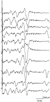
Dissociation of the pathways mediating ipsilateral and contralateral motor-evoked potentials in human hand and arm muscles
- PMID: 10420023
- PMCID: PMC2269467
- DOI: 10.1111/j.1469-7793.1999.0895p.x
Dissociation of the pathways mediating ipsilateral and contralateral motor-evoked potentials in human hand and arm muscles
Abstract
1. Growing evidence points toward involvement of the human motor cortex in the control of the ipsilateral hand. We used focal transcranial magnetic stimulation (TMS) to examine the pathways of these ipsilateral motor effects. 2. Ipsilateral motor-evoked potentials (MEPs) were obtained in hand and arm muscles of all 10 healthy adult subjects tested. They occurred in the finger and wrist extensors and the biceps, but no response or inhibitory responses were observed in the opponens pollicis, finger and wrist flexors and the triceps. 3. The production of ipsilateral MEPs required contraction of the target muscle. The threshold TMS intensity for ipsilateral MEPs was on average 1.8 times higher, and the onset was 5.7 ms later (in the wrist extensor muscles) compared with size-matched contralateral MEPs. 4. The corticofugal pathways of ipsilateral and contralateral MEPs could be dissociated through differences in cortical map location and preferred stimulating current direction. 5. Both ipsi- and contralateral MEPs in the wrist extensors increased with lateral head rotation toward, and decreased with head rotation away from, the side of the TMS, suggesting a privileged input of the asymmetrical tonic neck reflex to the pathway of the ipsilateral MEP. 6. Large ipsilateral MEPs were obtained in a patient with complete agenesis of the corpus callosum. 7. The dissociation of the pathways for ipsilateral and contralateral MEPs indicates that corticofugal motor fibres other than the fast-conducting crossed corticomotoneuronal system can be activated by TMS. Our data suggest an ipsilateral oligosynaptic pathway, such as a corticoreticulospinal or a corticopropriospinal projection as the route for the ipsilateral MEP. Other pathways, such as branching of corticomotoneuronal axons, a transcallosal projection or a slow-conducting monosynaptic ipsilateral pathway are very unlikely or can be excluded.
Figures


Similar articles
-
Cortical motor representation of the ipsilateral hand and arm.Exp Brain Res. 1994;100(1):121-32. doi: 10.1007/BF00227284. Exp Brain Res. 1994. PMID: 7813640 Clinical Trial.
-
Different effects of fatiguing exercise on corticospinal and transcallosal excitability in human hand area motor cortex.Exp Brain Res. 2004 Dec;159(4):530-6. doi: 10.1007/s00221-004-1978-y. Epub 2004 Jul 13. Exp Brain Res. 2004. PMID: 15249989 Clinical Trial.
-
Spread of electrical activity at cortical level after repetitive magnetic stimulation in normal subjects.Exp Brain Res. 2002 Nov;147(2):186-92. doi: 10.1007/s00221-002-1237-z. Epub 2002 Sep 20. Exp Brain Res. 2002. PMID: 12410333
-
Ia presynaptic inhibition in human wrist extensor muscles: effects of motor task and cutaneous afferent activity.J Physiol Paris. 1999 Sep-Oct;93(4):395-401. doi: 10.1016/s0928-4257(00)80067-4. J Physiol Paris. 1999. PMID: 10574128 Review.
-
Functional studies of motor cortex.Ciba Found Symp. 1987;132:83-97. doi: 10.1002/9780470513545.ch6. Ciba Found Symp. 1987. PMID: 3123172 Review.
Cited by
-
Ipsilateral Motor Evoked Potentials as a Measure of the Reticulospinal Tract in Age-Related Strength Changes.Front Aging Neurosci. 2021 Mar 3;13:612352. doi: 10.3389/fnagi.2021.612352. eCollection 2021. Front Aging Neurosci. 2021. PMID: 33746734 Free PMC article.
-
Minimum Electromyographic Burst Duration in Healthy Controls: Implications for Electrodiagnosis in Movement Disorders.Mov Disord Clin Pract. 2020 Aug 26;7(7):827-833. doi: 10.1002/mdc3.13044. eCollection 2020 Oct. Mov Disord Clin Pract. 2020. PMID: 33033737 Free PMC article.
-
White matter tract disruption is associated with ipsilateral hand impairment in subacute stroke: a diffusion MRI study.Neuroradiology. 2022 Aug;64(8):1605-1615. doi: 10.1007/s00234-022-02927-8. Epub 2022 Mar 28. Neuroradiology. 2022. PMID: 35344052
-
Electrical stimulation driving functional improvements and cortical changes in subjects with stroke.Exp Brain Res. 2004 Feb;154(4):450-60. doi: 10.1007/s00221-003-1695-y. Epub 2003 Nov 15. Exp Brain Res. 2004. PMID: 14618287 Clinical Trial.
-
Modulation of transcallosal inhibition by bilateral activation of agonist and antagonist proximal arm muscles.J Neurophysiol. 2014 Jan;111(2):405-14. doi: 10.1152/jn.00322.2013. Epub 2013 Oct 23. J Neurophysiol. 2014. PMID: 24155008 Free PMC article.
References
-
- Aiello I, Rosati G, Sau GF, Patraskakis S, Bissakou M, Traccis S. Tonic neck reflexes on upper limb flexor tone in man. Experimental Neurology. 1988;101:41–49. - PubMed
-
- Aizawa H, Mushiake H, Inase M, Tanji J. An output zone of the monkey primary motor cortex specialized for bilateral hand movement. Experimental Brain Research. 1990;82:219–221. - PubMed
-
- Basu AP, Turton A, Lemon RN. Activation of ipsilateral upper limb muscles by transcranial magnetic stimulation. The Journal of Physiology. 1994;479.P:144P.
-
- Benecke R, Meyer BU, Freund HJ. Reorganisation of descending motor pathways in patients after hemispherectomy and severe hemispheric lesions demonstrated by magnetic brain stimulation. Experimental Brain Research. 1991;83:419–426. - PubMed
-
- Brasil-Neto JP, Cohen LG, Panizza M, Nilsson J, Roth BJ, Hallett M. Optimal focal transcranial magnetic activation of the human motor cortex: Effects of coil orientation, shape of the induced current pulse, and stimulus intensity. Journal of Clinical Neurophysiology. 1992;9:132–136. - PubMed
Publication types
MeSH terms
LinkOut - more resources
Full Text Sources

