. 1999 Jul 15;13(14):1822-33.
doi: 10.1101/gad.13.14.1822.

The von Hippel-Lindau tumor suppressor protein is a component of an E3 ubiquitin-protein ligase activity

Affiliations

The von Hippel-Lindau tumor suppressor protein is a component of an E3 ubiquitin-protein ligase activity

J Lisztwan et al. Genes Dev. .

Abstract

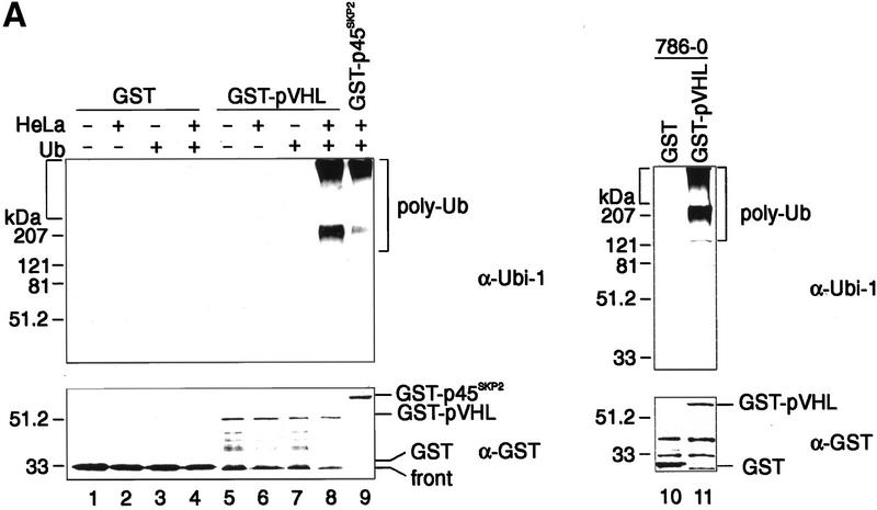
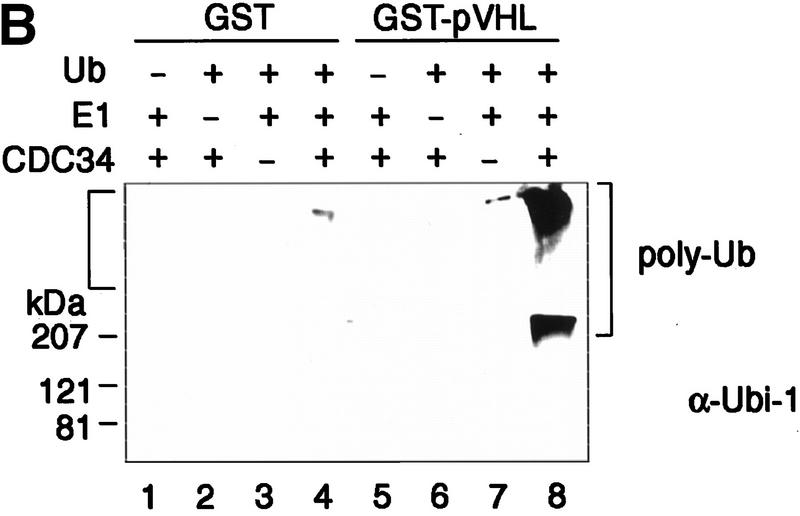
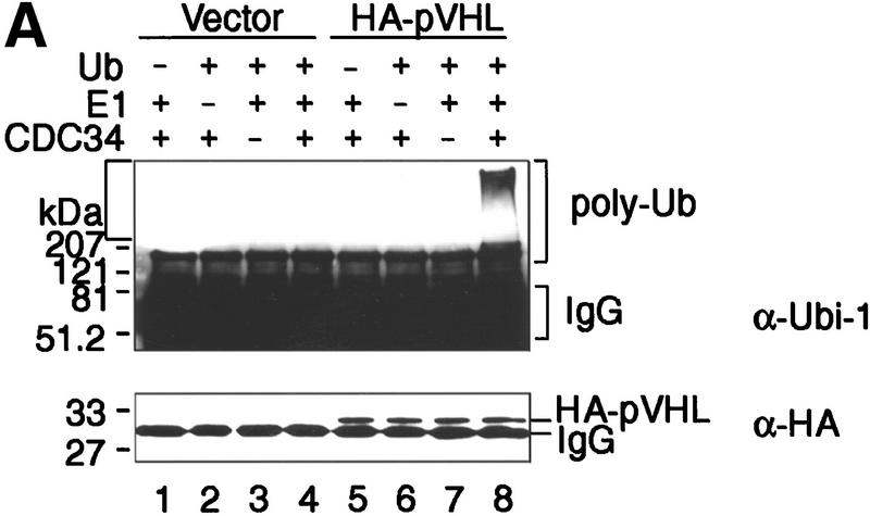
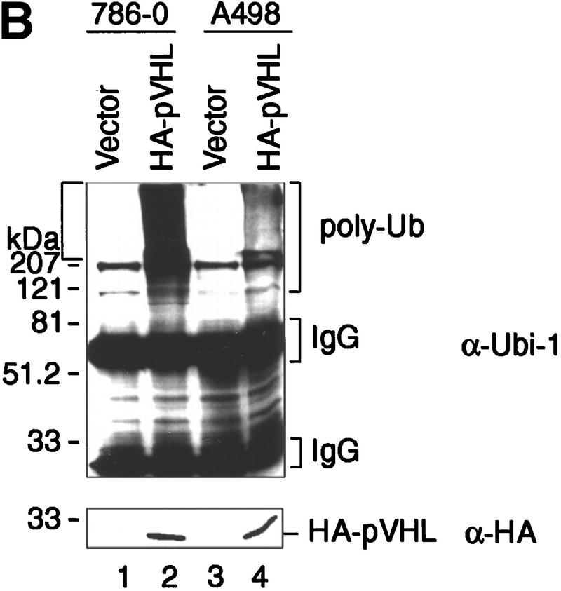
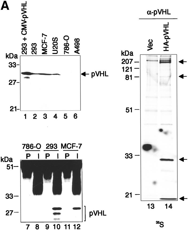
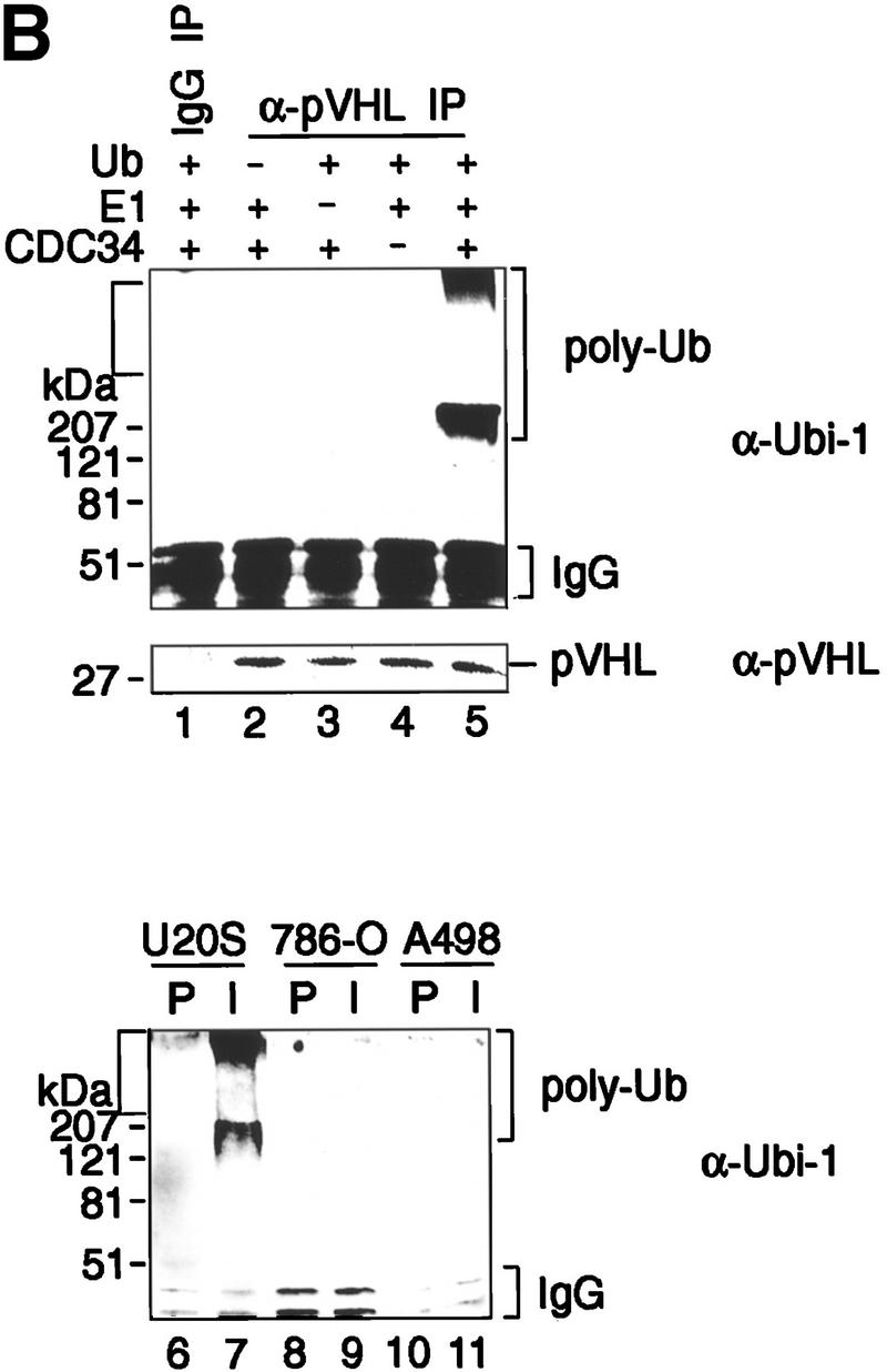
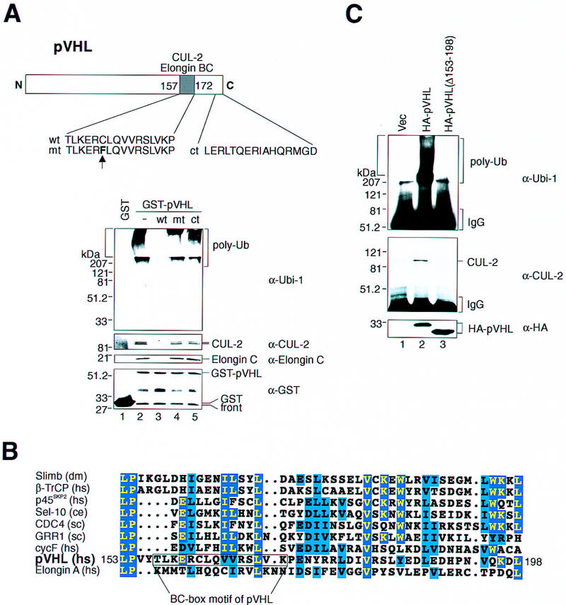
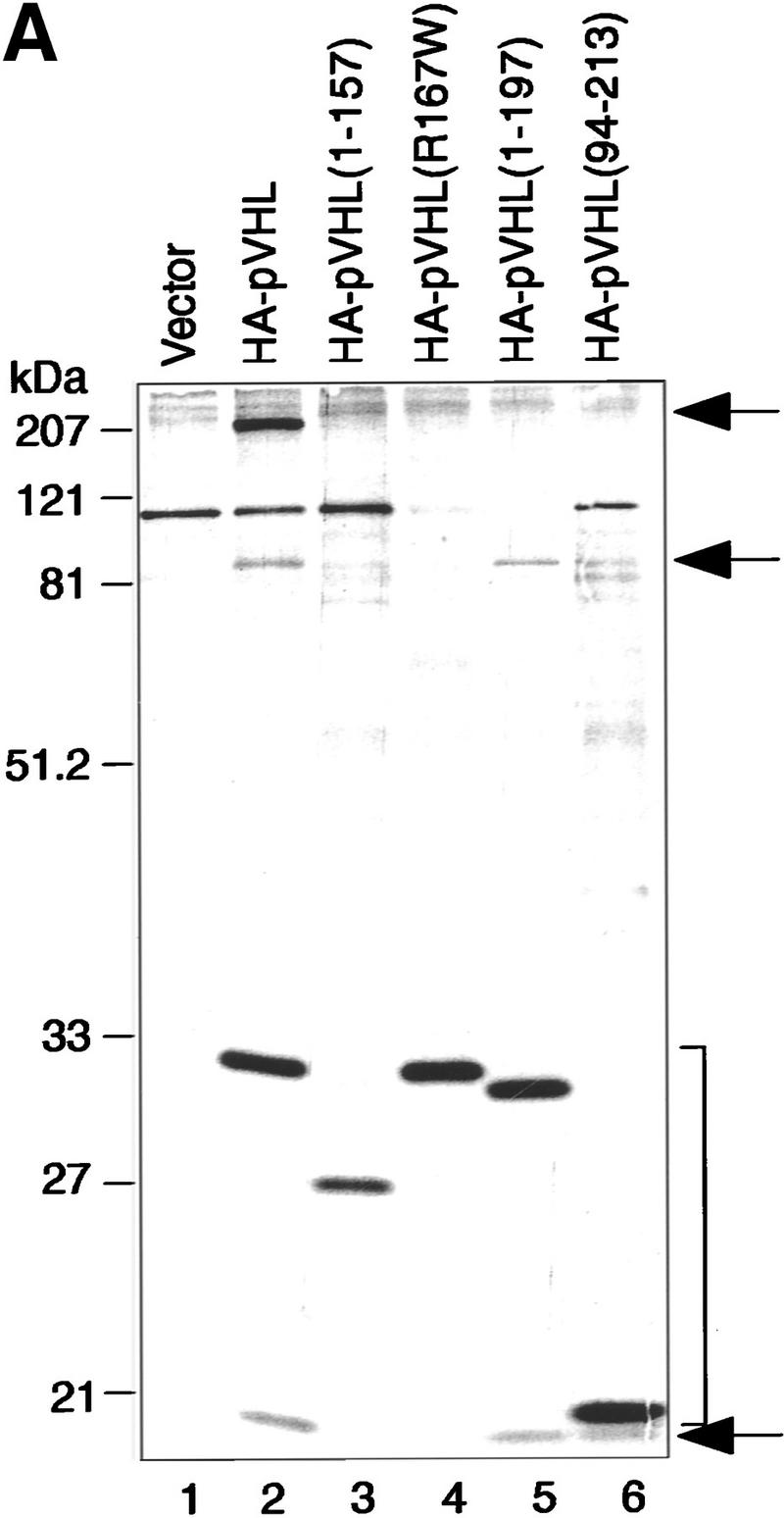
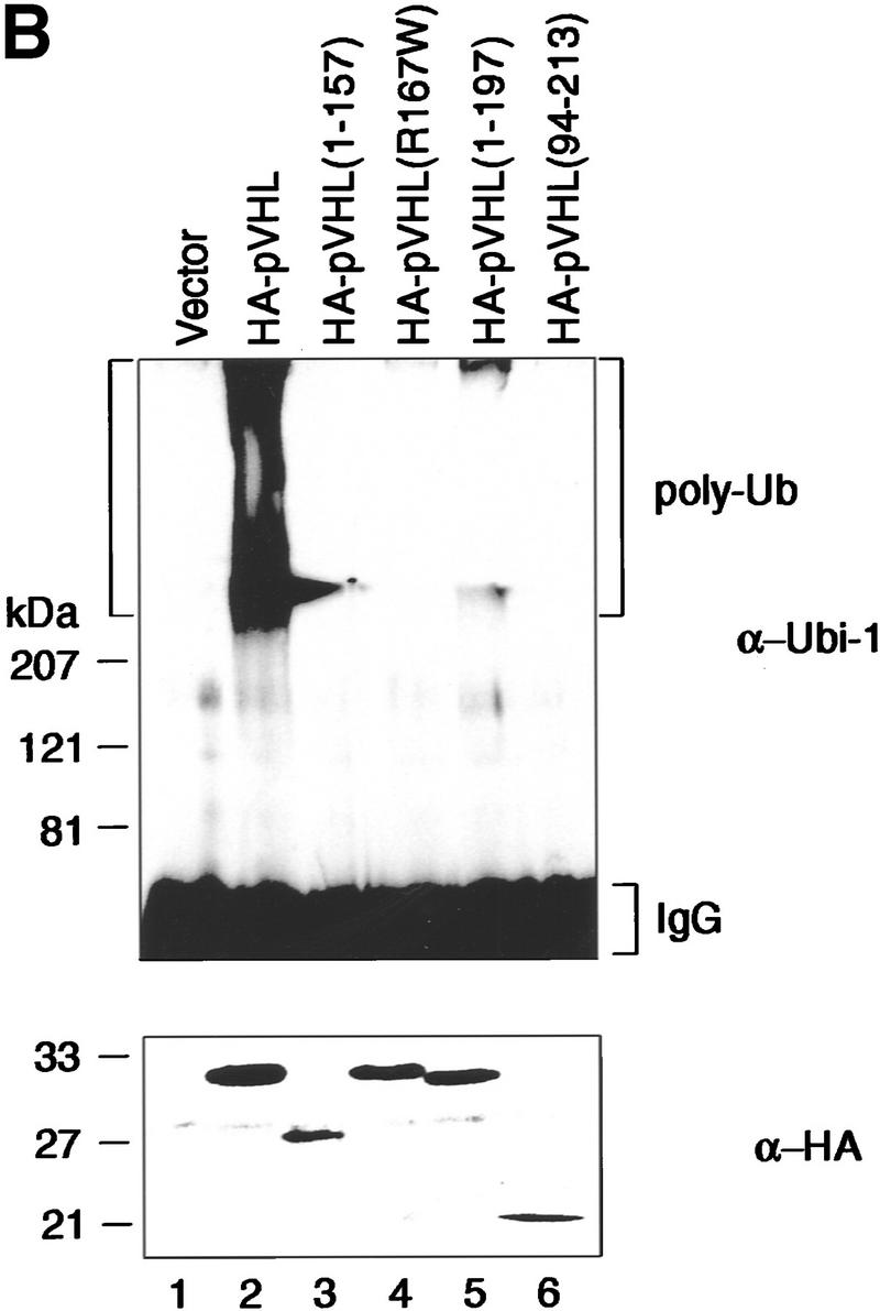
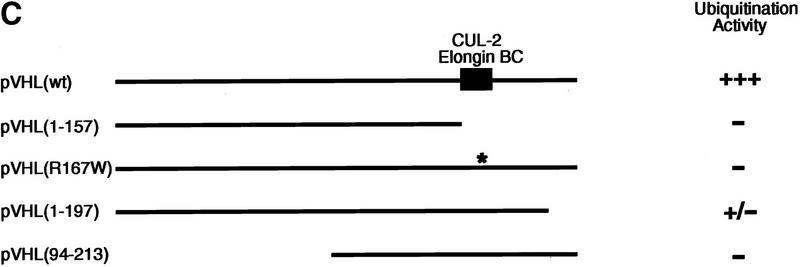
pVHL, the product of the VHL tumor suppressor gene, plays an important role in the regulation of cell growth and differentiation of human kidney cells, and inactivation of the VHL gene is the most frequent genetic event in human kidney cancer. The biochemical function of pVHL is unknown. Here we report that pVHL exists in vivo in a complex that displays ubiquitination-promoting activity in conjunction with the universally required components E1, E2, and ubiquitin. pVHL-associated ubiquitination activity requires, at a minimum, pVHL to bind elongin C and Cul-2, relatives of core components of SCF (Skp1-Cdc53/Cul-1-F-box protein) E3 ligase complexes. Notably, certain tumor-derived mutants of pVHL demonstrate loss of associated ubiquitination promoting activity. These results identify pVHL as a component of a potential SCF-like E3 ubiquitin-protein ligase complex and suggest a direct link between pVHL tumor suppressor and the process of ubiquitination.


Figures

Figure 1
Figure 1
Recombinant pVHL purifies ubiquitination-promoting activity from human cell extracts. (A) GST (lanes 14,10), GST–pVHL (lanes 58,11), or GST–p45Skp2 (lane 9) was bound to glutathione–Sepharose beads and either directly exposed to the ubiquitination reaction as described in Materials and Methods in the absence (lanes 1,2,5,6) or presence (lanes 3,4,7,8,9) of ubiquitin (Ub) or were first incubated overnight in HeLa cell lysates as indicated by the plus and minus signs at top or in 786-O cell lysates (lanes 10,11). Reaction mixtures were analyzed on 10% SDS-gels and processed for Western blotting with anti-ubiquitin antibody (Ubi-1) (top) or reprobing with anti-GST antibody (bottom). (B) GST (lanes 14) or GST–pVHL (lanes 58) was bound to glutathione–Sepharose beads, incubated in HeLa lysates and exposed to the ubiquitination assay conditions as described in A in the absence or presence of either Ub, E1, or Cdc34 as indicated at top. Samples were analyzed on 10% SDS-gels and processed for Western blotting with anti-ubiquitin antibody (Ubi-1). Square bracket at left indicates the gel stack. Square bracket at right indicates the ladder of multi-ubiquitinated proteins.
Figure 1
Figure 1
Recombinant pVHL purifies ubiquitination-promoting activity from human cell extracts. (A) GST (lanes 14,10), GST–pVHL (lanes 58,11), or GST–p45Skp2 (lane 9) was bound to glutathione–Sepharose beads and either directly exposed to the ubiquitination reaction as described in Materials and Methods in the absence (lanes 1,2,5,6) or presence (lanes 3,4,7,8,9) of ubiquitin (Ub) or were first incubated overnight in HeLa cell lysates as indicated by the plus and minus signs at top or in 786-O cell lysates (lanes 10,11). Reaction mixtures were analyzed on 10% SDS-gels and processed for Western blotting with anti-ubiquitin antibody (Ubi-1) (top) or reprobing with anti-GST antibody (bottom). (B) GST (lanes 14) or GST–pVHL (lanes 58) was bound to glutathione–Sepharose beads, incubated in HeLa lysates and exposed to the ubiquitination assay conditions as described in A in the absence or presence of either Ub, E1, or Cdc34 as indicated at top. Samples were analyzed on 10% SDS-gels and processed for Western blotting with anti-ubiquitin antibody (Ubi-1). Square bracket at left indicates the gel stack. Square bracket at right indicates the ladder of multi-ubiquitinated proteins.
Figure 2
Figure 2
Ectopically produced pVHL displays associated ubiquitination-promoting activity. (A) VHL−/− 786-O cell lines stably transfected with a plasmid encoding HA-tagged pVHL (lanes 58) or with the backbone expression plasmid (lanes 14) were lysed and immunoprecipitated with anti-HA antibody. Immunoprecipitates were processed for ubiquitination assay reactions as described in Material and Methods, omitting Ub (lanes 1,5), E1 (lanes 2,6), or Cdc34 (lanes 3,7) from reaction mixtures. Samples were analyzed on 10% SDS gels and processed for Western blotting with anti-ubiquitin antibody (Ubi-1) (top) or anti-HA antibody (bottom). (B) VHL−/− 786-O and A498 cell lines stably transfected with a plasmid encoding HA-tagged pVHL (lanes 2,4) or with the backbone expression plasmid (lanes 1,3) were lysed, immunoprecipitated with anti-HA antibody and immunoprecipitates were processed for ubiquitination assay reactions and Western blot analysis as described above with anti-ubiquitin antibody (Ubi-1) (top) or anti-HA antibody (bottom).
Figure 2
Figure 2
Ectopically produced pVHL displays associated ubiquitination-promoting activity. (A) VHL−/− 786-O cell lines stably transfected with a plasmid encoding HA-tagged pVHL (lanes 58) or with the backbone expression plasmid (lanes 14) were lysed and immunoprecipitated with anti-HA antibody. Immunoprecipitates were processed for ubiquitination assay reactions as described in Material and Methods, omitting Ub (lanes 1,5), E1 (lanes 2,6), or Cdc34 (lanes 3,7) from reaction mixtures. Samples were analyzed on 10% SDS gels and processed for Western blotting with anti-ubiquitin antibody (Ubi-1) (top) or anti-HA antibody (bottom). (B) VHL−/− 786-O and A498 cell lines stably transfected with a plasmid encoding HA-tagged pVHL (lanes 2,4) or with the backbone expression plasmid (lanes 1,3) were lysed, immunoprecipitated with anti-HA antibody and immunoprecipitates were processed for ubiquitination assay reactions and Western blot analysis as described above with anti-ubiquitin antibody (Ubi-1) (top) or anti-HA antibody (bottom).
Figure 3
Figure 3
Immunoprecipitates of endogenous pVHL possess ubiquitination-promoting activity. (A) Specificity of anti-pVHL antibody. Whole cell extracts were prepared by the addition of SDS-sample buffer to 293 cells transfected with expression plasmids encoding untagged pVHL (lane 1) as well as to untransfected 293 (lane 2), MCF-7 (lane 3), U2-OS (lane 4), 786-O (lane 5), and A498 (lane 6) cells. Protein mixtures were separated by SDS-PAGE and processed for Western blotting with affinity-purified anti-pVHL antibody. The exposure time of lane 1 is ten times less than that of lanes 26. Note that anti-pVHL antibodies do not recognize pVHL, as expected, in VHL−/− 786-O or A498 cells. Whole cell extracts prepared from 786-O (lanes 7,8), 293 (lanes 9,10), or MCF-7 (lanes 11,12) cells were subjected to immunoprecipitation with either 15 μg of affinity-purified anti-pVHL antibody (lanes 8,10,12) or affinitypurified rabbit IgG (lanes 7,9,11) and processed for Western blotting with affinity-purified anti-pVHL antibody. VHL−/− 786-O cells ectopically producing HA-tagged pVHL wild-type (lane 14) or containing backbone expression plasmid (lane 13) were metabolically labeled with [35S]methionine, lysed, and immunoprecipitated with 10 μg of affinity-purified anti-pVHL antibodies. Bound proteins were resolved by SDS-PAGE and detected by fluorography. Arrows at right mark the positions of coimmunoprecipitating fibronection, Cul-2, HA–pVHL, and Elongin B (from top to bottom). Note that the pattern of coimmunoprecipitating proteins are similar to work published previously (for references see Results). (B) Lysates were prepared from 293 (lanes 15), U2-OS (lanes 6,7), 786-O (lanes 8,9), and A498 (lanes 10,11) cells and subjected to immunoprecipitations with 5 μg of either affinity-purified rabbit IgG (lanes 1,6,8,10) or affinity-purified anti-pVHL antibody (lanes 25,7,9,11) and processed as described in the legend to Fig. 2A. Samples were analyzed on 10% SDS gels and processed for Western blotting with anti-ubiquitin antibody (Ubi-1) or anti-pVHL antibody.
Figure 3
Figure 3
Immunoprecipitates of endogenous pVHL possess ubiquitination-promoting activity. (A) Specificity of anti-pVHL antibody. Whole cell extracts were prepared by the addition of SDS-sample buffer to 293 cells transfected with expression plasmids encoding untagged pVHL (lane 1) as well as to untransfected 293 (lane 2), MCF-7 (lane 3), U2-OS (lane 4), 786-O (lane 5), and A498 (lane 6) cells. Protein mixtures were separated by SDS-PAGE and processed for Western blotting with affinity-purified anti-pVHL antibody. The exposure time of lane 1 is ten times less than that of lanes 26. Note that anti-pVHL antibodies do not recognize pVHL, as expected, in VHL−/− 786-O or A498 cells. Whole cell extracts prepared from 786-O (lanes 7,8), 293 (lanes 9,10), or MCF-7 (lanes 11,12) cells were subjected to immunoprecipitation with either 15 μg of affinity-purified anti-pVHL antibody (lanes 8,10,12) or affinitypurified rabbit IgG (lanes 7,9,11) and processed for Western blotting with affinity-purified anti-pVHL antibody. VHL−/− 786-O cells ectopically producing HA-tagged pVHL wild-type (lane 14) or containing backbone expression plasmid (lane 13) were metabolically labeled with [35S]methionine, lysed, and immunoprecipitated with 10 μg of affinity-purified anti-pVHL antibodies. Bound proteins were resolved by SDS-PAGE and detected by fluorography. Arrows at right mark the positions of coimmunoprecipitating fibronection, Cul-2, HA–pVHL, and Elongin B (from top to bottom). Note that the pattern of coimmunoprecipitating proteins are similar to work published previously (for references see Results). (B) Lysates were prepared from 293 (lanes 15), U2-OS (lanes 6,7), 786-O (lanes 8,9), and A498 (lanes 10,11) cells and subjected to immunoprecipitations with 5 μg of either affinity-purified rabbit IgG (lanes 1,6,8,10) or affinity-purified anti-pVHL antibody (lanes 25,7,9,11) and processed as described in the legend to Fig. 2A. Samples were analyzed on 10% SDS gels and processed for Western blotting with anti-ubiquitin antibody (Ubi-1) or anti-pVHL antibody.
Figure 4
Figure 4
Effects of BC-box peptides on pVHL-associated ubiquitination activity. (A) Structure of pVHL(wt). The schematic depicts the carboxy-terminal BC-box motif that serves as an interaction site for Elongin BC/Cul-2 complexes. The amino acid sequence of peptides encoding the wild-type (wt) and mutant (mt) BC-box domain and the most carboxy-terminal 16 amino acids (ct) are displayed below. The vertical arrow highlights the single point mutation present in the mt peptide. GST (lane 1) or GST–pVHL (lanes 25) was bound to glutathione–Sepharose beads, incubated in HeLa lysates in the absence (lanes 1,2) or presence of either wt (lane 3), mt (lane 4), or ct (lane 5) peptides and exposed to ubiquitination assay conditions as described in the legend to Fig. 1A. Reaction mixtures were analyzed on 10% SDS gels and processed for Western blotting with either anti-ubiquitin (Ubi-1) (top panel), anti-Cul-2 (second panel), anti-Elongin C (third panel), or anti-GST (bottom panel) antibodies. Note, that HeLa Cul-2 and Elongin C failed to associate with GST–pVHL in the presence of wt but not mt peptide. (B) Sequence alignment of the main region of homology between pVHL and the F-box motif of selected F-box proteins of different origins. Identical residues are indicated by yellow letters on blue boxes and functionally similar residues are in black letters on blue boxes. The BC-box motif of pVHL comprising 15 amino acid residues is shown. (C) VHL−/− 786-O cells producing the indicated HA-tagged pVHL species (lanes 2,3) or containing backbone expression plasmid (lane 1) were lysed and immunoprecipitated with anti-HA antibody and immunoprecipitates were processed for ubiquitination assay reactions and Western blot analysis with anti-ubiquitin antibody (Ubi-1) (top) or anti-Cul-2 antibody (middle). Aliquots of whole cell extracts of the aforementioned cell lines were processed for Western blotting with anti-HA antibody (bottom).
Figure 5
Figure 5
Tumor-derived mutations of pVHL fail to display ubiquitination-promoting activity. (A) VHL−/− 786-O cells ectopically producing the indicated HA-tagged pVHL species (lanes 26) or containing backbone expression plasmid (lane 1) were metabolically labeled with [35S]methionine, lysed, and immunoprecipitated with anti-HA antibody. Bound proteins were resolved by SDS-PAGE and detected by fluorography. Arrows at right mark the positions of coimmunoprecipitating Fibronection, Cul-2, and Elongin B (from top to bottom). Note that the pattern of coimmunoprecipitating proteins are similar to previously published work (for references see Results). The bracket indicates the positions of the various pVHL species. Elongin C is not visible on this SDS gel because it migrated with the front. (B) VHL−/− 786-O cell lines analyzed in A were lysed, immunoprecipitated with anti-HA antibody, and processed for ubiquitination assay reactions as described in the legend to Fig. 3A. Samples were analyzed on 10% SDS gels and processed for Western blotting with anti-ubiquitin antibody (Ubi-1) (top) or anti-HA antibody (bottom). (C) Schematic representation of the various pVHL mutants tested and their corresponding ubiquitination activity (+ and −). (█) The BC box in pVHL; (*) the location of the point mutation.
Figure 5
Figure 5
Tumor-derived mutations of pVHL fail to display ubiquitination-promoting activity. (A) VHL−/− 786-O cells ectopically producing the indicated HA-tagged pVHL species (lanes 26) or containing backbone expression plasmid (lane 1) were metabolically labeled with [35S]methionine, lysed, and immunoprecipitated with anti-HA antibody. Bound proteins were resolved by SDS-PAGE and detected by fluorography. Arrows at right mark the positions of coimmunoprecipitating Fibronection, Cul-2, and Elongin B (from top to bottom). Note that the pattern of coimmunoprecipitating proteins are similar to previously published work (for references see Results). The bracket indicates the positions of the various pVHL species. Elongin C is not visible on this SDS gel because it migrated with the front. (B) VHL−/− 786-O cell lines analyzed in A were lysed, immunoprecipitated with anti-HA antibody, and processed for ubiquitination assay reactions as described in the legend to Fig. 3A. Samples were analyzed on 10% SDS gels and processed for Western blotting with anti-ubiquitin antibody (Ubi-1) (top) or anti-HA antibody (bottom). (C) Schematic representation of the various pVHL mutants tested and their corresponding ubiquitination activity (+ and −). (█) The BC box in pVHL; (*) the location of the point mutation.

References

    1. Aso T, Lane WS, Conaway JW, Conaway RC. Elongin (SIII): A multisubunit regulator of elongation by RNA polymerase II. Science. 1995;269:1439–1443. - PubMed
    1. Aso T, Haque D, Barstead RJ, Conaway RC, Conaway JW. The inducible elongin A elongation activation domain: Structure, function and interaction with the elongin BC complex. EMBO J. 1996;15:5557–5566. - PMC - PubMed
    1. Bai C, Sen P, Hofmann K, Ma L, Goebl M, Harper JW, Elledge SJ. SKP1 connects cell cycle regulators to the ubiquitin proteolysis machinery through a novel motif, the F-box. Cell. 1996;86:263–274. - PubMed
    1. Banerjee A, Gregori L, Xu Y, Chau V. The bacterially expressed yeast CDC34 gene product can undergo autoubiquitination to form a multiubiquitin chain-linked protein. J Biol Chem. 1993;268:5668–5675. - PubMed
    1. Brodsky JL, McCracken AA. ER-associated proteasome-mediated protein degradation: How two topologically restricted events come together. Trends Cell Biol. 1997;7:151–156. - PubMed

Publication types

MeSH terms

LinkOut - more resources