. 1999 Jul 26;146(2):273-84.
doi: 10.1083/jcb.146.2.273.

Expression of the 180-kD ribosome receptor induces membrane proliferation and increased secretory activity in yeast

Affiliations

Expression of the 180-kD ribosome receptor induces membrane proliferation and increased secretory activity in yeast

F Becker et al. J Cell Biol. .

Abstract

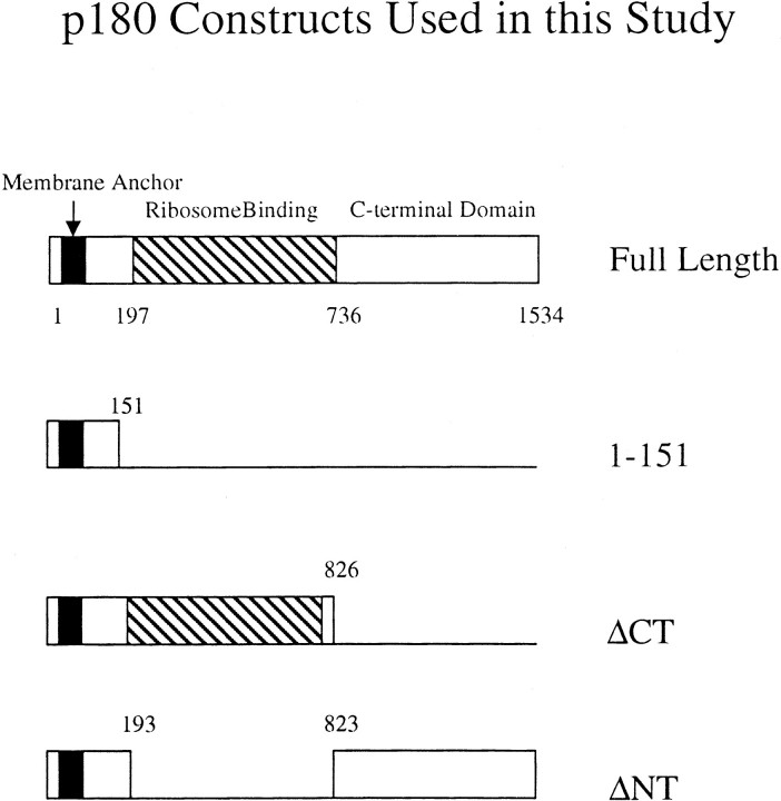
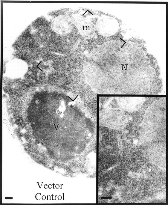
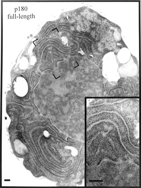
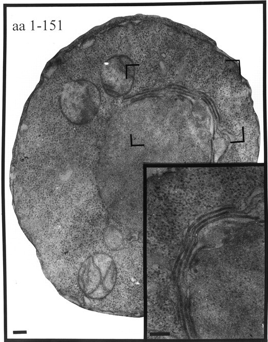
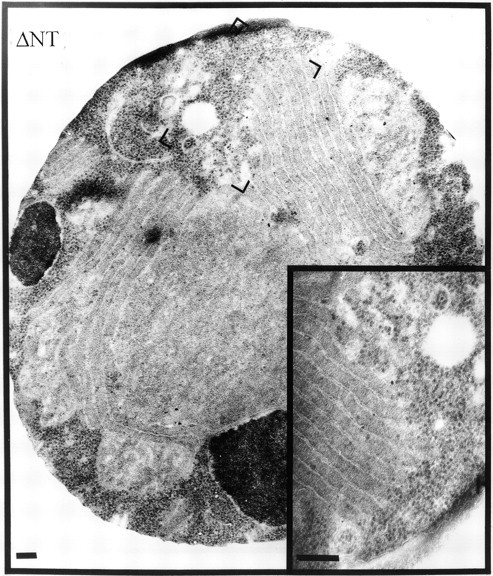
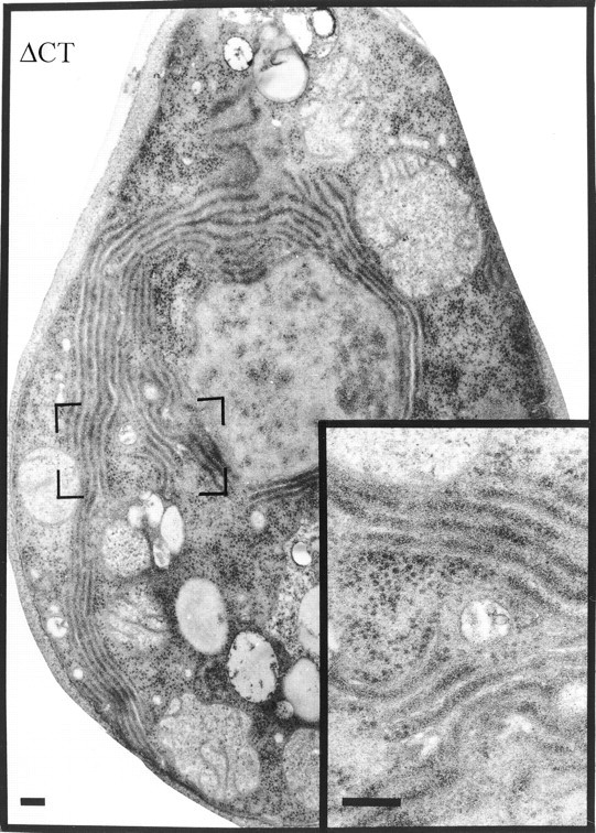
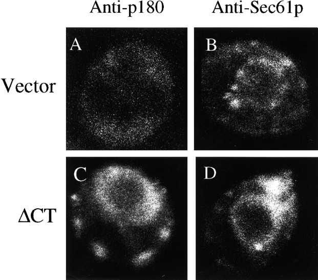
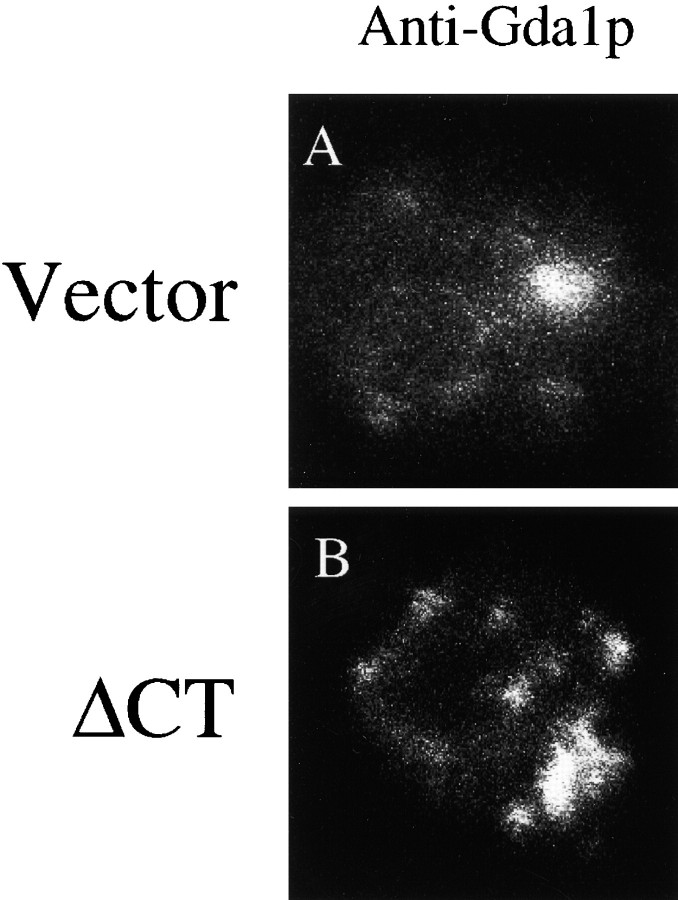
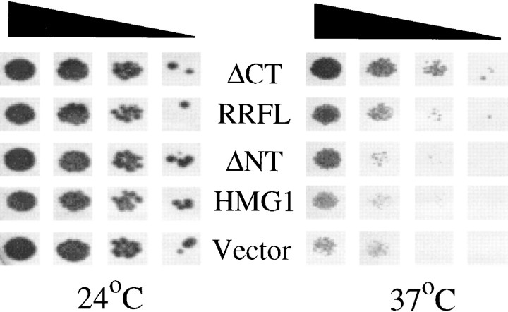
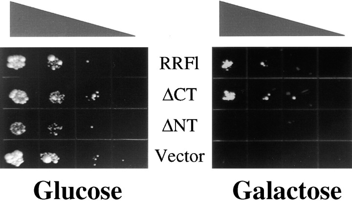
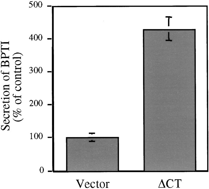
Expression of the canine 180-kD ribosome receptor (p180) in yeast cells resulted in a marked proliferation of intracellular membranes. The type of membranes observed varied with the expression of specific portions of p180. Rough membranes predominated when the ribosome binding domain of p180 was present, whereas expression constructs lacking this region resulted in smooth membranes. Northern analysis indicated that expression of the NH(2)-terminal 767 amino acids (DeltaCT), which include the ribosome binding domain, upregulated the transcription and translation of genes involved in exocytosis. The membranes that were proliferated were functional as these cells overcame a temperature-sensitive translocation defect. Most significantly, cells that overexpressed DeltaCT and proliferated rough endoplasmic reticulum exhibited severalfold higher levels of secretion of an ectopically expressed secretory protein. We conclude that p180 expression triggers a cascade of events leading to an increase in secretory potential akin to the terminal differentiation of mammalian secretory cells and tissues.


Figures

Figure 1
Figure 1
Constructs used in this study. A description of cloning strategies can be found in Materials and Methods. The striped area, encoding a domain with 54 tandem decapeptides, has been shown previously to be essential for ribosome binding (Wanker et al. 1995). Numbers shown correspond to amino acids in the primary structure. In the text, the full-length construct is referred to as p180FL. When p1-151 is expressed, a protein is produced that comprises the NH2-terminal 151 amino acids of p180. The Membrane Anchor designation refers to amino acids 6–33, which are uncharged and mainly hydrophobic, and whose deletion leads to the production of a soluble form of p180.
Figure 2
Figure 2
Vector-only control yeast cells have few intracellular membranes. Cells were transformed with pYEX-BX. Transformants were selected and grown in Cu2+-containing medium for 5 h before preparation for electron microscopy. Visible are mitochondria, the nucleus, the vacuole, and substantial quantities of free ribosomes in the cytosol. Bars, 200 nm.
Figure 3
Figure 3
Expression of full-length p180 induces rough membrane proliferation. Cells were transformed with pYEX-BX containing full-length p180 cDNA. Transformants were selected and grown in Cu2+-containing medium for 5 h before preparation for electron microscopy. Visible are extensive arrays of ribosome-studded membranes. Bars, 200 nm.
Figure 4
Figure 4
Expression of amino acids 1–151 of p180 induces membrane proliferation having a karmellar morphology. Cells were transfected with pYEX-BX-AA1-151. Transformants were selected and grown in Cu2+-containing medium for 5 h before preparation for electron microscopy. Visible are perinuclear membranes similar in appearance to karmellae. Bars, 200 nm.
Figure 5
Figure 5
Expression of p180 without its ribosome binding domain produces extensive smooth membrane proliferation different from karmellae. Cells were transfected with pYEX-BX containing the ΔNT version of p180. Transformants were selected and grown in Cu2+-containing medium for 5 h before preparation for electron microscopy. Visible are parallel arrays of smooth membranes. Note the exclusion of cytoplasmic ribosomes from the areas where membranes are evident, and their high density in membrane-free zones. Bars, 200 nm.
Figure 6
Figure 6
Expression of the NH2-terminal half of p180 induces extensive, closely spaced rough membranes. Cells were transfected with pYEX-BX containing the ΔCT version of p180. Transformants were selected and grown in Cu2+-containing medium for 5 h before preparation for electron microscopy. Visible are closely opposed rough membranes with a lower density of ribosomes in membrane-free zones (compared with cells shown in Fig. 4). Bars, 200 nm.
Figure 7
Figure 7
Expression of ΔCT results in a proliferation of Sec61p-containing membranes. Cells were transfected with pYEX-BX containing the ΔCT version of p180 cDNA. Transformants were selected and grown in Cu2+-containing medium for 5 h before preparation for immunofluorescence microscopy. (A and C) Vector-only and ΔCT-expressing cells stained with anti-p180 antibodies. (B and D) Vector-only and ΔCT-expressing cells stained with anti-Sec61p antibodies.
Figure 9
Figure 9
Expression of ΔCT results in a proliferation of Gda1p-containing membranes. Cells were grown and prepared as described in Fig. 7. Cells were stained with antibodies against the Golgi marker protein Gda1p.
Figure 8
Figure 8
Induced membrane proliferation rescues a temperature sensitive translocation defect. Cells harboring the sec63/ptl1ts mutation were transformed with the constructs shown in the middle of the figure (RRFL is the same as p180FL). Transformants were serially diluted (10-fold steps) onto Cu2+-containing medium and grown at either the permissive (24°C) or nonpermissive (37°C) temperature. The triangle above the figure indicates decreasing cell concentrations that were plated.
Figure 10
Figure 10
Expression of full-length p180 or ΔCT rescues BPTI-induced growth arrest. Cells transformed with the constructs indicated in the middle of the figure (RRFl is the same as p180FL) were subsequently transformed with a plasmid directing the expression of BPTI under galactose control, and serially diluted (10-fold steps) onto Cu2+-containing growth medium that included either glucose or galactose as the carbon source. Triangle above figure indicates decreasing cell concentrations that were plated.
Figure 11
Figure 11
Expression of ΔCT significantly increases BPTI secretion. Control and ΔCT-expressing cells, prepared as described in the legend to Fig. 10, were grown on galactose and copper-containing medium. BPTI secreted into the medium was determined by the colorimetric assay described in Materials and Methods.

References

    1. Adams A.E.M., Pringle J.R. Relationship of actin and tubulin distribution to bud growth in wild-type and morphogenic-mutant Saccharomyces cerevisiae . J. Cell Biol. 1984;98:934–945. - PMC - PubMed
    1. Adelman M.R., Sabatini D.D., Blobel G. Ribosome-membrane interaction. Nondestructive disassembly of rat liver rough microsomes into ribosomal and membranous components. J. Cell Biol. 1973;56:206–229. - PMC - PubMed
    1. Anderson R.G., Orci L., Brown M.S., Garcia S.L., Goldstein J.L. Ultrastructural analysis of crystalloid endoplasmic reticulum in UT-1 cells and its disappearance in response to cholesterol. J. Cell Sci. 1983;63:1–20. - PubMed
    1. Basson C.T., MacRae C.A., Schoenberg-Fejzo M., Morton C.C., Spinner N.B., Genin A., Krug E., Seidman J.G., Seidman C.E. Identification, characterization and chromosomal localization of the human homolog (hES) of ES/130. Genomics. 1996;35:628–631. - PubMed
    1. Borgese N., Mok W., Kreibich G., Sabatini D.D. Ribosomal-membrane interactionin vitro binding of ribosomes to microsomal membranes. J. Mol. Biol. 1974;88:559–580. - PubMed

Publication types

MeSH terms