. 1999 Jul 26;146(2):313-20.
doi: 10.1083/jcb.146.2.313.

N-Glycans mediate the apical sorting of a GPI-anchored, raft-associated protein in Madin-Darby canine kidney cells

Affiliations

N-Glycans mediate the apical sorting of a GPI-anchored, raft-associated protein in Madin-Darby canine kidney cells

J H Benting et al. J Cell Biol. .

Abstract

Glycosyl-phosphatidylinositol (GPI)- anchored proteins are preferentially transported to the apical cell surface of polarized Madin-Darby canine kidney (MDCK) cells. It has been assumed that the GPI anchor itself acts as an apical determinant by its interaction with sphingolipid-cholesterol rafts. We modified the rat growth hormone (rGH), an unglycosylated, unpolarized secreted protein, into a GPI-anchored protein and analyzed its surface delivery in polarized MDCK cells. The addition of a GPI anchor to rGH did not lead to an increase in apical delivery of the protein. However, addition of N-glycans to GPI-anchored rGH resulted in predominant apical delivery, suggesting that N-glycans act as apical sorting signals on GPI-anchored proteins as they do on transmembrane and secretory proteins. In contrast to the GPI-anchored rGH, a transmembrane form of rGH which was not raft-associated accumulated intracellularly. Addition of N-glycans to this chimeric protein prevented intracellular accumulation and led to apical delivery.


Figures

Figure 1
Figure 1
Schematic of fusion proteins used in this study. SS, signal sequence; rGH0, nonglycosylated rGH; rGH12, rGH with two artificial N-glycosylation sites represented by the trees; DAF, GPI anchor signal of decay acceleration factor; TMD, transmembrane domain of human LDL-R; CT12, truncated cytoplasmic tail of human LDL-R comprising 12 amino acids following the TMD lacking all basolateral sorting information.
Figure 2
Figure 2
Localization of rGH0-DAF and rGH12-DAF in polarized MDCK cells. Polarized filter-grown MDCK cells were fixed 18 h after infection with adenovirus expressing rGH0-DAF (A and B) or rGH12-DAF (C and D). Cells were stained with the anti-rGH antibody and analyzed by confocal immunofluorescence microscopy. Apical optical sections (11 μm above the filter in A and C) and basolateral optical sections (5 μm above the filter in B and D) are shown. Note that rGH0-DAF localizes to the apical and the basolateral side of MDCK cells, whereas rGH12-DAF is predominantly found at the apical surface. Bar, 10 μm.
Figure 4
Figure 4
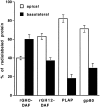
Quantification of biosynthetic surface delivery of rGH0-DAF, rGH12-DAF, PLAP, and gp80. The surface delivery of [35S]methionine–labeled rGH0-DAF, rGH12-DAF, and PLAP was analyzed after a 40-min chase as described and quantified by PhosphoImaging. Apical and basolateral secretion of gp80 was analyzed by immunoprecipitation from the apical and basolateral medium of adenovirus-infected MDCK cells expressing rGH-DAF.
Figure 3
Figure 3
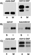
(A) Steady-state distribution of rGH0-DAF and rGH12-DAF. Adenovirus-infected filter-grown MDCK cells expressing rGH0-DAF or rGH12-DAF were surface-biotinylated from the apical (a) or basolateral (bl) surface. Biotinylated proteins were precipitated with streptavidin-agarose. The presence of precipitated rGH0-DAF and rGH12-DAF was analyzed on a Western blot. (B) Both the apical and the basolateral pools of rGH0-DAF are raft-associated. Polarized filter-grown MDCK cells were either biotinylated from the apical or basolateral side. The cells were extracted with TX-100 and DIGs were floated by Optiprep gradient centrifugation. The presence of rGH0-DAF in the soluble fraction (S, 40% Optiprep) and the insoluble floated material (I, 5–30% Optiprep interface) was analyzed on a Western blot. (C) Biosynthetic surface delivery of rGH0-DAF and rGH12-DAF. Filter-grown MDCK cells infected with adenovirus were pulse labeled with [35S]methionine and chased for 40 min as described. Surface proteins were biotinylated from the apical (a) or basolateral (bl) side and rGH-DAF was immunoprecipitated. After elution from the protein A beads, the biotinylated fraction of rGH-DAF was precipitated with streptavidin-agarose and analyzed by SDS-PAGE and autoradiography.
Figure 5
Figure 5
rGH-LDL-R fusion proteins are not raft-associated. TX-100 extraction and floatation of MDCK cell lines expressing rGH0-LDL-R (lanes 1 and 2) or rGH12-LDL-R (lanes 3 and 4). The presence of the fusion proteins and of caveolin-1 (Cav 1) in the soluble fraction (S, 40% Optiprep, lanes 1 and 3) and the insoluble floated material (I, 5–30% Optiprep interface, lanes 2 and 4) was analyzed by Western blot. The bands corresponding to monoglycosylated rGH-LDL-R and doubly glycosylated rGH-LDL-R are marked with 1 and 2, respectively (lanes 3 and 4).
Figure 6
Figure 6
Localization of rGH0-LDL-R and rGH12-LDL-R in unpolarized MDCK cells by immunofluorescence microscopy (A and B). rGH0-LDL-R is detected mainly in internal perinuclear structures (A) whereas rGH12-LDL-R shows a clear surface staining (B). Confocal immunofluorescence microscopy on filter-grown MDCK cells expressing rGH12-LDL-R (C and D). An apical confocal section 11.5 μm above the filter (C) and a basolateral section (D, 6.5 μm above the filter) are shown. Note that rGH12-LDL-R was almost exclusively detected at the apical cell surface. Bar, 10 μm.
Figure 7
Figure 7
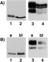
Intracellular accumulation of rGH0-LDL-R (A) and apical localization of rGH12-LDL-R at steady state (B). (A) The majority of rGH0-LDL-R accumulates intracellularly, whereas rGH12-LDL-R is mainly at the cell surface. Total surface biotinylation of filter-grown MDCK cell lines expressing rGH0-LDL-R (lanes 1 and 2) or rGH12-LDL-R (lanes 3 and 4). Biotinylated surface proteins were precipitated with streptavidin-agarose and are loaded in lanes 1 and 3. Nonbiotinylated (intracellular) proteins left in the supernatant after depletion of biotinylated proteins are shown in lanes 2 and 4. The presence of the rGH-fusion proteins in the fractions was analyzed on a Western blot. (B) Steady-state distribution of rGH0-LDL-R and rGH12-LDL-R. Surface biotinylation of filter-grown MDCK cell lines expressing rGH0-LDL-R (lanes 1 and 2) or rGH12-LDL-R (lanes 3 and 4) from the apical (a) or basolateral side (bl). Biotinylated proteins were precipitated with streptavidin-agarose. The presence of precipitated rGH0-LDL-R and rGH12-LDL-R was analyzed on a Western blot.

References

    1. Ahmed S.N., Brown D.A., London E. On the origin of sphingolipid-cholesterol rich detergent-insoluble domains in cell membranesphysiological concentrations of cholesterol and sphingolipid induce formation of a detergent-insoluble, liquid-ordered lipid phase in model membranes. Biochemistry. 1997;36:10944–10953. - PubMed
    1. Alonso M.A., Fan L., Alarcon B. Multiple sorting signals determine apical localization of a non-glycosylated integral membrane protein. J. Biol. Chem. 1997;272:30748–30752. - PubMed
    1. Arreaza G., Brown D.A. Sorting and intracellular trafficking of a glycosylphosphatidylinositol-anchored protein and two hybrid transmembrane proteins with the same ectodomain in Madin-Darby canine kidney epithelial cells. J. Biol. Chem. 1995;270:23641–23647. - PubMed
    1. Brown D.A., London E. Functions of lipid rafts in biological membranes. Annu. Rev. Cell Dev. Biol. 1998;14:111–136. - PubMed
    1. Brown D.A., Rose J.K. Sorting of GPI-anchored proteins to glycolipid-enriched membrane subdomains during transport to the apical cell surface. Cell. 1992;68:533–544. - PubMed

Publication types

MeSH terms