. 1999 Jul 26;146(2):427-37.
doi: 10.1083/jcb.146.2.427.

Establishment of the dorsal-ventral axis in Xenopus embryos coincides with the dorsal enrichment of dishevelled that is dependent on cortical rotation

Affiliations

Establishment of the dorsal-ventral axis in Xenopus embryos coincides with the dorsal enrichment of dishevelled that is dependent on cortical rotation

J R Miller et al. J Cell Biol. .

Abstract

Examination of the subcellular localization of Dishevelled (Dsh) in fertilized Xenopus eggs revealed that Dsh is associated with vesicle-like organelles that are enriched on the prospective dorsal side of the embryo after cortical rotation. Dorsal enrichment of Dsh is blocked by UV irradiation of the vegetal pole, a treatment that inhibits development of dorsal cell fates, linking accumulation of Dsh and specification of dorsal cell fates. Investigation of the dynamics of Dsh localization using Dsh tagged with green fluorescent protein (Dsh-GFP) demonstrated that Dsh-GFP associates with small vesicle-like organelles that are directionally transported along the parallel array of microtubules towards the prospective dorsal side of the embryo during cortical rotation. Perturbing the assembly of the microtubule array with D(2)O, a treatment that promotes the random assembly of the array and the dorsalization of embryos, randomizes translocation of Dsh-GFP. Conversely, UV irradiation of the vegetal pole abolishes movement of Dsh-GFP. Finally, we demonstrate that overexpression of Dsh can stabilize beta-catenin in Xenopus. These data suggest that the directional translocation of Dsh along microtubules during cortical rotation and its subsequent enrichment on the prospective dorsal side of the embryo play a role in locally activating a maternal Wnt pathway responsible for establishing dorsal cell fates in Xenopus.


Figures
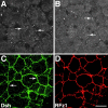
Figure 1
Figure 1
Localization of Dsh in animal cap explants and its relocalization in response to Frizzled expression. (A and B) Blastula stage animal cap explants were stained with anti–Dvl-1 antibodies and the distribution of Dsh was determined by confocal microscopy. Dsh associates with intracellular vesicle-like organelles (arrows) and is also found diffusely throughout the cytoplasm. (C and D) RNA encoding RFz1-myc was injected into the animal pole of 4-cell stage embryos and the distribution of Dsh (C) and RFz1-myc (D) was determined by confocal microscopy. In response to RFz1-myc expression Dsh accumulated at the plasma membrane (arrows in C). RFz1-myc is also localized to the plasma membrane in animal cap cells. Bar, 20 μm.
Figure 2
Figure 2
Dsh is enriched dorsally after cortical rotation. Eggs at 0.9 of the first cell cycle were fixed, manually dissected into dorsal and ventral halves and then stained with anti–Dvl-1 (A and B) or affinity-purified anti–Xenopus Dsh (C and D) antibodies. The distribution of Dsh in dorsal and ventral equatorial zones of the same embryo was then determined by confocal microscopy. After cortical rotation, Dsh localized to vesicle-like organelles similar to that seen in animal cap cells and these organelles were highly enriched in dorsal regions (A and C) relative to ventral regions (B and D). Images shown represent a 65 × 65-μm region at a depth of 4–8 μm from the cell surface. Bar, 20 μm.
Figure 3
Figure 3
Dsh-GFP moves along microtubule tracks to the prospective dorsal side during cortical rotation. (A) Vegetal pole of an egg during the main period of cortical rotation at time 0. (B) Same egg as in A, after 15 s. White arrows point to Dsh-GFP particles whose motion was tracked successively through confocal time-lapse movie frames captured at 1.5-s intervals for at least 15 s. See Video 1 for an example (available at http://www.jcb.org/cgi/content/full/146/2/427/F3/DC1). As an indication of the subcortical rotation (a global displacement of core yolk platelets), a white arrow labeled with a Y tracks the motion of a representative subcortical yolk platelet. The field of view shown for all panels is 35 × 35 μm at a focal plane viewing into subcortical rotation shear zone, 4–8 μm in from the egg's vegetal surface. The total field of view that was recorded was broader, ∼75 × 95 μm of the vegetal hemisphere region. (C) Summary of the displacement of representative Dsh-GFP particles from A and B. (D) Displacement vectors of representative Dsh-GFP particles plotted from a common origin (t = 26 s). The open arrowhead (Y) shows the movement of the egg's yolky core. Dsh-GFP particles move uniformly in the opposite direction from that of the yolk platelets. Since yolk platelets move towards the ventral side these data demonstrate that Dsh-GFP translocates towards the dorsal side of the embryo.
Figure 4
Figure 4
Quantitative analysis of Dsh-GFP in normal and microtubule-disrupted eggs. (A) Cortical rotation: this plot shows the angles of subcortical rotation for 44 matured and prick-activated oocytes observed in these experiments, relative to an arbitrary stable reference point on the viewing dish platform that oocytes were placed into for confocal imaging. As expected, the rotation angles were random with respect to this arbitrary reference point. (B) Dsh-GFP. 12 oocytes from 3 female frogs and 110 GFP-tagged organelle saltations were observed. The average orientation of Dsh-GFP saltations was 180.6°, relative to subcortical rotation displacement being at 0°. The range of displacement angles was from 135.1° to 223.5°. Thus displacements tended to occur in a direction 180° ± 46°, approximately opposite the direction of subcortical rotation. (C) D2O-treated Dsh-GFP. 10 oocytes from 3 female frogs were analyzed. The orientation of Dsh-GFP saltations were random relative to subcortical displacement.
Figure 5
Figure 5
Translocation of Dsh-GFP is randomized in D2O-treated eggs. (A) Egg treated with D2O at time 0. (B) Same egg as in A, after 15 s. White arrows point to Dsh-GFP particles whose motion was tracked successively through confocal time-lapse movie frames captured at 1.5-s intervals for at least 15 s. See Video 2 for an example (available at http://www.jcb.org/cgi/content/full/146/2/427/F5/DC1). (C) Summary of the displacement of representative Dsh-GFP particles from A and B. (D) Displacement vectors of Dsh-GFP particles plotted from a common origin demonstrating that D2O treatment results in the random translocation of Dsh-GFP particles (t = 26 s).
Figure 6
Figure 6
Endogenous Dsh is present at higher levels in dorsal blastomeres relative to ventral blastomeres and this asymmetry is dependent on cortical rotation. Lysates from dorsal and ventral regions of control (A) and UV-irradiated (B) 64–128-cell stage embryos were immunoblotted with anti–Dvl-1 antibodies. (A) In control embryos, steady state levels of Dsh are higher in dorsal regions relative to ventral regions. (B) After UV irradiation, this asymmetry is abolished. Lysates were also probed with anti–α-fodrin antibodies to control for total protein content and gel loading (lower panels). α-Fodrin levels do not exhibit dorsal–ventral differences in either control or UV-irradiated embryos.
Figure 7
Figure 7
Overexpression of Dsh stabilizes β-catenin. Immunoblots of lysates prepared from embryos injected with β-catenin-myc RNA (25 pg) in combination with either GFP RNA (1 ng) or Dsh-GFP RNA (1 ng) reveal that overexpression of Dsh-GFP increases the stability of β-catenin. Lysates were also immunoblotted with anti–α-fodrin antibodies to control for total protein content and gel loading.
Figure 8
Figure 8
Model of the mechanism of localized Wnt pathway activation during dorsal–ventral axis specification in Xenopus. Dsh associates with a specific class of vesicles at the vegetal pole and these vesicles are transported dorsally along the subcortical microtubule array during cortical rotation. This translocation contributes to the asymmetric distribution of Dsh along the dorsal–ventral axis and the localized activation of a maternal Wnt signaling pathway. Activation of Wnt signaling leads to the downregulation of GSK-3 activity thereby promoting the stabilization of β-catenin. β-catenin then accumulates in dorsal nuclei where in combination with XTcf-3 it activates transcription of dorsal-specific regulatory genes. See text for details.

Similar articles

Cited by

References

    1. Aberle H., Bauer A., Stappert J., Kispert A., Kemler R. β-catenin is a target for the ubiquitin-proteasome pathway. EMBO (Eur. Mol. Biol. Organ.) J. 1997;16:3797–3804. - PMC - PubMed
    1. Axelrod J.D., Matsuno K., Artavanis-Tsakonas S., Perrimon N. Interaction between Wingless and Notch signaling pathways mediated by Dishevelled. Science. 1996;271:1826–1832. - PubMed
    1. Axelrod J.D., Miller J.R., Shulman J.M., Moon R.T., Perrimon N. Differential recruitment of Dishevelled provides signaling specificity in the planar cell polarity and Wingless signaling pathways. Genes Dev. 1998;12:2610–2622. - PMC - PubMed
    1. Brannon M., Gomperts M., Sumoy L., Moon R.T., Kimelman D. A β-catenin/XTcf-3 complex binds to the siamois promoter to regulate dorsal axis specification in Xenopus . Genes Dev. 1997;11:2359–2370. - PMC - PubMed
    1. Brannon M., Brown J.D., Bates R., Kimelman D., Moon R.T. XCtBP is a XTcf-3 co-repressor with roles throughout Xenopus development. Development. 1999;126:3159–3170. - PubMed

Publication types

MeSH terms