. 1999 Jul 26;146(2):453-64.
doi: 10.1083/jcb.146.2.453.

DAip1, a Dictyostelium homologue of the yeast actin-interacting protein 1, is involved in endocytosis, cytokinesis, and motility

Affiliations

DAip1, a Dictyostelium homologue of the yeast actin-interacting protein 1, is involved in endocytosis, cytokinesis, and motility

A Konzok et al. J Cell Biol. .

Abstract

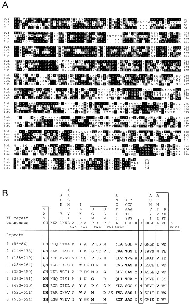
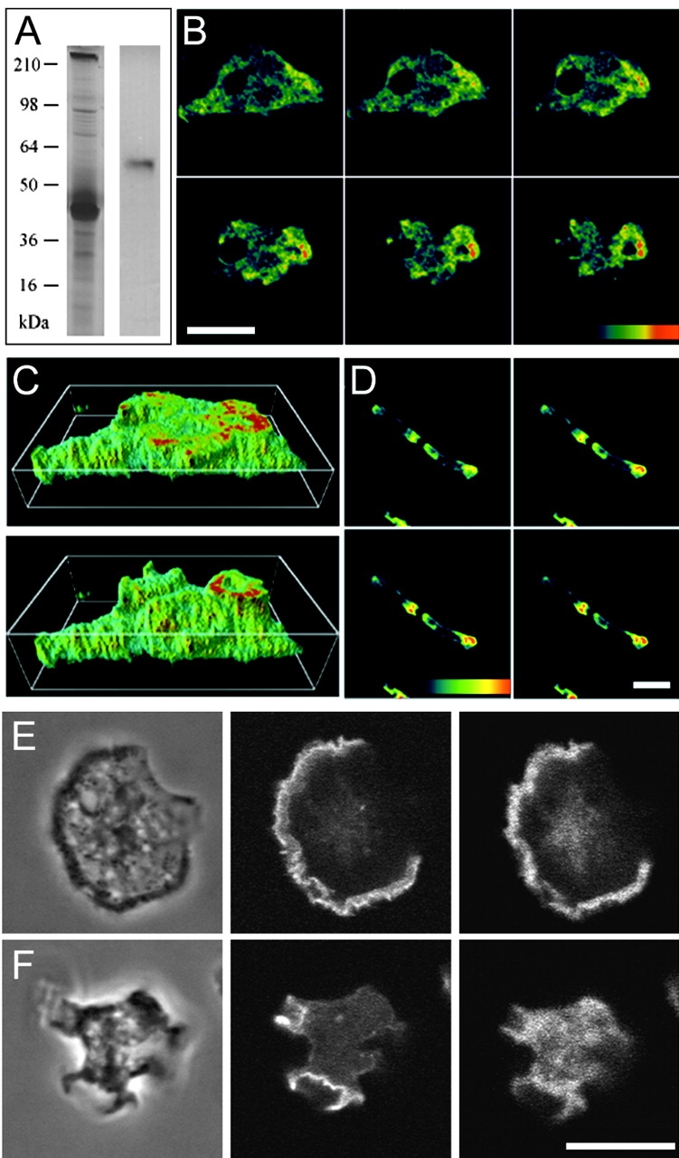
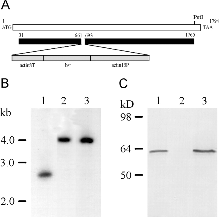
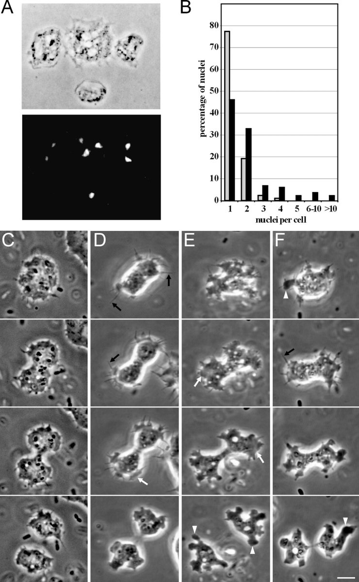
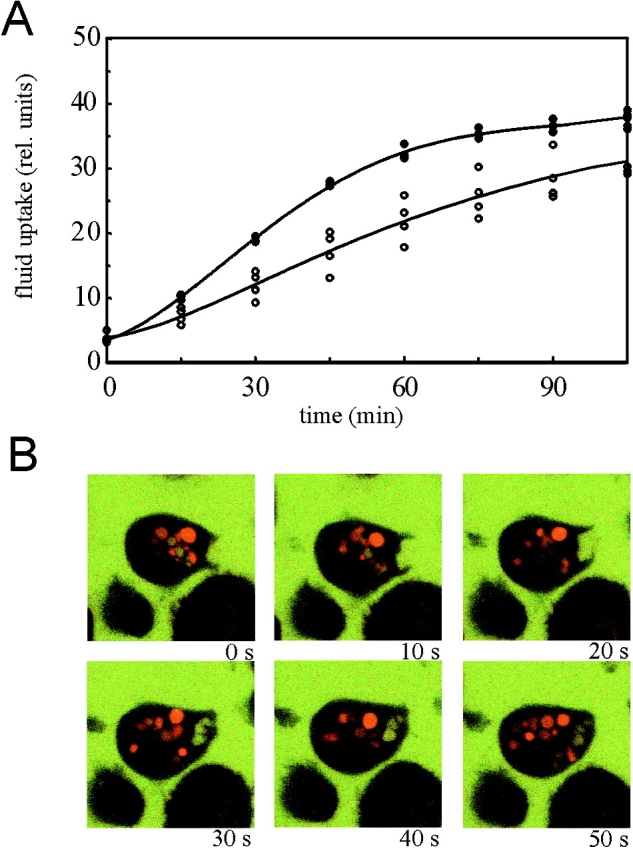
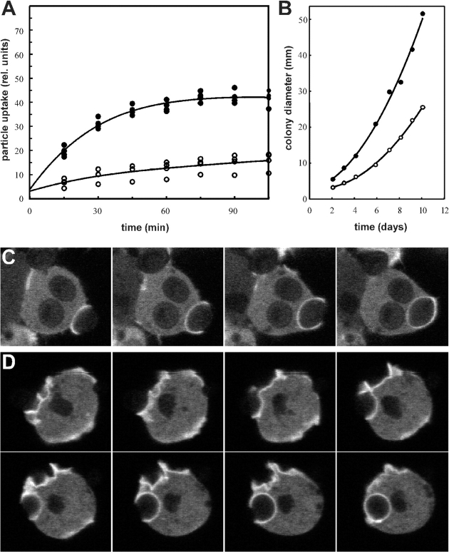
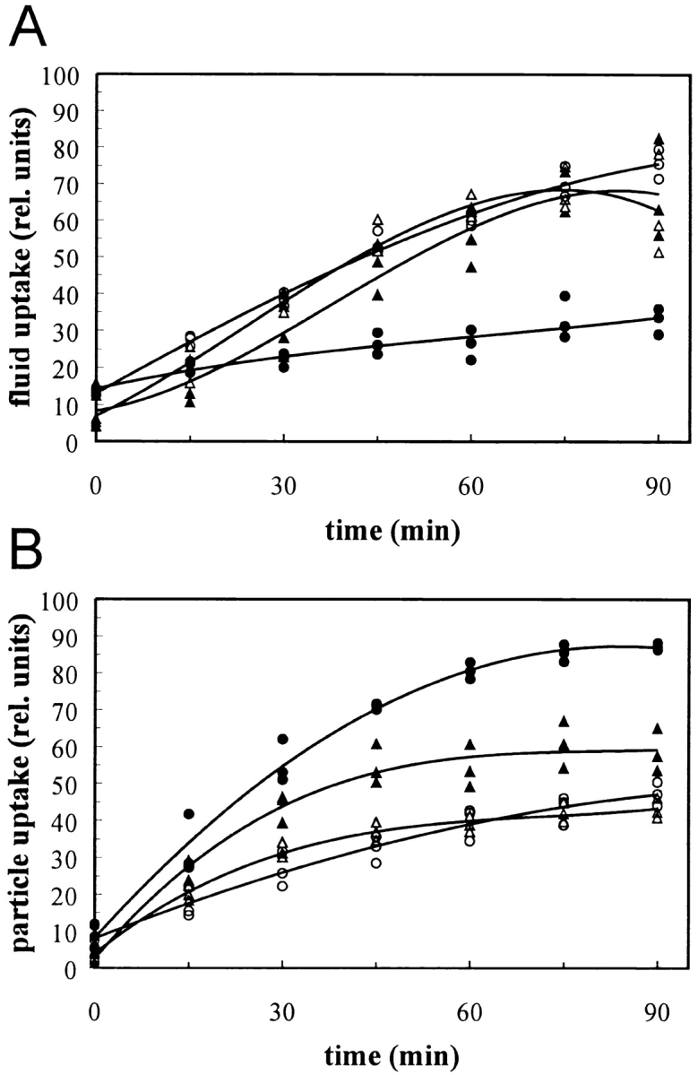
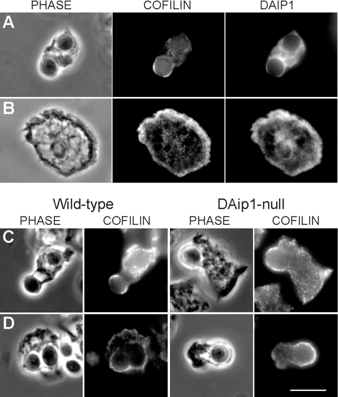
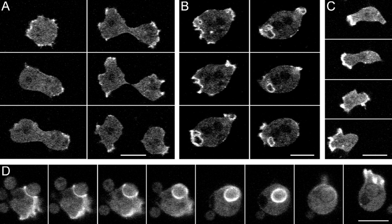
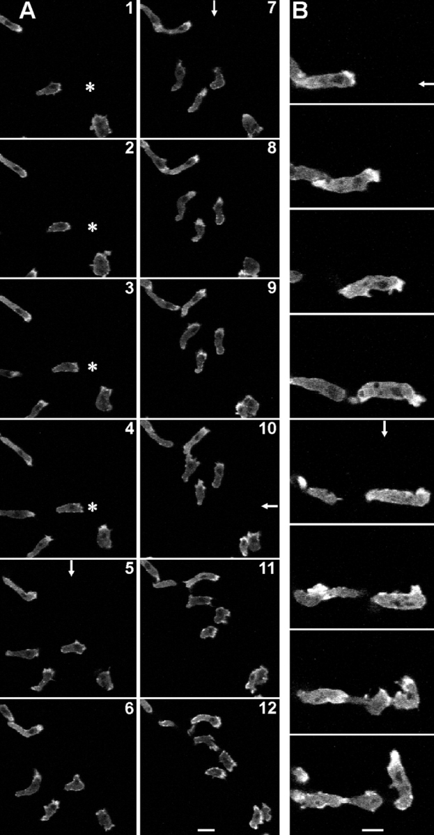
The 64-kD protein DAip1 from Dictyostelium contains nine WD40-repeats and is homologous to the actin-interacting protein 1, Aip1p, from Saccharomyces cerevisiae, and to related proteins from Caenorhabditis, Physarum, and higher eukaryotes. We show that DAip1 is localized to dynamic regions of the cell cortex that are enriched in filamentous actin: phagocytic cups, macropinosomes, lamellipodia, and other pseudopodia. In cells expressing green fluorescent protein (GFP)-tagged DAip1, the protein rapidly redistributes into newly formed cortical protrusions. Functions of DAip1 in vivo were assessed using null mutants generated by gene replacement, and by overexpressing DAip1. DAip1-null cells are impaired in growth and their rates of fluid-phase uptake, phagocytosis, and movement are reduced in comparison to wild-type rates. Cytokinesis is prolonged in DAip1-null cells and they tend to become multinucleate. On the basis of similar results obtained by DAip1 overexpression and effects of latrunculin-A treatment, we propose a function for DAip1 in the control of actin depolymerization in vivo, probably through interaction with cofilin. Our data suggest that DAip1 plays an important regulatory role in the rapid remodeling of the cortical actin meshwork.


Figures

Figure 1
Figure 1
Sequence analysis of D. discoideum DAip1. (A) Amino acid sequence alignment of DAip1 (D.d.) (GenBank accession number U36936) with the putative homologues Aip1p from S. cerevisiae (S.c.) (GenBank accession numbers S54451 and U35666), CO4F6.4 from C. elegans (C.e.) (GenBank accession number U42835), and P. polycephalum (P.p.) (Matsumoto et al. 1998). Alignment was performed using PILEUP (Genetics Computer Group) and the output was shaded using PRETTYBOX. Identical residues are shaded black, homologous residues are gray. The program used marks only homologous residues appearing within conserved blocks. Amino acid residues are numbered on the right. (B) Comparison of the amino acid sequences of the WD-repeat consensus described by Neer et al. 1994 (upper) and of DAip1 (lower). WD-repeat proteins are defined as having at least one unit that matches the consensus with zero or one mismatch and at least one other unit that has three or fewer mismatches. X means that any amino acid can be found at that position, and the numbers underneath give the range over which that symbol may be repeated. Numbers in brackets on the left give the positions of the individual repeats.
Figure 2
Figure 2
Localization of DAip1 in a purified actin-myosin complex by Western blotting, and in the cell cortex and surface projections of Dictyostelium cells by immunofluorescence. (A) A purified actin-myosin complex was separated by SDS-PAGE and stained with Coomassie blue (left lane), or blotted and probed with DAip1-specific mAb 246-153-2 (right lane). (B) Confocal sections of growth-phase AX2 wild-type cells labeled with DAip1-specific mAb 246-466-6. (C) Three-dimensional image reconstruction from confocal sections shown in B. (D) Confocal sections of aggregation-competent AX2 cells labeled with mAb 246-466-6. The color code represents relative fluorescence intensities as indicated by the colored scales. Distance between sections in B and D is 0.5 μm. (E and F) Confocal sections of two growth-phase AX2 cells double-labeled with DAip1-specific mAb 246-153-2 (right) and TRITC-phalloidin (middle) to visualize F-actin. Typical for the immunofluorescence labeling of DAip1 is the diffuse cytoplasmic distribution reflecting the presence of a cytoplasmic pool of the protein. Phase-contrast images are shown on the left. Bars, 10 μm.
Figure 3
Figure 3
Generation of DAip1-null cells by gene replacement. (A) Map of the DAip1 gene, and the construct used for the gene replacement. The blasticidin resistance gene (bsr) is transcribed from the actin-15 promoter (actin15P) and terminates in the actin-8 terminator (actin8T). (B) Southern blot analysis of genomic DNA. DNA of the wild-type AX2 (lane 1), or of DAip1-null mutants 9.1 (lane 2) and 10.10 (lane 3), was digested with EcoRI, and subjected to Southern blot analysis by hybridizing with a COOH-terminal fragment comprising nucleotides 1577–1791 of the DAip1 coding region. (C) Western blot analysis of DAip1 expression. Total cellular proteins from AX2 wild-type cells (lane 1), from 9.1 DAip1-null cells (lane 2), and from HG1569 coronin-null cells (lane 3) were separated by SDS-PAGE and subjected to immunoblot analysis using mAb 246-153-1.
Figure 4
Figure 4
DAip1-null cells are defective in cytokinesis. (A) Phase-contrast image (top), and fluorescence image after DAPI staining (bottom), of DAip1-null cells. (B) Histogram illustrating a quantitation of nuclei in AX2 wild-type (gray) and in DAip1-null cells (black). Cells cultivated for 2 d on glass coverslips in nutrient medium were fixed, the nuclei were stained with DAPI, and several hundred cells were counted. With cells grown in shaking culture, analogous results were obtained. (C) Shape of a wild-type AX2 cell during cytokinesis. Typically, only moderate ruffling at the polar regions is observed. (D–F) Cell shape of DAip1-null mutants during cytokinesis. Prominent cell protrusions appear as filopodia (black arrows in D and F), lamellipodia (white arrows in D and E), and pseudopodia (white arrowheads in E and F). The three forms convert into each other, yet filopodia and lamellipodia are predominant in the earlier stages, whereas pseudopodia are most frequent in the final stage of cytokinesis. The position of the incipient cleavage furrow is often not clearly discernible at early stages and remains covered with protrusions until the furrow has already ingressed substantially (E and F). Time intervals between consecutive frames: (C) 60, 70, and 40 s; (D) 60, 30, and 50 s; (E) 110, 110, and 140 s; (F) 80, 60, and 90 s. Bar, 10 μm.
Figure 5
Figure 5
Reduced fluid-phase uptake in DAip1-null mutant cells. (A) Endocytosis of TRITC-dextran was measured in AX2 wild-type cells (filled circles) and DAip1-null mutant cells (open circles). The data from four independent experiments were averaged to calculate the curves. (B) Confocal sections through single DAip1-null cells undergoing macropinocytosis. To distinguish older, acidified endosomes (red) from freshly formed macropinosomes (yellow), cells were incubated with a mixture of FITC-labeled and TRITC-labeled dextrans as described (Maniak 1999). Time points are in seconds.
Figure 6
Figure 6
Reduced growth and phagocytosis rate of DAip1-null cells. (A) Uptake of TRITC-labeled yeast particles was determined in AX2 wild-type (filled circles) and in DAip1-null cells (open circles). The data from three independent experiments were averaged to calculate the curves. (B) Growth of AX2 wild-type (filled circles) and of DAip1-null mutant (open circles), on a lawn of K. aerogenes. For each time point, the diameter of 10 colonies was measured and averaged. (C and D) Monitoring of phagocytic cup formation from attachment until the complete engulfment of a yeast particle in wild-type (C), and DAip1-null cells (D), expressing actin-GFP. Images in C and D were taken in 10-s intervals.
Figure 7
Figure 7
Effects of Lat-A and of DAip1 overexpression on fluid-phase and particle uptake. (A) Fluid-phase uptake of TRITC-dextran was first determined in the AX2 wild-type (open triangles), and in DAip1-overexpressing cells (filled triangles). Fluid-phase uptake was independently determined for AX2 cells treated with a representative concentration of 0.3 μM Lat-A (filled circles) and an untreated control (open circles). The curves were plotted into a single graph to obtain maximal overlap between the wild-type data and the untreated control (open symbols). The data from three independent experiments were used to calculate the curves. (B) Uptake of TRITC-labeled yeast particles. Data acquisition and symbols as in A.
Figure 8
Figure 8
Localization of cofilin in Dictyostelium wild-type and DAip1-null cells by immunofluorescence microscopy. (A and B) Colocalization of cofilin and DAip1 in Dictyostelium wild-type cells in phagocytic cups (A) and in lamellipodia (B) by double label immunofluorescence microscopy. (C and D) Wild-type and DAip1-mutant cells were labeled with cofilin-specific antibodies during early (C), and late stages (D) of phagocytosis of yeast particles. Bar, 10 μm.
Figure 9
Figure 9
Localization of GFP-(N)-DAip1 fusion protein expressed in DAip1-null cells during cytokinesis (A), pinocytosis (B), cell movement (C), and phagocytosis (D). (A) GFP-(N)-DAip1 is enriched in the cortical protrusions at the onset and in the course of cell division. Time intervals between consecutive frames: 110, 70, 40, 20, and 30 s. (B) GFP-(N)-DAip1 is enriched in nascent macropinosomes. At the time point of the next recorded frame, the macropinosome was internalized and the GFP label disappeared (not shown). Time interval between consecutive frames: 10 s. (C) GFP-(N)-DAip1 is enriched at the leading edge of moving cells. Time intervals between consecutive frames: 40, 40, and 60 s. (D) GFP-(N)-DAip1 is enriched at the sites of enclosure and internalization of yeast particles. Approximately 1 min after the particle was engulfed, GFP-(N)-DAip1 was released from the phagosome. Shortly afterwards, the fusion protein was redistributed to the leading edge as cell movement commenced. Time interval between consecutive frames was 10 s up to the fourth frame and 30 s afterwards. Bars, 10 μm.
Figure 10
Figure 10
Rapid redistribution of GFP-(N)-DAip1 fusion protein expressed in DAip1-null cells moving in chemotactic gradients. The asterisk marks the position of the micropipette tip filled with the chemoattractant cAMP. Arrows indicate direction of the negative cAMP concentration gradient when the pipette was placed outside of the displayed field of view. An arrow is drawn only in those frames where the position of the pipette was changed. (A) Interval between frames, 40 s. Bar, 20 μm. (B) Intervals between frames, 60 s. Bar, 10 μm.

Similar articles

Cited by

References

    1. Adler H.J., Winnicki R.S., Gong T.-W.L., Lomax M.I. A gene upregulated in the acoustically damaged chick basilar papilla encodes a novel WD40 repeat protein. Genomics. 1999;56:59–69. - PubMed
    1. Aizawa H., Sutoh K., Tsubuki S., Kawashima S., Ishii A., Yahara I. Identification, characterization, and intracellular distribution of cofilin in Dictyostelium discoideum . J. Biol. Chem. 1995;270:10923–10932. - PubMed
    1. Aizawa H., Sutoh K., Yahara I. Overexpression of cofilin stimulates bundling of actin filaments, membrane ruffling, and cell movement in Dictyostelium . J. Cell Biol. 1996;132:335–344. - PMC - PubMed
    1. Aizawa H., Fukui Y., Yahara I. Live dynamics of Dictyostelium cofilin suggests a role in remodeling actin latticework into bundles. J. Cell Sci. 1997;110:2333–2344. - PubMed
    1. Aizawa H., Katadae M., Maruya M., Sameshima M., Murakami-Murofushi K., Yahara I. Contraction of actin bundles induced in Dictyostelium by overexpression of cofilin in response to hyperosmotic stress Mol. Biol. Cell. 9Suppl.1998. 93

Publication types

MeSH terms