. 1999 Jul 26;146(2):465-75.
doi: 10.1083/jcb.146.2.465.

ZASP: a new Z-band alternatively spliced PDZ-motif protein

Affiliations

ZASP: a new Z-band alternatively spliced PDZ-motif protein

G Faulkner et al. J Cell Biol. .

Abstract

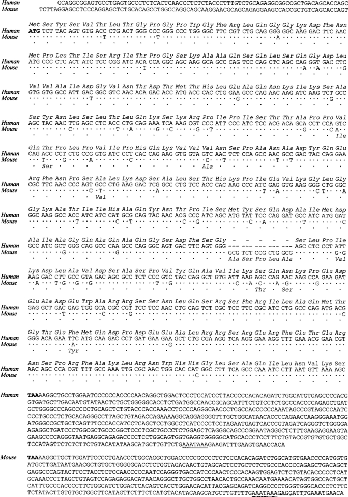
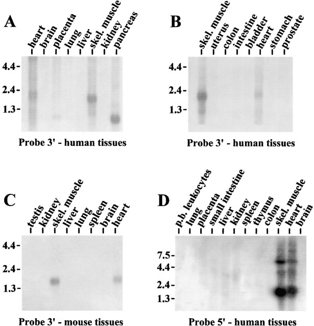
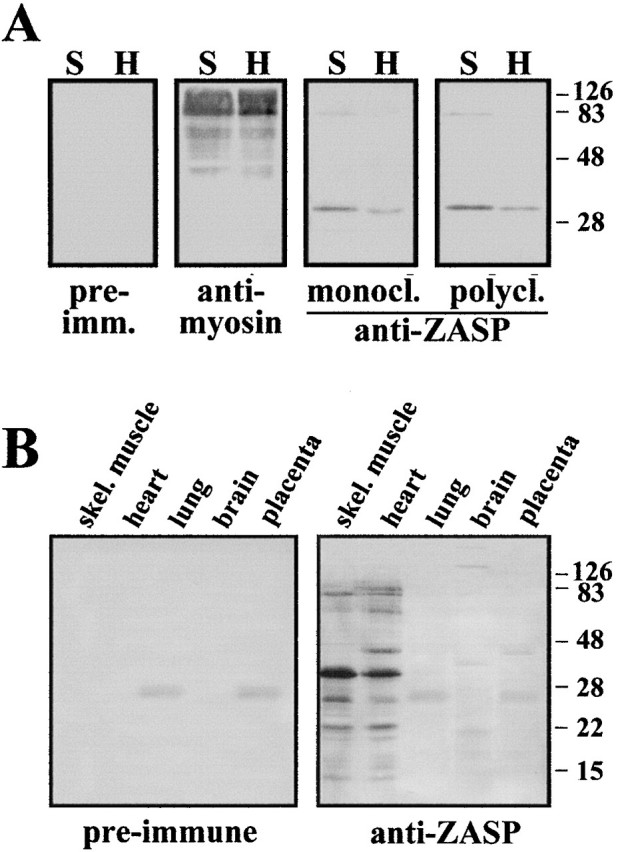
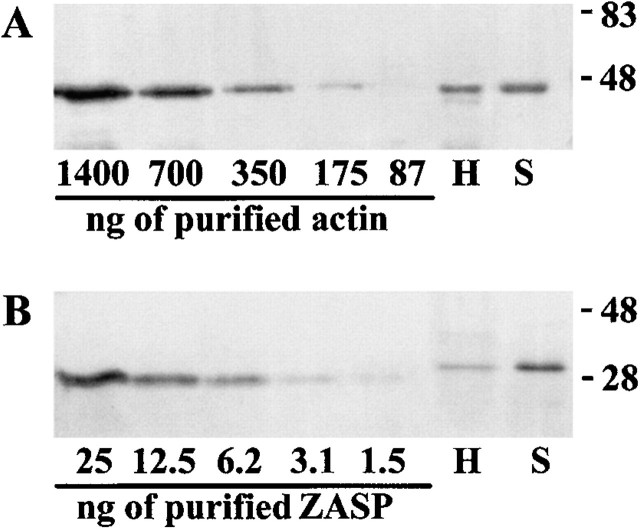
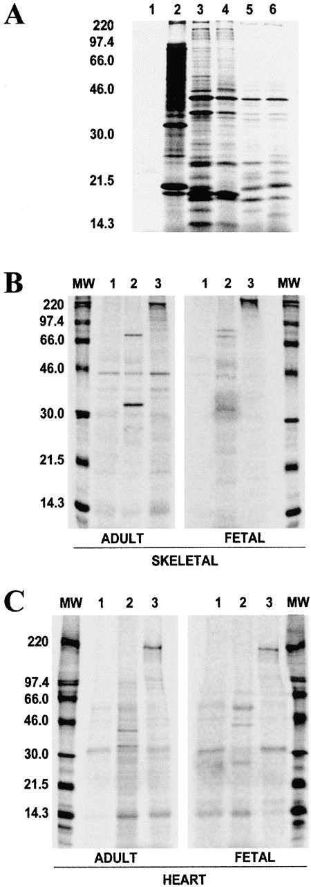
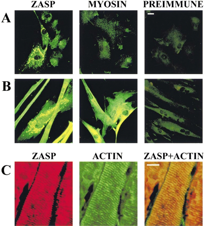
PDZ motifs are modular protein-protein interaction domains, consisting of 80-120 amino acid residues, whose function appears to be the direction of intracellular proteins to multiprotein complexes. In skeletal muscle, there are a few known PDZ-domain proteins, which include neuronal nitric oxide synthase and syntrophin, both of which are components of the dystrophin complex, and actinin-associated LIM protein, which binds to the spectrin-like repeats of alpha-actinin-2. Here, we report the identification and characterization of a new skeletal muscle protein containing a PDZ domain that binds to the COOH-terminal region of alpha-actinin-2. This novel 31-kD protein is specifically expressed in heart and skeletal muscle. Using antibodies produced to a fragment of the protein, we can show its location in the sarcomere at the level of the Z-band by immunoelectron microscopy. At least two proteins, 32 kD and 78 kD, can be detected by Western blot analysis of both heart and skeletal muscle, suggesting the existence of alternative forms of the protein. In fact, several forms were found that appear to be the result of alternative splicing. The transcript coding for this Z-band alternatively spliced PDZ motif (ZASP) protein maps on chromosome 10q22.3-10q23.2, near the locus for infantile-onset spinocerebellar ataxia.


Figures

Figure 1
Figure 1
cDNA and amino acid sequences of human and mouse ZASP. The start and stop codons are in bold characters and the polyadenylation sites are underlined. In the coding part of the mouse sequence, the conserved nucleotides and amino acids are represented by dots. Dashes indicate gaps that have been inserted to align the two sequences. The sequence data are available from Genbank/EMBL/DDBJ under the accession number AJ133766 and AJ005621, respectively, for human and mouse. The two variant forms of ZASP, described in the other sections of this paper, are available under accession number AJ133768 and AJ133767, as well as at http://grup. bio.unipd.it/muscle.
Figure 2
Figure 2
Northern blot analysis of human (A, B, and D) and mouse (C) tissues demonstrating patterns of expression of ZASP mRNAs. Blots containing Poly(A)+ RNA from a variety of human and mouse tissues were probed with 3′ untranslated region of ZASP (A–C) or with the 5′ region (D) as indicated. The ZASP transcript is expressed primarily in skeletal muscle and heart. The numbers on the side indicate size in kb.
Figure 3
Figure 3
(A) Western blot analysis of heart and skeletal muscle tissue with antibodies to myosin, ZASP, and preimmune sera. Equal amounts of proteins were run in each lane (10 μg) on a 15% SDS-polyacrylamide gel and then blotted onto Immobilon-P membrane. The ZASP mAb was used undiluted, whereas the pAb was used at 1/20,00 dilution, as were the preimmune and myosin sera. (B) Tissue distribution of ZASP as demonstrated by Western blotting. Protein extracts from human heart and skeletal muscle (10 μg), as well as from brain, lung, and placenta (60 μg) were loaded in each lane, run on a 15% SDS-polyacrylamide gel, and then blotted. The membrane was probed with mouse pAb specific for ZASP and preimmune mouse sera, both used at 1/200 dilution. Sigma Chemical Co. color molecular weight markers were used.
Figure 4
Figure 4
(A) Western blot analysis of known amounts of recombinant human α-actin (left) and known amounts of total protein (2.5 μg) from human heart and skeletal muscle tissue (right). Actin was detected using a mouse pAb specific for human α-actin. (B) Western blot analysis of known amounts of recombinant ZASP protein (left) and known amounts of total protein (10 μg) from human heart and skeletal muscle tissue (right). The recombinant protein lacks some amino acids from the NH2-terminal end of the ZASP sequence, therefore it is slightly smaller in size than the native muscle protein. Mouse pAb to ZASP was used to detect the protein. From densitometric analysis of the bands, it can be calculated that the actin signal from 2.5 μg of total muscle proteins (both heart or skeletal muscle) is approximately equivalent to 500 ng of purified α-actin, whereas for ZASP, there is less protein present in heart than skeletal muscle tissue: 4.5 ng, as opposed to 18 ng in 10 μg of total muscle proteins.
Figure 5
Figure 5
(A) Total protein obtained from in vitro translation of muscle and control mRNAs using the rabbit reticulocyte lysate system (Promega Corp.). Lanes: 1, negative control sample with water instead of mRNA; 2, positive control, BMV mRNA; 3, human adult skeletal muscle mRNA; 4, human fetal skeletal muscle mRNA; 5, human adult heart mRNA; and 6, human fetal heart mRNA. (B) Immunoprecipitation of in vitro translated human adult and fetal skeletal muscle proteins. (C) Immunoprecipitation of in vitro translated human adult and fetal heart. Equal amounts of [35S]methionine-labeled proteins were mixed with the appropriate antibody and immunoprecipitated using protein A–Sepharose, and were then run on SDS-polyacrylamide gels. In B and C, the total in vitro translated human heart and muscle proteins were immunoprecipitated with the antibodies denoted by the corresponding lane numbers: 1, preimmune sera; 2, antibody to ZASP; and 3, antibody to myosin. Numbers on the left of the figures indicate rainbow 14C-methylated protein molecular weight markers, in kD.
Figure 6
Figure 6
Indirect immunofluorescence of undifferentiated human myoblasts (A) and differentiated human myotubes (B). Preimmune sera and ZASP pAb were used at a dilution of 1/50; myosin mAb (MF 20) was used at 1/100 dilution. FITC-conjugated anti-mouse immunoglobulin (Sigma Chemical Co.) was used as the second antibody. (C) Indirect immunofluorescence of skeletal muscle tissue sections using mouse pAb to ZASP (1/30 dilution) and rabbit antiactin antibody (1/40). FITC-labeled goat anti–rabbit Ig (green) and TRITC-labeled goat anti–mouse Ig (red) were used as second antibodies. Bars, 10 μm.
Figure 7
Figure 7
Localization of ZASP in human skeletal muscle by immunoelectron microscopy. Immunoelectron microscopy with ZASP pAb as the primary antibody and anti-mouse IgG whole molecule conjugated with 5-nm gold particles (Sigma Chemical Co.; G7527) as the secondary antibody. (A) Low magnification of a section of skeletal muscle. The gold particles were detected in the Z-band. (B) Higher magnification of the same image, rotated 90°. The ZASP protein is seen throughout the Z-band. Bars, 0.1 μm.
Figure 8
Figure 8
Schematic representation of the ZASP transcript, two alternative muscle variants, and brain transcript KIAA0613. Boxes (not always in scale) represent the coding region and the numbers inside each box indicate the length in bases. The 3′ and 5′ untranslated regions are represented by the terminal lines. The original KIAA0613 sequence begins just a few bases in front of the first ATG starting codon. However, the box has been marked as 322 bases long to facilitate the interpretation.
Figure 9
Figure 9
Schematic representation of the positive α-actinin-2 clones found using the yeast two-hybrid system. The domains of α-actinin-2 are shown at the top, whereas the remaining part of the figure shows the coding regions of the α-actinin-2, which are involved in a positive interaction with the PDZ domain of ZASP. The numbers at the side of the bars indicate the starting amino acid. The coding regions of all the α-actinin-2 clones reach the true stop codon of the protein at position 894.

References

    1. Adams M.E., Butler M.H., Dwyer T.M., Peters M.F., Murnane A.A., Froehner S.C. Two forms of mouse syntrophin, a 58-kd dystrophin-associated protein, differ in primary structure and tissue distribution. Neuron. 1993;11:531–540. - PubMed
    1. Agatep, R., R.D. Kirkpatrick, D.L. Parchaliuk, R.A. Woods, and R.D. Gietz. 1998. Transformation of Saccharomyces cerevisiae by the lithium acetate/single-stranded carrier DNA/polyethylene glycol protocol. Technical Tips Online. (http://tto.biomednet.com) 01525.
    1. Altschul S.F., Madden T.S., Schaffer A.A., Zhang J., Zhang Z., Miller W., Lipman D.J. Gapped BLAST and PSI-BLASTa new generation of protein database search programs. Nucleic Acids Res. 1997;25:3389–3402. - PMC - PubMed
    1. Ausubel F.M., Brent R., Kingston R.E., Moore D.D., Seidman J.G., Smith J.A., Struhl K. Current Protocols in Molecular Biology. Greene Publishing Associates and Wiley-Interscience; New York: 1994.
    1. Bateman A., Birney E., Rubin R., Eddy S.R., Finn R.D., Sonnhammer E.L. Pfam3.11313 multiple alignments match the majority of proteins. Nucleic Acids Res. 1999;27:260–262. - PMC - PubMed

Publication types

MeSH terms

Associated data

Grants and funding