. 1999 Aug;104(3):229-37.
doi: 10.1172/JCI5705.

Independent impairment of osteoblast and osteoclast differentiation in klotho mouse exhibiting low-turnover osteopenia

Affiliations

Independent impairment of osteoblast and osteoclast differentiation in klotho mouse exhibiting low-turnover osteopenia

H Kawaguchi et al. J Clin Invest. 1999 Aug.

Abstract

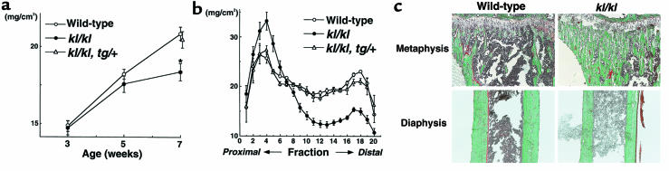
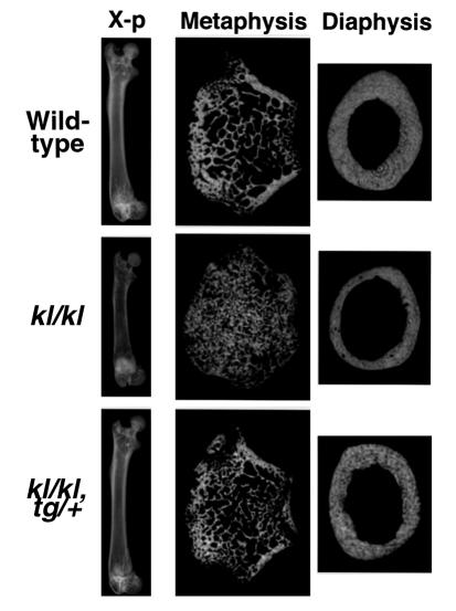
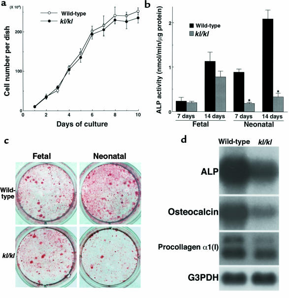
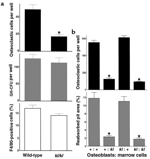
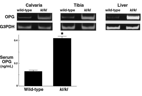
We recently identified a new gene, klotho, which is involved in the suppression of multiple aging phenotypes. The mouse homozygous for a disruption of the klotho locus (kl/kl) exhibited multiple pathological conditions resembling human aging. Histomorphometric analysis revealed low-turnover osteopenia in kl/kl mice. The decrease in bone formation exceeded that of bone resorption, resulting in a net bone loss. The number of osteoblast progenitors determined by ex vivo bone marrow cultures was reduced in kl/kl mice. In addition, cultured osteoblastic cells derived from kl/kl mice showed lower alkaline phosphatase activity and matrix nodule formation than those from wild-type mice. Osteoclastogenesis in the coculture of marrow cells and osteoblastic cells was decreased only when marrow cells originated from kl/kl mice independently of the origin of osteoblastic cells. We also found that the expression of osteoprotegerin, an osteoclastogenesis inhibitor, was significantly upregulated in kl/kl mice. We conclude that a defect in the klotho gene expression causes the independent impairment of both osteoblast and osteoclast differentiation, leading to low-turnover osteopenia. Because this state represents a characteristic feature of senile osteoporosis in humans, kl/kl mice can be regarded as a useful model for investigating cellular and molecular mechanisms of age-related bone loss.


Figures

Figure 1
Figure 1
In vivo findings of the tibiae of wild-type, klotho (kl/kl), and rescued klotho (kl/kl, tg/+) mice. (a) BMD of the whole tibiae at 3, 5, and 7 weeks of age. *Significantly different from wild-type mice (P < 0.01). Data are expressed as mean (symbols) ± SEM (error bars) for 8 bones per group for wild-type and kl/kl, and 5 bones for kl/kl, tg/+. (b) BMD of each of 20 equal longitudinal divisions of tibiae from 7-week-old mice. (c) Histological features of the proximal metaphysis and the diaphysis of the tibiae of 7-week-old wild-type and kl/kl mice. Mineralized bone is stained green and unmineralized osteoid stained red by the Villanueva-Goldner method. Original magnification: ×20. Histomorphometric parameters are shown in Table 1.
Figure 2
Figure 2
Plain x-rays and CT images of the femora of 7-week-old wild-type, kl/kl, and kl/kl, tg/+ mice. The length of the femora, as well as the tibiae, of kl/kl mice was 20–30% shorter than those of wild-type mice, reflecting growth retardation in kl/kl mice. CT was taken at 0.2 mm above the growth plate at the distal metaphysis and at the midportion at the diaphysis.
Figure 3
Figure 3
Cultures of osteoblastic cells from wild-type and kl/kl mice. (a) Growth curve of cultured osteoblastic cells from neonatal mouse calvariae during 10 days of culture. Data are expressed as mean (symbols) ± SEM (error bars) of pooled data from 3 independent experiments (6 dishes per group). (b) ALP activity of cultured osteoblastic cells from fetal (17 days after coitus) and neonatal (5–6 days old) wild-type or kl/kl mouse calvariae at 7 and 14 days of culture. Data are expressed as mean (bars) ± SEM (error bars) for 8 wells per group. *Significantly different from wild-type mice (P < 0.01). (c) Representative pictures of alizarin red–stained matrix nodule formation by cultured osteoblastic cells from fetal and neonatal wild-type or kl/kl mouse calvariae at 21 days of culture. Areas of positively stained matrix nodule were 35.3 ± 4.1 mm2 for fetal wild-type, 31.1 ± 2.5 mm2 for fetal kl/kl, 53.4 ± 6.3 mm2 for neonatal wild-type, and 8.7 ± 3.1 mm2 for neonatal kl/kl (all mean ± SEM for 6 wells per group). Significant difference (P < 0.01) between wild-type and kl/kl was seen in cultured osteoblastic cells from neonatal mice, but not in those from fetal mice. (d) Steady-state mRNA levels of ALP, osteocalcin, and procollagen α1 (I) in cultured osteoblastic cells from neonatal wild-type or kl/kl mouse calvariae at 7 days of culture, as determined by Northern blot analysis.
Figure 4
Figure 4
Osteoclastogenesis in ex vivo bone marrow cell cultures (a) and in the coculture of osteoblastic cells and bone marrow cells (b) from wild-type mice and kl/kl mice. (a) The number of TRAP-positive osteoclastic cells (top) and GM-CFU (middle) at 8 days of culture of bone marrow cells from 7-week-old wild-type or kl/kl mice. Data are expressed as mean (bars) ± SEM (error bars) for 8 and 6 wells per group, respectively. *Significantly different from wild-type cells (P < 0.01). The percentage of F4/80-positive monocyte macrophages in bone marrow cells was measured by flow cytometric analysis using FITC-labeled F4/80 (bottom). (b) The number of TRAP-positive osteoclastic cells formed at 8 days of coculture (top) and the pit area resorbed by osteoclastic cells by an additional 48 hours of culture on a dentine slice (bottom; percent of whole dentine). The origins (+ for wild-type mice and kl for kl/kl mice) of the cells are indicated as “Osteoblasts: marrow cells.” Data are expressed as mean (bars) ± SEM (error bars) for 8 wells per group. *Significantly different from wild-type cultures (P < 0.01).
Figure 5
Figure 5
Messenger RNA level and serum protein level of OPG in wild-type and kl/kl mice with mRNA levels of OPG in calvariae, tibiae, and liver of 7-week-old wild-type and kl/kl mice analyzed by RT-PCR (collected from 8 mice per group). Serum OPG levels in wild-type and kl/kl mice were measured by ELISA. Data are expressed as mean (bars) ± SEM (error bars) for 5 mice per group. *Significantly different from wild-type mice (P < 0.01).

References

    1. Riggs BL, Melton LJ., III Involutional osteoporosis. N Engl J Med. 1986;314:1676–1686. - PubMed
    1. Manolagas SC, Jilka RL. Mechanisms of diseases: bone marrow, cytokines, and bone remodeling — emergency insights into the pathophysiology of osteoporosis. N Engl J Med. 1995;332:305–311. - PubMed
    1. Kuro-o M, et al. Mutation of the mouse klotho gene leads to a syndrome resembling aging. Nature. 1997;390:45–51. - PubMed
    1. Shiraki-Iida T, et al. Structure of the mouse klotho gene and its two transcripts encoding membrane and secreted protein. FEBS Lett. 1998;424:6–10. - PubMed
    1. Matsumura Y, et al. Identification of the human klotho gene and its two transcripts encoding membrane and secreted Klotho protein. Biochem Biophys Res Commun. 1998;242:626–630. - PubMed

Publication types

MeSH terms