. 1999 Aug;104(3):281-90.
doi: 10.1172/JCI6656.

Mixed chimerism induced without lethal conditioning prevents T cell- and anti-Gal alpha 1,3Gal-mediated graft rejection

Affiliations

Mixed chimerism induced without lethal conditioning prevents T cell- and anti-Gal alpha 1,3Gal-mediated graft rejection

H Ohdan et al. J Clin Invest. 1999 Aug.

Abstract

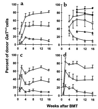
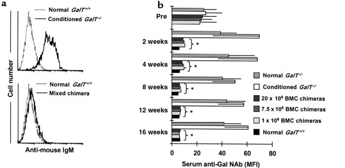
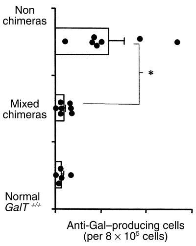
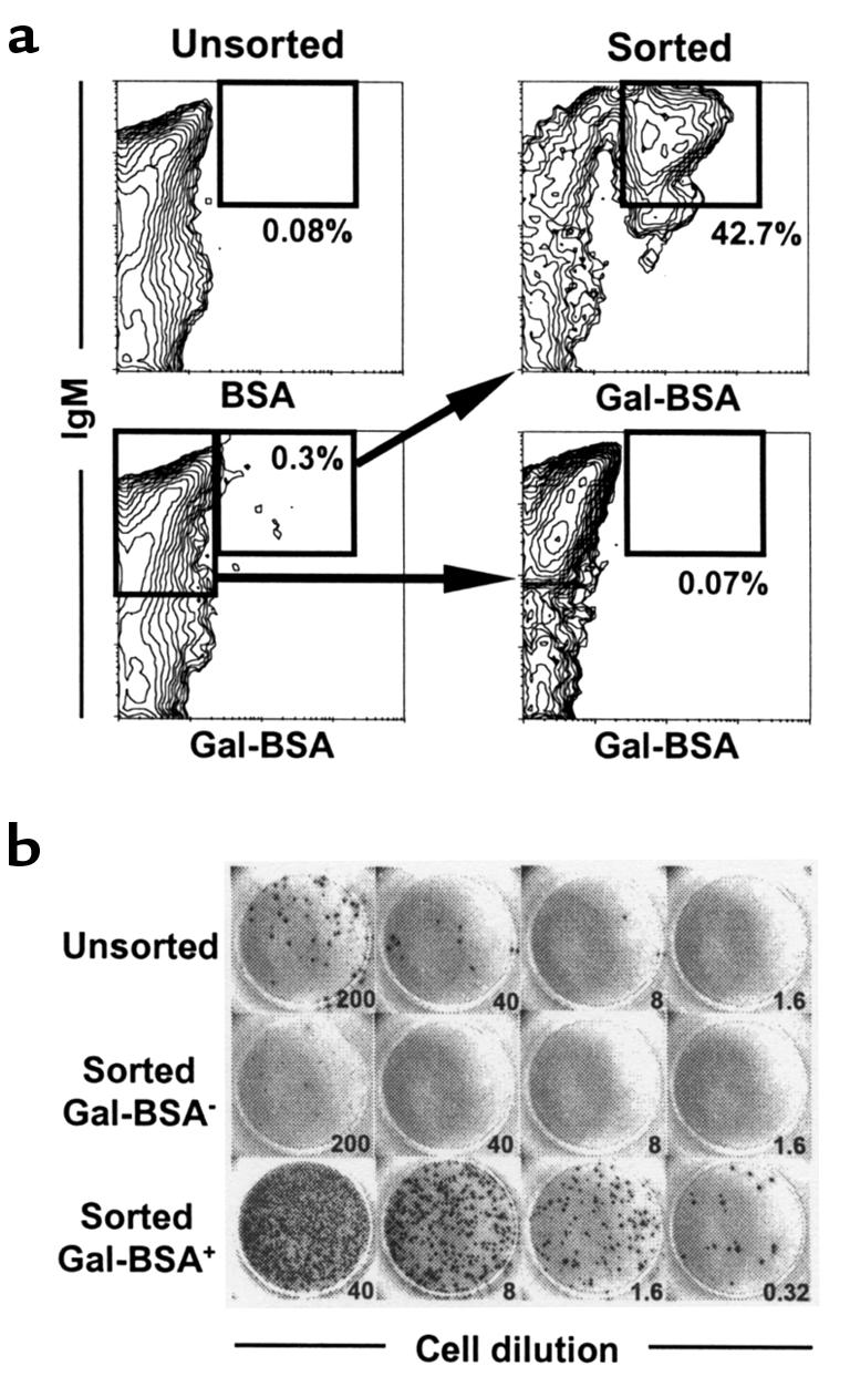
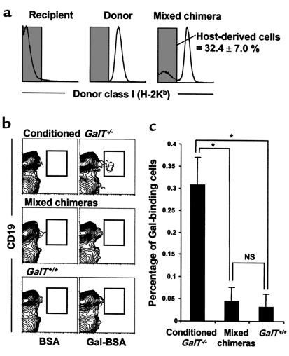
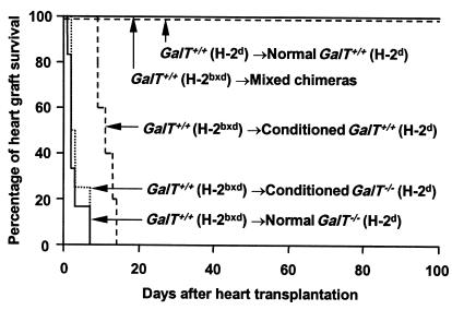
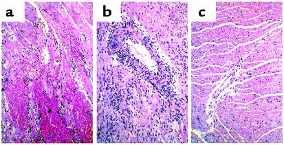
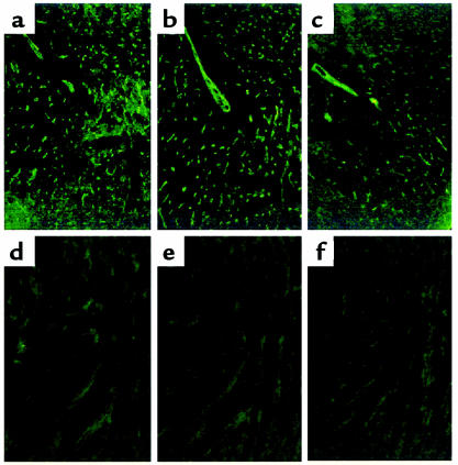
Gal alpha 1,3Gal-reactive (Gal-reactive) antibodies are a major impediment to pig-to-human xenotransplantation. We investigated the potential to induce tolerance of anti-Gal-producing cells and prevent rejection of vascularized grafts in the combination of alpha 1,3-galactosyltransferase wild-type (GalT(+/+)) and deficient (GalT(-/-)) mice. Allogeneic (H-2 mismatched) GalT(+/+) bone marrow transplantation (BMT) to GalT(-/-) mice conditioned with a nonmyeloablative regimen, consisting of depleting CD4 and CD8 mAb's and 3 Gy whole-body irradiation and 7 Gy thymic irradiation, led to lasting multilineage H-2(bxd) GalT(+/+) + H-2(d) GalT(-/-) mixed chimerism. Induction of mixed chimerism was associated with a rapid reduction of serum anti-Gal naturally occurring antibody levels. Anti-Gal-producing cells were undetectable by 2 weeks after BMT, suggesting that anti-Gal-producing cells preexisting at the time of BMT are rapidly tolerized. Even after immunization with Gal-bearing xenogeneic cells, mixed chimeras were devoid of anti-Gal-producing cells and permanently accepted donor-type GalT(+/+) heart grafts (>150 days), whereas non-BMT control animals rejected these hearts within 1-7 days. B cells bearing receptors for Gal were completely absent from the spleens of mixed chimeras, suggesting that clonal deletion and/or receptor editing may maintain B-cell tolerance to Gal. These findings demonstrate the principle that induction of mixed hematopoietic chimerism with a potentially relevant nonmyeloablative regimen can simultaneously lead to tolerance among both T cells and Gal-reactive B cells, thus preventing vascularized xenograft rejection.


Figures

Figure 1
Figure 1
Long-lasting multilineage GalT+/+GalT–/– mixed chimerism in recipients prepared with a nonmyeloablative conditioning regimen. Peripheral WBCs were stained with FITC-conjugated anti-CD4, anti-CD8, B220, and anti-MAC1 mAb’s, together with biotinylated anti–donor mouse H-2Kb 5F1 mAb and PE-streptavidin. Cells staining with 5F1 were identified as donor-derived cells. Kinetics of donor reconstitution (mean ± SEM) among B cells (a), T cells (b; open symbols: CD4+ cells; closed symbols: CD8+ cells), monocytes (c), and granulocytes (d) in WBCs. GalT–/– (H-2d) mice received anti-CD4 and anti-CD8 mAb’s on day –5, followed by whole-body irradiation (3 Gy), thymic irradiation (7 Gy), and T cell–depleted GalT+/+ (H-2bxd) BMCs (circles: 20 × 106 BMCs [n = 5]; squares: 7.5 × 10⁶ BMCs [n = 5]; triangles: 1 × 10⁶ BMCs [n = 3]) on day 0.
Figure 2
Figure 2
Reduced anti-Gal NAb levels in sera of GalT+/+GalT–/– mixed chimeras. (a) Representative histograms obtained by FCM analysis show an absence of anti-Gal NAb’s in GalT+/+GalT–/– mixed chimeras. C.B.-17 scid mouse (GalT+/+) spleen and BMCs were stained with sera from normal GalT+/+, from control conditioned GalT–/– mice, or from BMT recipients; NAb’s were detected using rat anti-mouse IgM-FITC as secondary mAb. Representative histogram appearances from sera obtained at 4 weeks after conditioning/BMT are shown (10 μL of undiluted serum per 1 × 10⁶ C.B.-17 scid cells was used). (b) Kinetics of serum anti-Gal NAb levels measured by FCM analysis. The anti-Gal NAb levels are presented as median fluorescence intensity (MFI). Average values ± SEM for the individual groups are shown. Number of animals in each group: normal GalT–/– mice, n = 6; conditioned GalT–/– mice, n = 5; chimeras receiving 20 × 106 BMCs, n = 5; chimeras receiving 7.5 × 10⁶ BMCs, n = 5; chimeras receiving 1 × 10⁶ BMCs, n = 3; and normal GalT+/+ mice, n = 5. *P < 0.05 compared with similarly conditioned GalT–/– control mice that did not receive BMT. There was no statistical difference between BMT chimeras and normal GalT+/+ control mice after 4 weeks.
Figure 3
Figure 3
Absence of anti-Gal–producing cells in GalT+/+GalT–/– mixed chimeras (19–20 weeks after BMT). (a) ELISPOT detection of anti-Gal–producing (IgM + IgG) cells. Spleen cells (SPL), BMCs (BM), and peritoneal cavity cells (Per C), prepared from normal and conditioned GalT–/– mice, normal GalT+/+ mice, and mixed chimeras 8 days after immunization with rabbit erythrocytes, were used in ELISPOT assay. The frequency of anti-Gal–producing cells was determined as average of red plaque number in triplicate wells of serially diluted cells. The results shown are the average values ± SEM for the individual groups. Number of animals in each group: normal GalT–/– mice, n = 4; normal GalT+/+ mice, n = 4; conditioned GalT–/– mice, n = 5; GalT+/+GalT–/– mixed chimeras, n = 7 (results are combined for recipients of 20 × 106, 7.5 × 10⁶, and 1 × 10⁶ BMCs). *P < 0.05 compared with normal control GalT–/– mice and similarly conditioned GalT–/– control mice that did not receive BMT. There was no statistically significant difference between mixed chimeras and normal GalT+/+ control mice in any tissue. (b) Serum levels of anti-Gal antibodies after immunization with Gal-bearing xenogeneic cells. Normal and conditioned GalT–/– mice, normal GalT+/+ mice, and mixed chimeras were immunized with rabbit erythrocytes, and serum anti-Gal levels were measured by ELISA assay 8 days after immunization. Average values ± SEM for the individual groups are shown. Number of animals in each group: normal GalT–/– mice, n = 5; conditioned GalT–/– mice, n = 4; mixed chimeras, n = 13 (results are combined for recipients of 20 × 106, 7.5 × 10⁶, and 1 × 10⁶ BMCs); normal GalT+/+ mice, n = 5. *P < 0.05 compared with normal control GalT–/– mice and similarly conditioned GalT–/– control mice that did not receive BMT. There was no statistical difference between mixed chimeras and normal GalT+/+ control mice at any serum dilution in either anti-Gal IgM or IgG levels.
Figure 4
Figure 4
Absence of anti-Gal–producing cells in mixed chimeras 2 weeks after BMT (ELISPOT detection of anti-Gal IgM/IgG–producing cells). Spleen cells from conditioned GalT–/– mice receiving 20 × 106 GalT+/+ BMCs (mixed chimeras), from conditioned GalT–/– mice receiving 20 × 106 30 Gy–irradiated GalT+/+ BMCs (nonchimeras), and from normal GalT+/+ mice were used in ELISPOT assay. The frequency of anti-Gal–producing cells was determined as the average of red plaque numbers in triplicate wells of 8 × 10⁵ cells. Average values ± SEM for the individual groups are shown. Each point represents an individual mouse. *P < 0.05 compared with conditioned GalT–/– mice receiving irradiated BMCs.
Figure 5
Figure 5
B cells bearing receptors for Gal detected by Gal-BSA comprise the anti-Gal–producing population in the spleens of GalT–/– mice. Spleen cells were prepared from 5 normal GalT–/– mice (12 weeks of age) 8 days after immunization by intraperitoneal injection of 1 × 10⁹ rabbit erythrocytes. The pooled cells were stained with FITC-conjugated Gal-BSA or control FITC-conjugated BSA, together with biotinylated anti-mouse IgM mAb and PE-streptavidin. The populations of Gal-BSA–binding and –nonbinding B cells (IgM+) were sorted as described in Methods. (a) FCM results of Gal-BSA–binding spleen cells. Sorted cells were reanalyzed for purity; 30,000 cells were analyzed for each contour plot. Percentages given are of total spleen cells. (b) ELISPOT detection of anti-Gal (IgM)–producing cells. The frequencies of anti-Gal–producing cells were determined for unsorted and sorted cells by ELISPOT assay. Numbers refer to the total cells seeded per well (×103). Anti-Gal–producing cells were greatly enriched in the sorted Gal-BSA–binding B-cell population. The calculated frequencies of anti-Gal–producing cells were 0.1/103, 0.005/103, and 56/103 in the unsorted, sorted Gal-BSA/IgM+, and Gal-BSA+/IgM+ populations, respectively.
Figure 6
Figure 6
Absence of B cells bearing receptors for Gal in the spleens of mixed chimeras (22 weeks after BMT). Spleen cells were prepared from conditioned GalT–/– mice (H-2d), normal GalT+/+ mice (H-2d), and mixed chimeras (H-2bxd→H-2d) 8 days after immunization with rabbit erythrocytes. The cells were stained with FITC-conjugated Gal-BSA or control FITC-conjugated BSA together with PE-conjugated anti-CD19 mAb and biotinylated anti–donor mouse H-2Kb 5F1 mAb + CyChrome-streptavidin. (a) In mixed chimeras, the host-derived H-2Kb–negative cells were selected by gating and analyzed for the frequency of Gal-BSA–binding B cells. The percentage of host-derived cells in the spleens of mixed chimeras is indicated as average value ± SEM. (b) Representative contour plots obtained by FCM analysis show an absence of Gal-BSA–binding B cells in the spleens of mixed chimeras. To ensure statistical significance, data on 100,000 host-derived cells were collected for each sample. (c) The frequency of Gal-BSA–binding B cells was calculated by subtracting the percentage of CD19+ cells staining with control FITC-conjugated BSA from the percentage of CD19+ cells staining with FITC-conjugated Gal-BSA. Percentages of total host-derived spleen cells are shown. Average values ± SEM for the individual groups are shown (*P < 0.01; NS = no statistical difference). Number of animals in each group: conditioned GalT–/– mice that did not receive BMT, n = 4; mixed chimeras, n = 4; normal GalT+/+ mice, n = 4.
Figure 7
Figure 7
Permanent acceptance of GalT+/+ donor-type hearts in GalT+/+GalT–/– mixed chimeras. The hearts from donor-type GalT+/+ (H-2bxd) mice were heterotopically transplanted into mixed chimeras (n = 6; including 2 chimeras transplanted with 20 × 106, 7.5 × 10⁶, or 1 × 10⁶ GalT+/+ BMCs) 19–20 weeks after BMT, as well as into conditioned control GalT–/– (H-2d) (n = 6) and GalT+/+ mice (H-2d) (n = 5) and untreated control GalT–/– mice (H-2d) (n = 4). As an H-2–identical control, hearts from GalT+/+ (H-2d) mice were transplanted into GalT+/+ (H-2d) mice (n = 3). To enhance anti-Gal NAb production, all recipient animals were immunized by intraperitoneal injection of 1 × 10⁹ rabbit erythrocytes 8 or 9 days before heart transplantation. Survival curves of the grafted hearts are shown. P < 0.005 normal GalT–/–, conditioned GalT–/–, or conditioned GalT+/+ vs. mixed chimeras. P < 0.005 normal GalT–/– or conditioned GalT–/– vs. conditioned GalT+/+.
Figure 8
Figure 8
Histology of transplanted GalT+/+ mouse hearts (H&E staining). (a) Histological findings of rejected GalT+/+ mouse heart 2 days after transplantation into control conditioned GalT–/– mouse that did not receive BMT, with interstitial hemorrhage and neutrophil infiltration. (b) Histological findings of rejected GalT+/+ mouse heart 12 days after transplantation into control conditioned GalT+/+ mouse, showing diffuse mononuclear cell infiltration. (c) Histological findings of GalT+/+ mouse heart 165 days after transplantation into GalT+/+GalT–/– mixed chimera. The graft shows no evidence of any type of rejection.
Figure 9
Figure 9
Immunofluorescence staining for IgM, IgG, and C3 of transplanted GalT+/+ mouse hearts. Rejected GalT+/+ mouse heart 2 days after transplantation into control conditioned GalT–/– mouse that did not receive BMT shows endothelial deposition of IgM (a), IgG (b), and C3 (c). GalT+/+ mouse heart 165 days after transplantation into GalT+/+GalT–/– mixed chimera shows no deposition of IgM (d), IgG (e), or C3 (f).

Comment in

References

    1. Platt JL. Xenotransplantation: recent progress and current perspectives. Curr Opin Immunol. 1996;8:721–728. - PubMed
    1. Sachs DH. The pig as a xenograft donor. Pathol Biol. 1994;42:217–219. - PubMed
    1. Oriol R, Ye Y, Koren E, Cooper DKC. Carbohydrate antigens of pig tissues reacting with human natural antibodies as potential targets for hyperacute vascular rejection in pig-to-man organ xenotransplantation. Transplantation. 1993;56:1433–1442. - PubMed
    1. Galili U. Interaction of the natural anti-Gal antibody with α-galactosyl epitopes: a major obstacle for xenotransplantation in humans. Immunol Today. 1993;14:480–482. - PubMed
    1. Sandrin M, Vaughan HA, Dabkowski PL, McKenzie IFC. Anti-pig IgM antibodies in human serum react predominantly with Galα1-3Gal epitopes. Proc Natl Acad Sci USA. 1993;90:11391–11395. - PMC - PubMed

Publication types

MeSH terms