. 1999 Aug;104(3):327-35.
doi: 10.1172/JCI6018.

Role of the tyrosine kinase pyk2 in the integrin-dependent activation of human neutrophils by TNF

Affiliations

Role of the tyrosine kinase pyk2 in the integrin-dependent activation of human neutrophils by TNF

M Fuortes et al. J Clin Invest. 1999 Aug.

Abstract

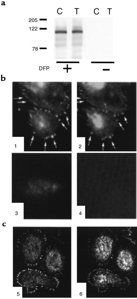
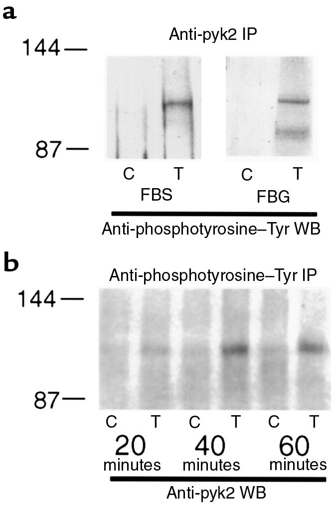
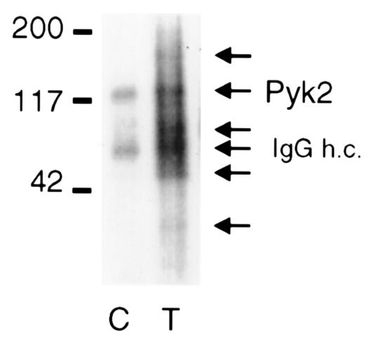
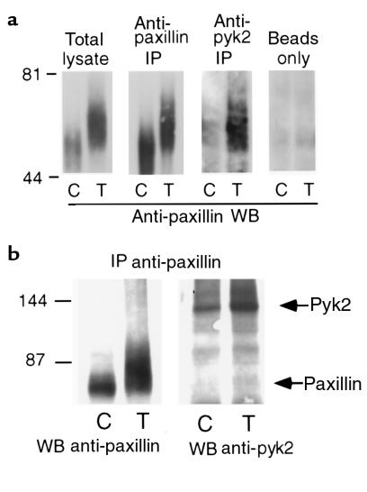
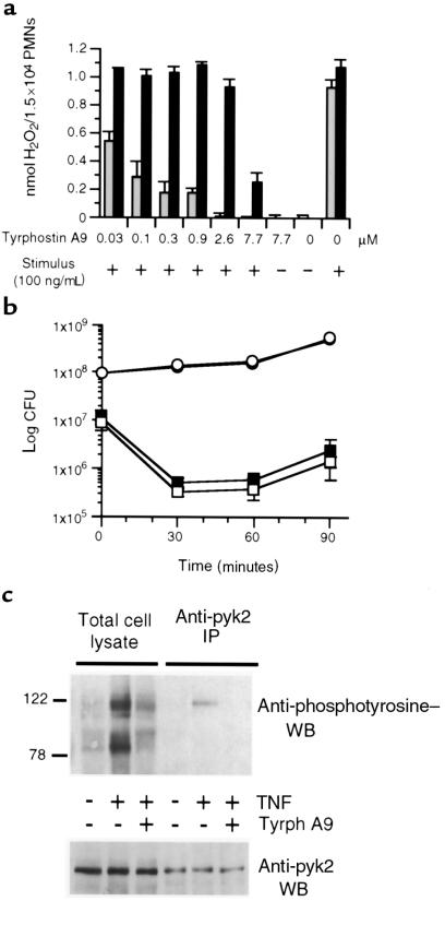
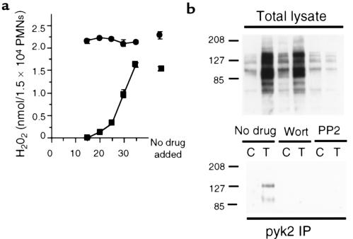
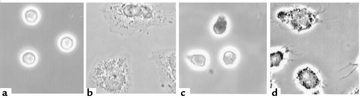
Secretion of inflammatory products from neutrophils can be induced by a combination of signals from ligated integrins and receptors for soluble, physiological agonists such as TNF. Here we identify pyk2 in primary human neutrophils; localize it to focal adhesions and podosomes; and demonstrate its tyrosine phosphorylation, activation, and association with paxillin during stimulation of adherent cells by TNF. Tyrphostin A9 emerged as the most potent and selective of 51 tyrosine kinase inhibitors tested against the TNF-induced respiratory burst. Tyrphostin A9 inhibited TNF-induced tyrosine phosphorylation of pyk2 without blocking the cells' bactericidal activity. Wortmannin, an inhibitor of phosphatidylinositol-3-kinase, potently blocked the TNF-induced respiratory burst and selectively inhibited tyrosine phosphorylation of pyk2. Thus, pyk2 appears to play an essential role in the ability of neutrophils to integrate signals from beta(2) integrins and TNF receptors.


Figures

Figure 1
Figure 1
Identification of pyk2 in human PMNs and its localization to adhesion structures. (a) Pyk2 is evident only in PMNs treated with diisopropylfluorophosphate before lysis (first 2 lanes), in addition to the other protease inhibitors used in the second 2 lanes (see text). PMNs were plated on FBS-coated plates and treated with buffer alone (control; C) or TNF (250 ng/mL; T) for 60 minutes. Identical amounts of protein (100 μg) were loaded in each lane. (b) Indirect immunofluorescence. PMNs adherent to FBS-coated glass coverslips were stimulated with TNF (100 ng/mL) for 60 minutes, fixed, permeabilized, and stained with rabbit anti-pyk2 antibody (panel 1) or mouse anti-vinculin mAb (panel 2), followed by Cy3-conjugated donkey anti-rabbit (panels 1 and 3) or Cy2-conjugated donkey anti-mouse IgG (panels 2 and 4). In panel 3, primary antibody was omitted. In panel 4, an irrelevant mAb was used. The photomicrograph is focused at the plane of cell contact with the substratum. (c) Confocal microscopy of cells prepared as in b and stained with rabbit anti-pyk2 antibody (panel 5) or mouse anti-vinculin mAb (panel 6). Sum of four 0.32-μm-thick images taken from the plane of contact with the substratum.
Figure 2
Figure 2
TNF-stimulated tyrosine phosphorylation of pyk2. (a) PMNs were plated on fetal bovine serum–coated (FBS) or fibrinogen-coated (FBG) plates and treated with buffer alone (control; C) or TNF (250 ng/mL; T) for 60 minutes (FBS) or 30 minutes (FBG). Cell lysates (200 μg) were immunoprecipitated (IP) with anti-pyk2 goat antibody. Proteins were separated by reducing SDS-PAGE, transferred to nitrocellulose, and Western blotted (WB) with horseradish peroxidase–conjugated (HRP-conjugated) anti-phosphotyrosine mAb followed by enhanced chemiluminescence (ECL) detection. (b) PMNs were plated on FBS-coated plates and treated with buffer alone (control; C) or TNF (250 ng/mL; T) for 20, 40, or 60 minutes as indicated. Cell lysates (500 μg) were immunoprecipitated (IP) with anti-phosphotyrosine mAb. Proteins were separated by reducing SDS-PAGE, transferred to nitrocellulose, and Western blotted (WB) with anti-pyk2 antibody followed by ECL detection. Molecular mass markers are indicated in kilodaltons.
Figure 3
Figure 3
Activation of pyk2 by TNF. PMNs plated on FBS-coated dishes were treated with buffer alone (control; C) or TNF (250 ng/mL; T) for 60 minutes. PMNs were lysed in modified RIPA buffer. Lysates (200 μg) were immunoprecipitated with anti-pyk2 antibody, and the immunoprecipitates were subjected to in vitro kinase assay, separated on reducing SDS-PAGE, transferred to a nylon membrane, and autoradiographed. Prominent species are marked by arrows, including IgG heavy chain (h.c.). Molecular mass markers are indicated in kilodaltons.
Figure 4
Figure 4
TNF-enhanced association of pyk2 with paxillin. (a) Coimmunoprecipitation of paxillin with pyk2. PMNs plated on FBS-coated dishes were treated with buffer alone (control; C) or TNF (250 ng/mL; T) for 60 minutes. PMNs were lysed in modified RIPA buffer. Lysates were immunoprecipitated (IP) with the indicated antibodies or with protein A-Sepharose beads alone, separated on reducing SDS-PAGE, transferred to nitrocellulose, and Western blotted (WB) with anti-paxillin mAb followed by anti-mouse HRP-conjugated antibody with ECL detection. Molecular mass markers are indicated in kilodaltons. (b) Coimmunoprecipitation of pyk2 with paxillin. PMNs plated on FBS-coated dishes were treated with buffer alone (control; C) or TNF (250 ng/mL; T) for 60 minutes and lysed in modified RIPA buffer. Lysates were immunoprecipitated with anti-paxillin mAb, separated on reducing SDS-PAGE, transferred to nitrocellulose, and Western blotted (WB) with anti-paxillin mAb or anti-pyk2 antibody followed by anti-mouse or anti-goat HRP-conjugated antibodies with ECL detection. Molecular mass markers are indicated in kilodaltons.
Figure 5
Figure 5
Tyrphostin A9 inhibits the TNF-induced respiratory burst of human PMNs and TNF-induced tyrosine phosphorylation of pyk2 but not their bactericidal activity. (a) Inhibition of TNF-stimulated H2O2 release. PMNs were plated in FBS-coated 96-well plates and preincubated for 30 minutes with the indicated concentrations of tyrphostin A9. Cells were then unstimulated (–) or stimulated (+) either with TNF (100 ng/mL; gray bars) or PMA (100 ng/mL; filled bars), and H2O2 release was recorded 60 minutes later as mean ± SEM of triplicates. (b) Lack of inhibition of bacterial killing. PMNs were preincubated (30 minutes at 4°C) with 0 (filled squares) or 5 μM (open squares) tyrphostin A9 and then infected with 10% autologous serum-opsonized Salmonella typhimurium. Cells were lysed at indicated times and CFUs were determined. Survival of bacteria without PMNs is shown with 0 (filled circles) or 5 μM (open circles) tyrphostin A9. Mean ± SEM of triplicates; most error bars fall within the symbols. (c) PMNs were plated on FBS-coated plates, preincubated for 30 minutes in the presence or absence of tyrphostin A9 (2 μM), and then stimulated with TNF (250 ng/mL) or left untreated for 60 minutes, as indicated. Cell lysates were immunoprecipitated (IP) with anti-pyk2 antibody. Total cell lysate and immunoprecipitates were separated by reducing SDS-PAGE, transferred to nitrocellulose, and Western blotted (WB) with anti-phosphotyrosine mAb (top) or anti-pyk2 antibody (bottom) followed by ECL detection. Molecular mass markers are indicated in kilodaltons.
Figure 6
Figure 6
Wortmannin inhibits the TNF-induced respiratory burst and tyrosine phosphorylation of pyk2. (a) H2O2 release from PMNs measured 120 minutes after the addition of TNF (100 ng/mL; squares) or PMA (100 ng/mL; circles). Wortmannin (10 nM) was added to separate sets of cells at the indicated number of minutes after TNF or PMA. Results are mean ± SEM for triplicates in 1 experiment of 3; some error bars fall within the symbols. (b) Tyrosine phosphorylation. PMNs were plated on FBS-coated plates and treated for 60 minutes with buffer alone or TNF (100 ng/mL) in the presence or absence of wortmannin (Wort) at 10 nM or the src kinase inhibitor PP2 (2.5 μM) as indicated. Cell lysates were immunoprecipitated (IP) with anti-pyk2 antibody. Total cell lysate (top) and anti-pyk2 immunoprecipitates (bottom) were separated by reducing SDS-PAGE, transferred to nitrocellulose, and Western blotted (WB) with anti-phosphotyrosine mAb followed by ECL detection. Molecular mass markers are indicated in kilodaltons.
Figure 7
Figure 7
Wortmannin and tyrphostin A9 inhibit TNF-induced neutrophil spreading. PMNs adherent to FBS-coated glass coverslips were left untreated (a) or stimulated with TNF (100 ng/mL) for 60 minutes (bd), either alone (b) or in the presence of wortmannin (10 nM) (c) or tyrphostin A9 (2 μM) (d), and then fixed and photographed with phase-contrast microscopy.

References

    1. Nathan CF, et al. Cytokine-induced respiratory burst of human neutrophils: dependence on extracellular matrix proteins and CD11/CD18 integrins. J Cell Biol. 1989;109:1341–1349. - PMC - PubMed
    1. Nathan CF. Neutrophil activation on biological surfaces. Massive secretion of hydrogen peroxide in response to products of macrophages and lymphocytes. J Clin Invest. 1987;80:1550–1560. - PMC - PubMed
    1. Nathan C, Sanchez E. Tumor necrosis factor and CD11/CD18 (β2) integrins act synergistically to lower cAMP in human neutrophils. J Cell Biol. 1990;111:2171–2181. - PMC - PubMed
    1. Graham IL, Anderson DC, Holers VM, Brown EJ. Complement receptor 3 (CR3, Mac-1, integrin alpha M beta 2, CD11b/CD18) is required for tyrosine phosphorylation of paxillin in adherent and nonadherent neutrophils. J Cell Biol. 1994;127:1139–1147. - PMC - PubMed
    1. Fuortes M, Jin WW, Nathan C. Adhesion-dependent protein tyrosine phosphorylation in neutrophils treated with tumor necrosis factor. J Cell Biol. 1993;120:777–784. - PMC - PubMed

Publication types

MeSH terms