. 1999 Aug 2;190(3):311-22.
doi: 10.1084/jem.190.3.311.

A role for dipeptidyl peptidase IV in suppressing the malignant phenotype of melanocytic cells

Affiliations

A role for dipeptidyl peptidase IV in suppressing the malignant phenotype of melanocytic cells

U V Wesley et al. J Exp Med. .

Abstract

Dipeptidyl peptidase IV (DPPIV) is a cell surface peptidase expressed by normal melanocytes, epithelial cells, and other cells. Malignant cells, including melanomas and carcinomas, frequently lose or alter DPPIV cell surface expression. Loss of DPPIV expression occurs during melanoma progression at a stage where transformed melanocytes become independent of exogenous growth factors for survival. Tetracycline-inducible expression vectors were constructed to express DPPIV in human melanoma cells. Reexpressing DPPIV in melanoma cells at or below levels expressed by normal melanocytes induced a profound change in phenotype that was characteristic of normal melanocytes. DPPIV expression led to a loss of tumorigenicity, anchorage-independent growth, a reversal in a block in differentiation, and an acquired dependence on exogenous growth factors for cell survival. Suppression of tumorigenicity and reversal of a block in differentiation were dependent on serine protease activity, assessed using mutant DPPIV molecules containing serine-->alanine substitutions. Surprisingly, dependence on exogenous growth factors was not dependent on serine protease activity. Reexpression of either wild-type or mutant DPPIV rescued expression of a second putative cell surface serine peptidase, fibroblast activation protein alpha, which can form a heterodimer with DPPIV. This observation suggests that rescue of fibroblast activation protein alpha may play a role in regulating growth of melanocytic cells. These results support the view that downregulation of DPPIV is an important early event in the pathogenesis of melanoma.


Figures
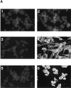
Figure 1
Figure 1
Expression of DPPIV in transfected melanoma cells. (A) Immunofluorescence microscopy showing expression of DPPIV. MEL-22a cells were cultured in the presence or absence of dox (2 μg/ml) for 48 h and stained with S27 mAb against DPPIV. Groups include untransfected MEL-22a cells (panel 1) and control vector–transfected cells (panel 2); cells transfected with wtDPPIV and grown in the absence of dox (panel 3) or induced with dox for 48 h (panel 4); cells transfected with mutDPPIV grown in the absence of dox (panel 5) or induced with dox for 48 h (panel 6). Original magnification 400. (B) Immunoprecipitation analysis of DPPIV expression in MEL-22a cells transfected with wt- or mutDPPIV. Cells were labeled with [35S]methionine for 18 h, lysed with 1% NP-40, immunoprecipitated with mAb S27 against DPPIV, analyzed by 9% SDS-PAGE, and visualized by autoradiography. The first and second lanes (from left) are untransfected MEL-22a and vector-transfected MEL-22a controls, respectively. The third and fourth lanes are two separate clones of transfected MEL-22a cells expressing either high (hi) or intermediate (med) levels of wtDPPIV when grown in the presence of dox. The fifth lane is MEL-22a cells transfected with mutDPPIV grown in the presence of dox. The sixth lane shows wtDPPIVhi-transfected MEL-22a clone grown in the absence of dox. Arrow, 110–120 kD band of DPPIV. −, not induced or +, induced with dox. (C) DPPIV activity in MEL-22a clones transfected with wtDPPIV, mutDPPIV, or control vector and in cultured normal foreskin melanocytes. Three different clones of MEL-22a cells transfected with wtDPPIV were analyzed expressing low (wtDPPIVlow), intermediate (wtDPPIVmed), and high (wtDPPIVhi) levels of enzyme activity. Open bars (−dox), enzyme activities in absence of dox; hatched bars (+dox), enzyme activity after induction with dox. Results shown are mean values ± 1 SD of triplicates.
Figure 1
Figure 1
Expression of DPPIV in transfected melanoma cells. (A) Immunofluorescence microscopy showing expression of DPPIV. MEL-22a cells were cultured in the presence or absence of dox (2 μg/ml) for 48 h and stained with S27 mAb against DPPIV. Groups include untransfected MEL-22a cells (panel 1) and control vector–transfected cells (panel 2); cells transfected with wtDPPIV and grown in the absence of dox (panel 3) or induced with dox for 48 h (panel 4); cells transfected with mutDPPIV grown in the absence of dox (panel 5) or induced with dox for 48 h (panel 6). Original magnification 400. (B) Immunoprecipitation analysis of DPPIV expression in MEL-22a cells transfected with wt- or mutDPPIV. Cells were labeled with [35S]methionine for 18 h, lysed with 1% NP-40, immunoprecipitated with mAb S27 against DPPIV, analyzed by 9% SDS-PAGE, and visualized by autoradiography. The first and second lanes (from left) are untransfected MEL-22a and vector-transfected MEL-22a controls, respectively. The third and fourth lanes are two separate clones of transfected MEL-22a cells expressing either high (hi) or intermediate (med) levels of wtDPPIV when grown in the presence of dox. The fifth lane is MEL-22a cells transfected with mutDPPIV grown in the presence of dox. The sixth lane shows wtDPPIVhi-transfected MEL-22a clone grown in the absence of dox. Arrow, 110–120 kD band of DPPIV. −, not induced or +, induced with dox. (C) DPPIV activity in MEL-22a clones transfected with wtDPPIV, mutDPPIV, or control vector and in cultured normal foreskin melanocytes. Three different clones of MEL-22a cells transfected with wtDPPIV were analyzed expressing low (wtDPPIVlow), intermediate (wtDPPIVmed), and high (wtDPPIVhi) levels of enzyme activity. Open bars (−dox), enzyme activities in absence of dox; hatched bars (+dox), enzyme activity after induction with dox. Results shown are mean values ± 1 SD of triplicates.
Figure 2
Figure 2
Effects of DPPIV expression on tumorigenicity. (A) Expression of wtDPPIV was associated with inhibition of tumor growth of melanoma cells MEL-22a in nude mice. Six different sets of nude mice (BALB/C nu/nu, n = 5–6 for each group) were challenged subcutaneously on day 0 with 3 × 10⁶ cells, either parental MEL-22a or transfected with wtDPPIV (with medium or high levels of expression induced by dox as presented in Fig. 1), mutDPPIV, or control vector. Cells were induced (+dox) or not induced (−dox) with dox for 7 d before tumor challenge. Untransfected (MEL-22a) or vector-transfected cells were used as controls. Tumor diameters were measured every 2–3 d. Results are presented as mean tumor diameter ± 1 SD. (B) A repeat of the experiment shown in A with 5–6 mice per group challenged with MEL-22a melanoma cells. The procedure was the same as in A. (C) Tumorigenicity of SK-MEL-29 melanoma cells, including parental SK-MEL-29 cells and cells transfected with wtDPPIV, mutDPPIV, or control vector. 5 × 10⁶ tumor cells were injected subcutaneously on day 0.
Figure 2
Figure 2
Effects of DPPIV expression on tumorigenicity. (A) Expression of wtDPPIV was associated with inhibition of tumor growth of melanoma cells MEL-22a in nude mice. Six different sets of nude mice (BALB/C nu/nu, n = 5–6 for each group) were challenged subcutaneously on day 0 with 3 × 10⁶ cells, either parental MEL-22a or transfected with wtDPPIV (with medium or high levels of expression induced by dox as presented in Fig. 1), mutDPPIV, or control vector. Cells were induced (+dox) or not induced (−dox) with dox for 7 d before tumor challenge. Untransfected (MEL-22a) or vector-transfected cells were used as controls. Tumor diameters were measured every 2–3 d. Results are presented as mean tumor diameter ± 1 SD. (B) A repeat of the experiment shown in A with 5–6 mice per group challenged with MEL-22a melanoma cells. The procedure was the same as in A. (C) Tumorigenicity of SK-MEL-29 melanoma cells, including parental SK-MEL-29 cells and cells transfected with wtDPPIV, mutDPPIV, or control vector. 5 × 10⁶ tumor cells were injected subcutaneously on day 0.
Figure 4
Figure 4
Phenotypic changes associated with DPPIV expression. (A) Morphology of MEL-22a clones. Untransfected (panel 1) and control vector–transfected cells (panel 2) showed short spindle-shaped and polygonal morphology and grew in unorganized clusters. MutDPPIV-transfected cells (panel 3) were morphologically similar to control cells. WtDPPIV-transfected cells (panel 4) showed long spindle bipolar morphology with more organized growth and sheet-like appearance. Original magnification 200. (B) Pigmentation of MEL-22a clones in cell pellets. Untransfected and control vector–transfected MEL-22a cells were not melanotic (first and second pellets from left). Minimal pigmentation was observed in mutDPPIV-transfected MEL-22a cells (third pellet). Expression of wtDPPIV led to brown pigmentation (fourth pellet). Dark brown pigmentation of normal foreskin melanocytes is shown in the fifth pellet. (C) Expression of human tyrosinase detected by Western blot analysis. Lysates (1% NP-40) of human melanoma cell line G-MEL or MEL-22a cells, either parental or transfected with wtDPPIV (med and hi), mutDPPIV, or control vector were separated on a 9% SDS-PAGE. After transfer to membrane, tyrosinase was detected as a broad, ∼75-kD band using rabbit anti-PEP7H antibody against human tyrosinase. Molecular mass markers are shown, left.
Figure 4
Figure 4
Phenotypic changes associated with DPPIV expression. (A) Morphology of MEL-22a clones. Untransfected (panel 1) and control vector–transfected cells (panel 2) showed short spindle-shaped and polygonal morphology and grew in unorganized clusters. MutDPPIV-transfected cells (panel 3) were morphologically similar to control cells. WtDPPIV-transfected cells (panel 4) showed long spindle bipolar morphology with more organized growth and sheet-like appearance. Original magnification 200. (B) Pigmentation of MEL-22a clones in cell pellets. Untransfected and control vector–transfected MEL-22a cells were not melanotic (first and second pellets from left). Minimal pigmentation was observed in mutDPPIV-transfected MEL-22a cells (third pellet). Expression of wtDPPIV led to brown pigmentation (fourth pellet). Dark brown pigmentation of normal foreskin melanocytes is shown in the fifth pellet. (C) Expression of human tyrosinase detected by Western blot analysis. Lysates (1% NP-40) of human melanoma cell line G-MEL or MEL-22a cells, either parental or transfected with wtDPPIV (med and hi), mutDPPIV, or control vector were separated on a 9% SDS-PAGE. After transfer to membrane, tyrosinase was detected as a broad, ∼75-kD band using rabbit anti-PEP7H antibody against human tyrosinase. Molecular mass markers are shown, left.
Figure 3
Figure 3
Anchorage-independent growth in soft agar. MEL-22a cells described in Fig. 2 A were cultured in the absence (−) or presence (+) of dox for 48 h, and 5,000 viable cells were plated in agar in triplicate as described in Materials and Methods. Results are mean number of colonies ± 1 SD at 14 d. Results are pooled from two separate experiments.
Figure 5
Figure 5
Cell cycle analysis of melanoma cells expressing DPPIV and FAPα. Expression of both wt- and mutDPPIV in MEL-22a cells was associated with changes in cell cycle. Parental MEL-22a cells and cells transfected with control vector, mutDPPIV, and wtDPPIV were assessed for cell cycle progression after 8 d of culture in serum-free media. Number of cells at each stage of the cell cycle were measured by flow cytometry (first peak was G0/G1, the intervening trough was S phase, and the second peak represented cells in G2/M) and analyzed by CellFIT and PC-LYSIS™ software.
Figure 6
Figure 6
Expression of DPPIV and FAPα. Expression of either wt- or mutDPPIV in MEL-22a cells rescued the expression of FAPα. Immunofluorescence staining and flow cytometry analysis of DPPIV-transfected cells was performed using mAbs against DPPIV and FAPα. Cell clones are described in the legends of Fig. 1 and Fig. 2. Y-axis, relative cell number; x-axis, log fluorescence intensity. Shaded curve, control IgG1 antibody; solid line, DPPIV expression; dotted line, FAPα expression.

Comment in

References

    1. Schrader W.P., Stacy A.R. Purification and subunit structure of adenosine deaminase from human kidney. J. Biol. Chem. 1977;252:6409–6415. - PubMed
    1. Houghton A.N., Albino A., Cordon-Cardo C., Davis L.J., Eisinger M. Cell surface antigens of human melanocytes and melanoma. Expression of adenosine deaminase binding protein is extinguished with melanocyte transformation. J. Exp. Med. 1988;167:197–212. - PMC - PubMed
    1. Morimoto C., Schlossman S. The structure and function of CD26 in the T-cell immune response. Immunol. Rev. 1998;61:55–70. - PubMed
    1. Schrader W.P., West C.A., Miczek A.D., Norton E.K. Characterization of the adenosine deaminase-adenosine deaminase complexing protein binding reaction. J. Biol. Chem. 1990;265:19312–19318. - PubMed
    1. Schrader W.P., Pollara B. Localization of an adenosine deaminase-binding protein in human kidney. J. Lab. Clin. Med. 1978;92:656–662. - PubMed

Publication types

MeSH terms