. 1999 Aug 2;190(3):355-66.
doi: 10.1084/jem.190.3.355.

Combination immunotherapy of B16 melanoma using anti-cytotoxic T lymphocyte-associated antigen 4 (CTLA-4) and granulocyte/macrophage colony-stimulating factor (GM-CSF)-producing vaccines induces rejection of subcutaneous and metastatic tumors accompanied by autoimmune depigmentation

Affiliations

Combination immunotherapy of B16 melanoma using anti-cytotoxic T lymphocyte-associated antigen 4 (CTLA-4) and granulocyte/macrophage colony-stimulating factor (GM-CSF)-producing vaccines induces rejection of subcutaneous and metastatic tumors accompanied by autoimmune depigmentation

A van Elsas et al. J Exp Med. .

Abstract

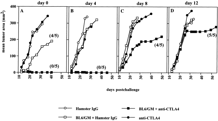
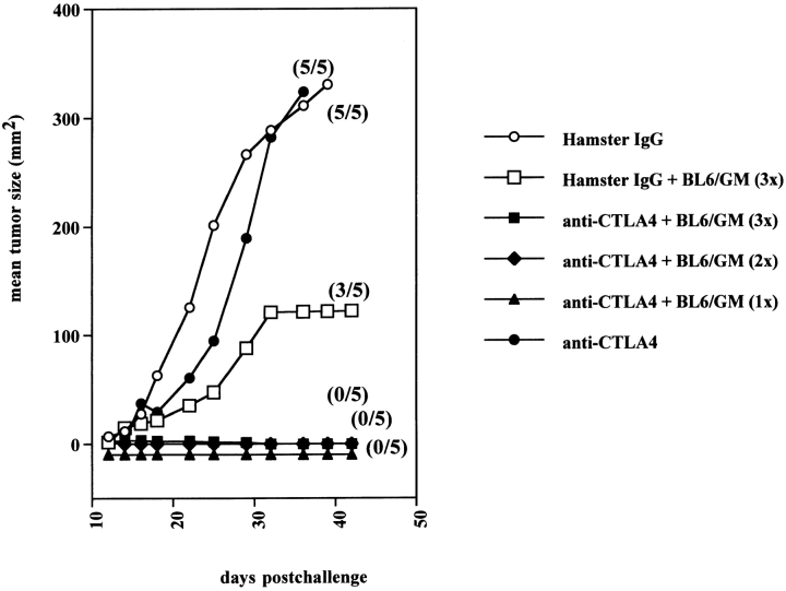
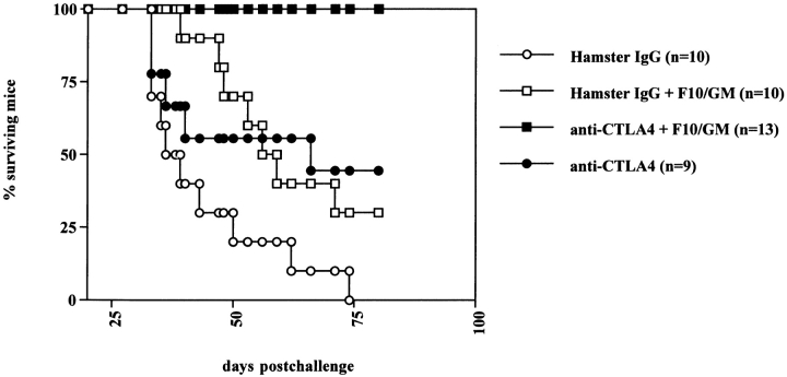
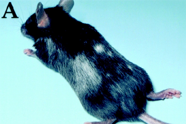
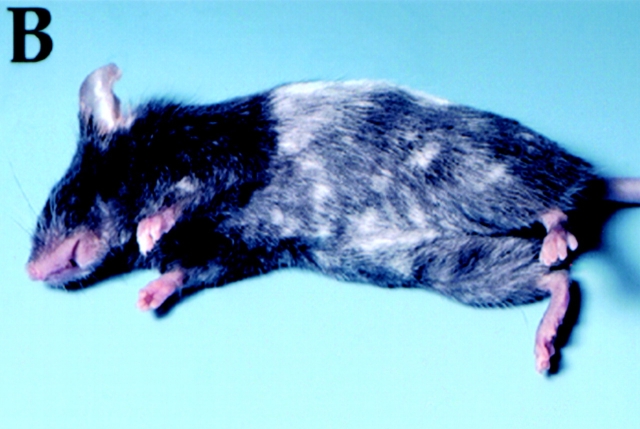
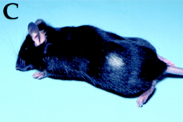
We examined the effectiveness of cytotoxic T lymphocyte-associated antigen 4 (CTLA-4) blockade, alone or in combination with a granulocyte/macrophage colony-stimulating factor (GM-CSF)-expressing tumor cell vaccine, on rejection of the highly tumorigenic, poorly immunogenic murine melanoma B16-BL6. Recently established tumors could be eradicated in 80% (68/85) of the cases using combination treatment, whereas each treatment by itself showed little or no effect. Tumor rejection was dependent on CD8(+) and NK1.1(+) cells but occurred irrespective of the presence of CD4(+) T cells. Mice surviving a primary challenge rejected a secondary challenge with B16-BL6 or the parental B16-F0 line. The same treatment regimen was found to be therapeutically effective against outgrowth of preestablished B16-F10 lung metastases, inducing long-term survival. Of all mice surviving B16-BL6 or B16-F10 tumors after combination treatment, 56% (38/68) developed depigmentation, starting at the site of vaccination or challenge and in most cases progressing to distant locations. Depigmentation was found to occur in CD4-depleted mice, strongly suggesting that the effect was mediated by CTLs. This study shows that CTLA-4 blockade provides a powerful tool to enhance T cell activation and memory against a poorly immunogenic spontaneous murine tumor and that this may involve recruitment of autoreactive T cells.


Figures

Figure 1
Figure 1
Successful treatment of preestablished B16-BL6 using anti–CTLA-4 and GM-CSF–producing BL6 vaccine. C57BL/6 female mice (five per group) were injected with 104 B16-BL6 cells subcutaneously on the back, on the same day (A) or 4, 8, or 12 d (B–D) before treatment was started. Treatment consisted of three consecutive injections (in a 6-d time frame as indicated in Materials and Methods) of anti–CTLA-4 antibody 9H10 intraperitoneally (•), control hamster IgG (100, 50, 50 μg; ○), or 106 irradiated BL6/g cells subcutaneously, in combination with 9H10 (▪) or hamster IgG (□). Tumor growth (mm2) was scored by measuring perpendicular diameters and was averaged for all mice within each group. In some treatment groups, only a fraction of the mice (indicated between brackets) developed a tumor.
Figure 2
Figure 2
A single dose of GM-CSF–producing vaccine cooperates with CTLA-4 blockade to induce 100% cure of B16-BL6. Mice were inoculated subcutaneously with 104 B16-BL6 cells. On the same day, combination treatment was initiated using triple BL6/g vaccine (days 0, 3, and 6) combined with either hamster IgG (100, 50, 50 μg on days 3, 6, and 9; □) or anti–CTLA-4 (▪). Control treatments consisted of antibody injections alone: hamster IgG (○) or anti–CTLA-4 (•). Also, anti–CTLA-4 treatment was combined with a single (▴) or double injection (♦) of the BL6/g vaccine. Average tumor size was calculated for all mice within a treatment group (mm2). The fraction of mice developing tumors is shown between brackets.
Figure 3
Figure 3
Anti–CTLA-4 enhances IFN-γ production by B16-specific T cells induced in vivo. Mice (four per group) were vaccinated with irradiated BL6/g (106 per mouse) and cotreated with control hamster IgG (A) or anti–CTLA-4 (B). After 4 wk, mice were challenged with 2 × 10⁴ B16-BL6, and 10 d later, splenocytes were pooled and restimulated in vitro using B16-BL6/B7.1 (open bars) or a mixture of B16-F10 and DC2.4 dendritic cells (filled bars). On day 8, cultures were tested for tumor-specific IFN-γ release as described in Materials and Methods. Targets included B16 sublines -F0, -F10, and -BL6, as well as unrelated H-2b tumors EL4 and MC38.
Figure 3
Figure 3
Anti–CTLA-4 enhances IFN-γ production by B16-specific T cells induced in vivo. Mice (four per group) were vaccinated with irradiated BL6/g (106 per mouse) and cotreated with control hamster IgG (A) or anti–CTLA-4 (B). After 4 wk, mice were challenged with 2 × 10⁴ B16-BL6, and 10 d later, splenocytes were pooled and restimulated in vitro using B16-BL6/B7.1 (open bars) or a mixture of B16-F10 and DC2.4 dendritic cells (filled bars). On day 8, cultures were tested for tumor-specific IFN-γ release as described in Materials and Methods. Targets included B16 sublines -F0, -F10, and -BL6, as well as unrelated H-2b tumors EL4 and MC38.
Figure 4
Figure 4
Mice bearing B16-F10 lung metastases show enhanced survival when treated with anti–CTLA-4 and F10/g vaccine. B16-F10 cells (5 × 10⁴ per mouse) were injected into the tail vein and 24 h later, treatment was started using control hamster IgG (10 mice, ○), anti–CTLA-4 antibody 9H10 (9 mice; •), irradiated F10/g (106 subcutaneously) in combination with hamster IgG (10 mice; □) or 9H10 (13 mice; ▪) on days 1, 4, and 7, according to the dosing schedule used for subcutaneous tumors (see Fig. 1 legend). Mice were followed for survival, and in some subjects death due to extensive pulmonary metastasis was confirmed by harvesting lungs postmortem.
Figure 5
Figure 5
B16-F10 metastases demonstrate lymphocytic infiltration after treatment with anti–CTLA-4 and F10/g vaccine. Mice injected with 105 B16-F10 intravenously and treated with control hamster IgG (A), 9H10 (B), or F10/g vaccine in combination with either hamster IgG (C) or 9H10 (D) on days 1, 4, and 7, as outlined in the Fig. 4 legend. On day 25, lungs were harvested, fixed in 10% neutral-buffered formalin, and processed for hematoxylin–eosin staining.
Figure 6
Figure 6
Rejection of B16-BL6 or B16-F10 as a result of treatment with anti–CTLA-4 and GM-CSF–producing vaccines causes autoimmune skin and hair depigmentation. After successful treatment for B16-BL6 subcutaneously or B16-F10 intravenously, C57Bl/6 mice developed skin and hair depigmentation. (A) Depigmentation of both sites of vaccination and challenge, after rejection of a day 0 tumor. (B) Progressive depigmentation found in a mouse rejecting a B16-BL6 subcutaneous tumor, established 8 d before treatment started. (C) Depigmentation at the site of vaccination of a mouse cured from preestablished B16-F10 lung metastases.
Figure 6
Figure 6
Rejection of B16-BL6 or B16-F10 as a result of treatment with anti–CTLA-4 and GM-CSF–producing vaccines causes autoimmune skin and hair depigmentation. After successful treatment for B16-BL6 subcutaneously or B16-F10 intravenously, C57Bl/6 mice developed skin and hair depigmentation. (A) Depigmentation of both sites of vaccination and challenge, after rejection of a day 0 tumor. (B) Progressive depigmentation found in a mouse rejecting a B16-BL6 subcutaneous tumor, established 8 d before treatment started. (C) Depigmentation at the site of vaccination of a mouse cured from preestablished B16-F10 lung metastases.

References

    1. Boon T., Cerottini J.C., Van den Eynde B., van der Bruggen P., Van Pel A. Tumor antigens recognized by T lymphocytes. Annu. Rev. Immunol. 1994;12:337–365. - PubMed
    1. Rosenberg S.A. Cancer vaccines based on the identification of genes encoding cancer regression antigens. Immunol. Today. 1997;18:175–182. - PubMed
    1. Liu G.Y., Fairchild P.J., Smith R.M., Prowle J.R., Kioussis D., Wraith D.C. Low avidity recognition of self-antigen by T cells permits escape from central tolerance. Immunity. 1995;3:407–415. - PubMed
    1. Poplonski L.B., Vukusic B., Pawling J., Clapoff S., Roder J., Hozumi N., Whither J. Tolerance is overcome in beef insulin-transgenic mice by activation of low-affinity autoreactive T cells. Eur. J. Immunol. 1996;26:601–606. - PubMed
    1. Morgan D.J., Kreuwel H.T.C., Fleck S., Levitsky H.I., Pardoll D.M., Sherman L.A. Activation of low avidity CTL for a self-epitope results in tumor rejection but not autoimmunity. J. Immunol. 1998;160:643–651. - PubMed

Publication types

MeSH terms